
Lophogaster typicus, M. Sars, 1857
|
publication ID |
https://doi.org/ 10.1093/zoolinnean/zlac083 |
|
DOI |
https://doi.org/10.5281/zenodo.7814234 |
|
persistent identifier |
https://treatment.plazi.org/id/D96287D1-4B73-321E-D452-12B1FBB71037 |
|
treatment provided by |
Plazi |
|
scientific name |
Lophogaster typicus |
| status |
|
LOPHOGASTER TYPICUS View in CoL View at ENA
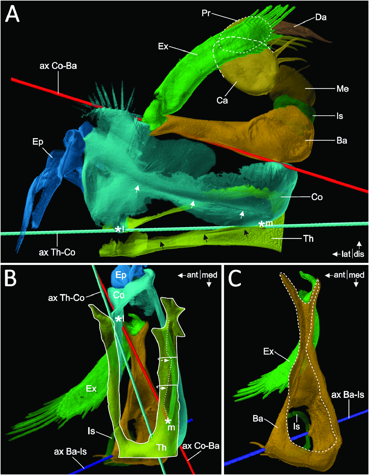
The first pair of thoracopods (Thp1) is highly modified compared with the posterior pairs (Thp2–8) of pediform thoracopods. Owing to this strong morphological difference, the following descriptions relate only to Thp1, based on a detailed examination of the right Thp1 of three adult males of L. typicus .
Cuticle and skeletal structures of Thp 1 in L. typicus
The more or less rectangular protopod (more than twice as wide as long) of L. typicus consists of the coxa (twothirds) and basis (one-third of the total length of the protopod). The thorax connects and articulates with the coxa by a cuticular half-ring, leaving the lateral area membranous (Th in Fig. 10A, B View Figure 10 ). Posteriorly, the cuticle of the thorax is invaginated as a lateromedial fold, forming a short roof-like apodeme (black arrows in Fig. 10A View Figure 10 ; partly dashed curved arrows and dashed line in Fig. 10B View Figure 10 ).
The coxa does not form a complete ring but has the medial to anteromedial area membranous (Co in Fig. 10A View Figure 10 ). A bicondylar articulation with the thorax is formed by an anterolateral and posteromedial articulation point, respectively (*l, *m and ax Th-Co in Fig. 10A View Figure 10 ). Posteriorly, the coxa forms an invagination, which appears as a linear lateromedial furrow (white arrows in Fig. 10A View Figure 10 ) that ends posterolaterally in a roundish depression. Distally, the podomere articulates anterolaterally and posteromedially with the basis (*l, *m and ax Co-Ba in Fig. 10B View Figure 10 ).
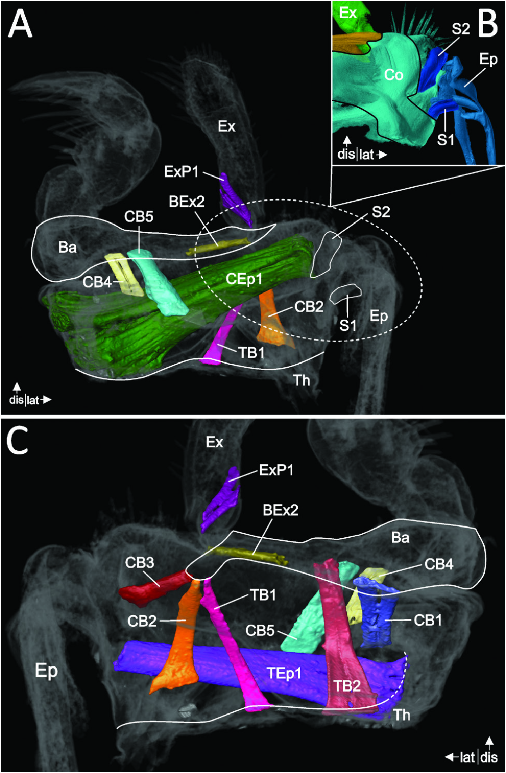
Laterally, a huge lamellar epipod (Ep in Fig. 10A View Figure 10 ) is attached to the coxa. It is broadly flattened anteroposteriorly and long, reaching at least double the length of the exopod (see below, this section). The epipod articulates with the coxa via two sclerites: anteriorly, a rod-shaped sclerite (S 1 in Fig. 11A, B View Figure 11 ) forms a dorsal articulation point, while a second ventral sclerite (S 2 in Fig. 11A, B View Figure 11 ) interlocks the epipod anteriorly on the coxa. The area between both sclerites is membranous. More distally on the epipod, several sclerites form the ‘frame’ of its lamellar distal portion (Ep in Figs 10A View Figure 10 , 11B View Figure 11 ).
The basis, from a dorsal perspective, appears roughly drop-shaped, with the medial side being the spherical part, narrowing towards the lateral side (Ba in Fig. 10C View Figure 10 ). Here, the proximal margin of the basis widens again into a V-shape, ending in two fingers, of which the posterior one serves as attachment site for three muscles (TB1, CB2 and CB3; Fig. 11C View Figure 11 ). The lateral wall appears membranous ( Fig. 10B, C View Figure 10 ). The distal opening of the basis into the endopod is found in the medial third of the podomere ( Fig. 10C View Figure 10 ). Here, a bicondylar articulation with the endopod (i.e. the ischium) is formed by an anteromedial and a posterior articulation point, respectively (ax Ba-Is in Fig. 10C View Figure 10 ).
A lamellar exopod attaches at the lateral membrane of the basis via a short proximal stem (Ex in Fig. 10A View Figure 10 ) that articulates with the anteromedial finger-like extension of the basis via a hook-like structure ( Fig. 10C View Figure 10 ). The stem is followed by a much longer distal portion (almost as long as the endopod), which is shaped as an anteroposteriorly flattened blade with a setose distal tip.
Table 3. Continued
The crescent-shaped endopod consists of the ischium, merus, carpus, propodus and dactylus. In relationship to the following podomeres, the ischium appears short (Is in Fig. 10A View Figure 10 ). An articulation with the merus is created by an anteromedial articulation point. Although the podomeres are in close contact posterolaterally, no clear articulation point can be made out. Nonetheless, this closeness might be sufficient to achieve the functionality of a bicondylar joint.
The merus (Me in Fig. 10A View Figure 10 ) measures about twice the length of the ischium. Its distal opening is not oriented distally but is displaced ~90° in the lateral direction. Anteromedially, the podomere is covered with strong, cuspidate setae. A bicondylar articulation with the carpus is formed by an anteromedial and posterolateral articulation point, respectively.
The carpus (Ca in Fig. 10A View Figure 10 ) is about twice the length of the merus. Four long cuspidate setae are attached medially. Its proximal margin (connected to the merus) faces medially, whereas its distal margin faces in the distal direction, articulating with the propodus by an anterior and posterolateral articulation point, respectively.
The propodus (Pr, dotted outline in Fig. 10A View Figure 10 ) is shorter than the carpus and roughly as long as the merus. Three long, cuspidate setae are attached medially. The distal margin of the propodus is slightly oriented in the medial direction, where the podomere is connected with the dactylus by an anterolateral and a posterior articulation point, respectively.
The dactylus (Da in Fig. 10A View Figure 10 ) is almost equal in length to the propodus. The podomere tapers towards its distal tip, where a strong and cuspidate seta is present, while four smaller cuspidate setae cover the dactylus medially.
No known copyright restrictions apply. See Agosti, D., Egloff, W., 2009. Taxonomic information exchange and copyright: the Plazi approach. BMC Research Notes 2009, 2:53 for further explanation.