
Aegla okora Páez & Teixeira, 2018
|
publication ID |
https://doi.org/10.11646/zootaxa.4527.3.3 |
|
publication LSID |
lsid:zoobank.org:pub:8EFC6F91-6268-4572-8B69-FA2C0541E528 |
|
DOI |
https://doi.org/10.5281/zenodo.5952110 |
|
persistent identifier |
https://treatment.plazi.org/id/DA188786-FFA8-C53B-13A9-FC21FEC8F9A6 |
|
treatment provided by |
Plazi |
|
scientific name |
Aegla okora Páez & Teixeira |
| status |
sp. nov. |
Aegla okora Páez & Teixeira View in CoL n. sp.
( Figs 2 View FIGURE 2 , 3 View FIGURE 3 , 4 View FIGURE 4 , 5 View FIGURE 5 , 6 View FIGURE 6 ,)
Type-material. Holotype: Male ( CLE 21.3 mm), Brazil, Paraná, Pinhão, Iguaçu River basin, Tapera River , 25°41’39.51”S, 51°40’13.23”W, G.M. Teixeira, F.P. Paez and R.A. Gregati coll., February 2018 ( MZUEL 250 ) GoogleMaps . Paratypes: 10 males ( CLE 10.4 ¯23.0 mm) and 10 females ( CLE 13.0¯18.0 mm), same data as holotype ( MZUEL 251 , genetic voucher: Bold Systems access AEGBR001-18 , AEGBR002-18 , AEGBR003-18 , AEGBR004-18 ) GoogleMaps .
Type-locality. Tapera River, city of Pinhão, Paraná, Brazil.
Geographical distribution. Known only from the type-locality, despite searching in five nearby streams within a radius of the 32 km.
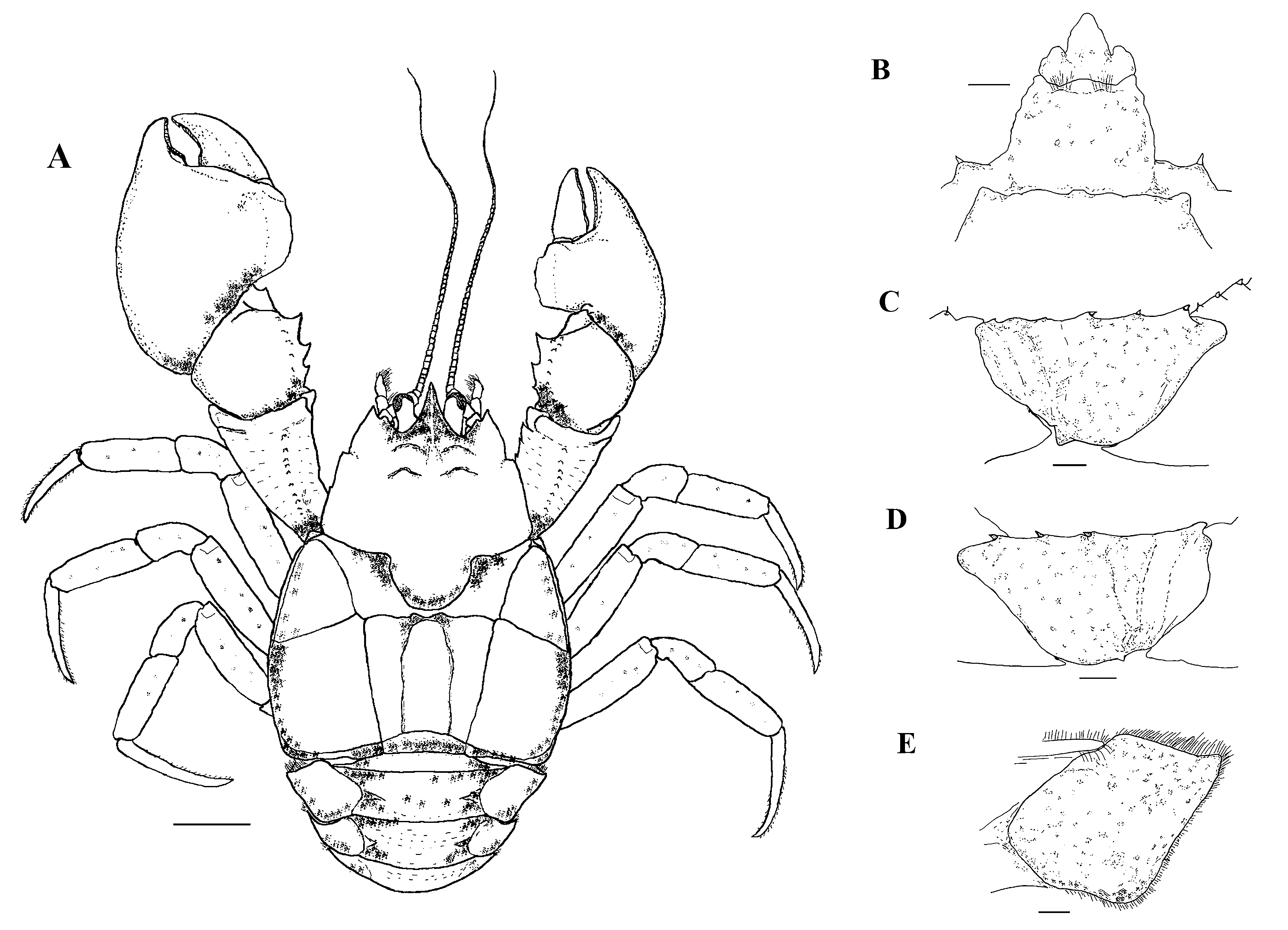
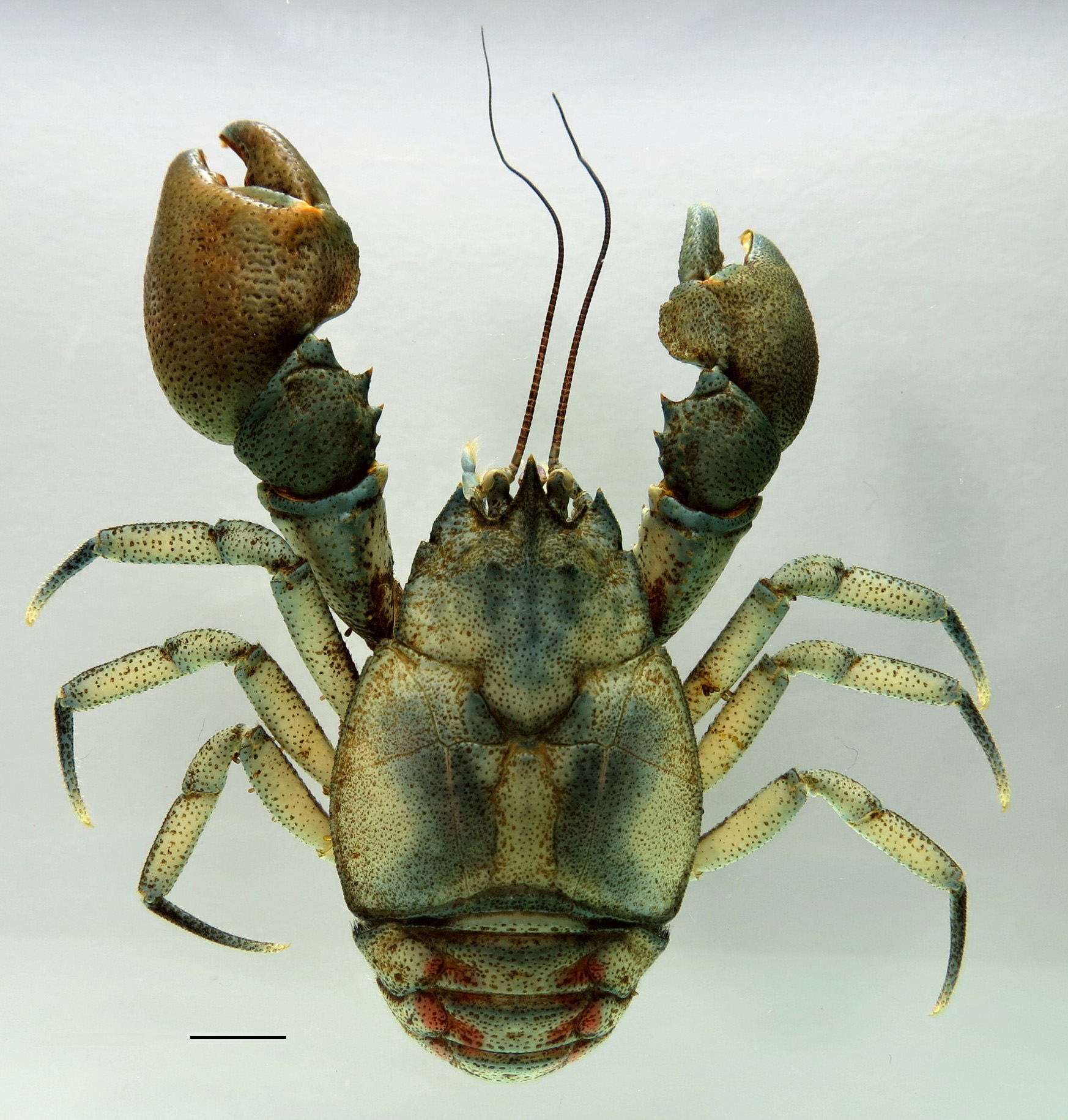
Diagnosis. Triangular rostrum with narrow base. Subrostral process developed, anteriorly oriented at a 45° with the rostrum. Epigastric prominences and protogastric lobes pronounced, with scales and small setae. Anterolateral spine reaching basal margin of cornea. Branchial region swollen. Areola trapezoidal. Cardiac area trapezoidal. Proximal dorsal margin of movable finger of cheliped without lobe. Palmar crest of major cheliped rectangular. Anterolateral angle of second abdominal epimeron unarmed, with setae. Ventromesial border of ischium of the cheliped ornate with three tubercles, one proximal, one median and one distal. Uropodal (endopods) wide.
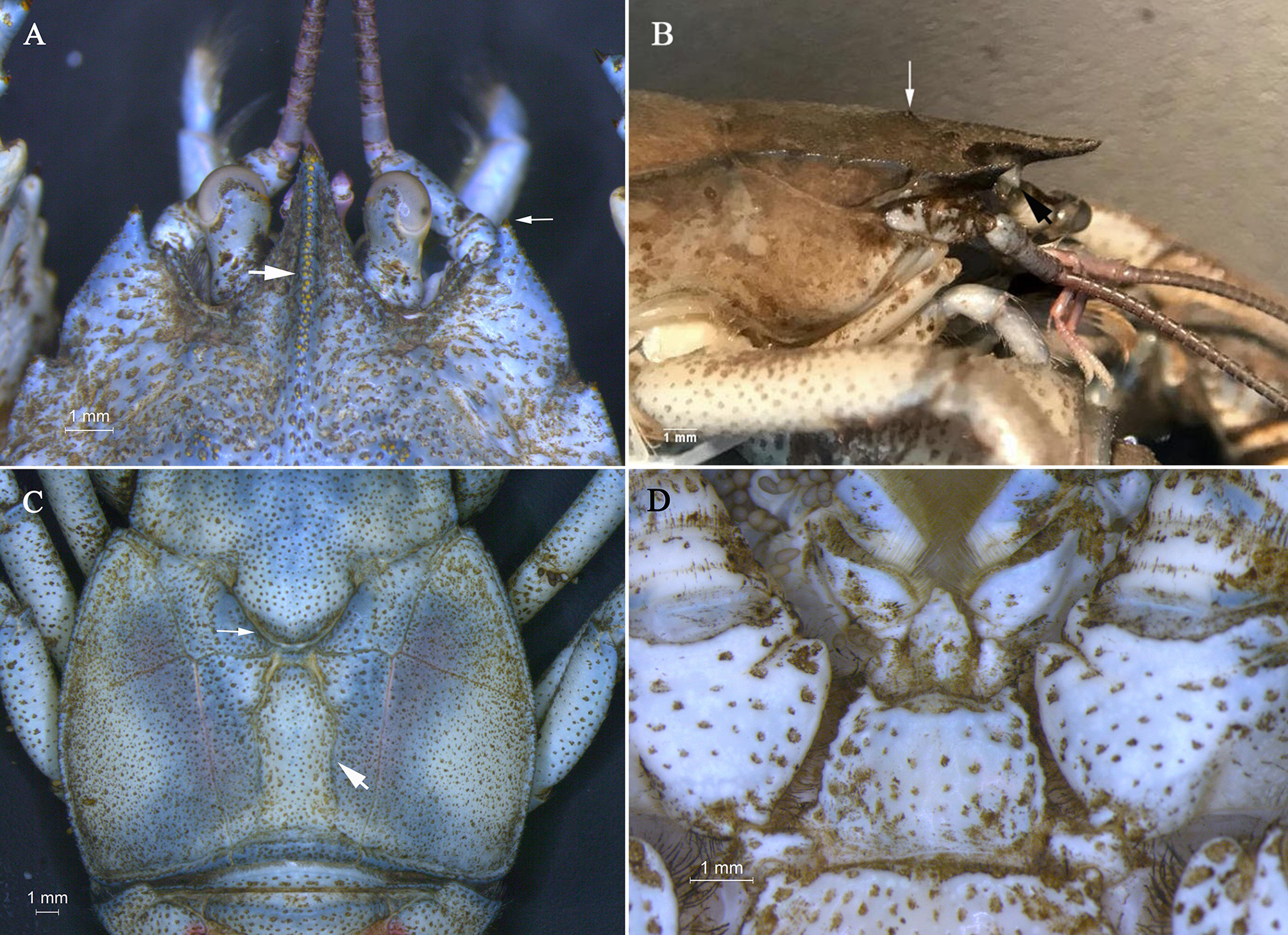
Description of male holotype. Carapace convex, branchial region swollen. Rostrum triangular, base narrow (RBW/LMR = 0.87), extending beyond distal apex of compound eyes, with scales on lateral margins. Rostral carina beginning at level of protogastric lobes, with two parallel rows of scales becoming one row on distal third near apex ( Fig. 2A View FIGURE 2 , 3 View FIGURE 3 , 4A View FIGURE 4 ). Subrostral process developed forming angle of 45° ( Fig. 4B View FIGURE 4 ).
Eyestalk and cornea well developed. Orbital and extra-orbital sinuses deep. Orbital sinus with scales. Orbital spines developed, rounded. Anterolateral spines with corneous scales on lateral margin of carapace, reaching basal margin of cornea ( Fig. 4A View FIGURE 4 ).
Epigastric prominences pronounced, with scales and short setae. Protogastric lobes pronounced, with scales. Gastric area prominently inflated in relation to hepatic lobe and rostrum in lateral view. Demarcation between hepatic lobes well defined. Lateral margins of first hepatic lobe with corneous scale, second and third hepatic lobes with sparse scales.
Cervical groove U-shaped ( Fig. 4C View FIGURE 4 ). Transverse dorsal linea slightly sinuous throughout its extension, sinuosity more pronounced in mesial section. Areola trapezoidal (APM/AAD = 2.5). Cardiac area trapezoidal (TDL/PMC = 1.53) ( Fig. 4C View FIGURE 4 ).
Epibranchial area with spine or well developed scales on apex. Lateral margin of anterior branchial area with distal spine, setae and scales, posterior area with setae and scales.
Anteromesial region of third thoracic sternite abrupt, with scattered setae on surface. Fourth thoracic sternite elevated in median region with setae, anterolateral angles well developed with spines ( Fig. 2B View FIGURE 2 , 4D View FIGURE 4 ).
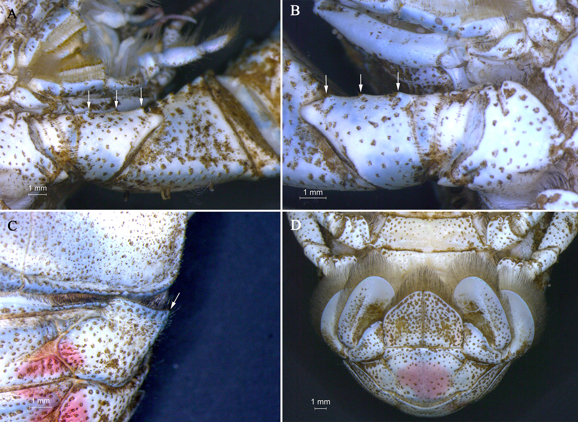
Chelipeds unequal in size ( Figs 2A View FIGURE 2 , 3 View FIGURE 3 ). Major cheliped (left) ( Fig. 5A View FIGURE 5 ). Dactylus: dorsal margin and outer surface granulate and ornamented with short scales. Pre-dactylar lobe absent. Proximal lobe on dorsal margin absent. Cutting margin with lobular basal tooth well developed proximally, with flattened corneous scales, followed by row of wide corneous scales up to distal end. Propodus: outer surface granulate. Palmar crest rectangular with outer surface excavated, margin with scales. Cutting margin of fixed finger with flattened corneous scales over its entire surface, with lobular basal tooth well developed proximally and acuminate corneous scale on distal end. Scattered tufts of long setae over inner surface, and alongside inner and outer surfaces next to cutting margin. Scattered scales and scales clustered into groups of 2 or 3 on inner surface. Carpus: dorsal margin with two tubercles proximally, two median spines with terminal corneous scale, one tubercle, internally displaced from the margin, distally, with terminal corneous scale, and sub-terminal lobe well defined, pointed, with corneous scales and setae apically. Inner surface with three tubercles with terminal corneous scale and setae. Outer surface with carpal ridge elevated along entire length, with scales clustered into groups of 3 – 5. Merus: dorsal margin with one tubercle. Dorsolateral edge with row of corneous scales and tubercles with corneous scales on distal third. Ventromesial edge with five tubercles decreasing in size proximally. Ventrolateral border with two tubercles distally, followed by row of scales clustered into groups of 2 – 3. Ischium: dorsolateral edge with distal spine with terminal corneous scale. Ventromesial border ornamented with one proximal tubercle, one median tubercle and one distal tubercle with one terminal corneous scale each ( Fig. 2C View FIGURE 2 , 6A View FIGURE 6 ).
Minor chelipeds (right) similar to major chelipeds except as noted hereafter ( Fig. 2D View FIGURE 2 , 5B View FIGURE 5 , 6B View FIGURE 6 ): Propodus: palmar crest rectangular to subdisciform. Merus: ventromesial edge with four tubercles decreasing in size proximally.
Second, third and fourth pereiopods morphologically similar; general surface of dactylus, propodus and carpus with longitudinal lines of short setae and scales; dorsal margin of merus and ischium with long tufts setae; ventral margin of ischium with tufts of setae.
Pleopods 2–5 absent.
Anterolateral angle of second abdominal epimeron unarmed, with small setae ( Fig. 2E View FIGURE 2 , 6C View FIGURE 6 ). Anterior margin of second abdominal epimeron slightly concave.
Uropods wide (WU/HWT = 1.18). Telson divided by longitudinal suture ( Fig. 6D View FIGURE 6 ).
Variations. Anterolateral angle of the carapace projected with a conical spine, protruding anteriorly, may just reach basal margin of the cornea. Of the 20 paratypes analyzed, the spine in 13 individuals is longer, extending beyond the basal margin of the cornea.
The shape of the cardiac area may vary in some specimens, being trapezoidal (n = 15) or subrectangular (n = 5).
The third thoracic sternite may vary from abrupt (n = 13) to tapered (n = 7).
Uropods may vary between narrow (n = 9) and wide (n = 11).
Biology. Unknown.
Etymology. The specific epithet “okora”, from the indigenous Kaingang language “ȍkor” means “pine cone seed in the water”, refers Pinhão City where the type-locality is located. It is a noun in apposition.
Molecular data. A total of 511 bp of COI were analyzed. No insertions, deletions or stop-codons were detected, indicating that all amplified regions correspond to a functional portion of the COI gene.
The genetic distance between Aegla okora n. sp. and the other species included in the analysis ranged from 0.017 to 0.041 ( Tab. 2). Aegla parana e A. schmitti presented the smallest genetic distance from A. okora n. sp., with a value of 0.017 for both. On the other hand, A. meloi was most divergent from A. okora n. sp., with a value of 0.041. The intra-populational distance ranged from 0.000 in A. parva to 0.002 in A. okora n. sp. However, A. parana and A. schmitti showed an intraspecific variation of up to 0.044 and 0.024, respectively ( Tab. 2).
The GMYC analysis suggests the presence of seven independent strains within the analyzed samples. All sequences of Aegla okora n. sp. were grouped in a single clade, indicating a single species. On the other hand, the disjunct distribution of the sequences of two species analyzed ( A. parana and A. schmitti ) suggests merophyletic clusters ( Fig. 7 View FIGURE 7 ).


| CLE |
Tullie House Museum |
No known copyright restrictions apply. See Agosti, D., Egloff, W., 2009. Taxonomic information exchange and copyright: the Plazi approach. BMC Research Notes 2009, 2:53 for further explanation.
|
Kingdom |
|
|
Phylum |
|
|
Class |
|
|
Order |
|
|
Family |
|
|
Genus |