
Attheyella (Canthosella) chocoensis, Gaviria, Santiago & Defaye, Danielle, 2012
|
publication ID |
https://doi.org/ 10.5281/zenodo.209217 |
|
DOI |
https://doi.org/10.5281/zenodo.5659227 |
|
persistent identifier |
https://treatment.plazi.org/id/DB104374-FFA3-FF8D-2B93-88F4FEBE774E |
|
treatment provided by |
Plazi |
|
scientific name |
Attheyella (Canthosella) chocoensis |
| status |
sp. nov. |
Attheyella (Canthosella) chocoensis n. sp.
( Figs. 1 View FIGURE 1 –3, 5–10, 11A, 11C, 11D, 12–13)
Holotype. ICN-MHN-CR 2220. Female, dissected on 1 slide, coll. M. Wolf and J. Betancur, 19 February 1999, from a phytotelm of Calathea sp. ( Marantaceae ) in Nuquí, Jurubida, Chocó, Colombia.
Allotype. ICN-MHN-CR 2221. Male, dissected on 1 slide, same collectors, date and locality as holotype.
Other paratypes. ICN-MHN-CR 2223, 5 females and 4 males, undissected, ethanol preserved; NHMW 20682, 1 female dissected on 1 slide; NHMW 20683 and 20684, 2 males each dissected on 1 slide; MNHN- Cp2296, 1 female dissected on 1 slide and MNHN-Cp2297, 1 female dissected on 1 slide, same locality, collectors and date as holotype. Phytotelm of Calathea sp. in Utría, Chocó, Colombia, same collectors, 17 February 1999: ICN-MHN-CR 2222, 1 male, dissected on 1 slide; NHHW 20560, 1 female, dissected on 1 slide; NHMW 20561, 1 male dissected on 1 slide; NHMW 20562, 8 females and 4 males, undissected, ethanol preserved; MNHN-Cp2299, 1 female, dissected on 1 slide; MNHN-Cp2298, 1 male, dissected on 1 slide; MNHN-Cp2300, 9 females and 8 males, undissected, ethanol preserved. Additionally, 1 female was treated for SEM photography and its morphology studied for comparison.
Etymology. The new species is named after the Colombian State “Chocó”, where the sampling localities Nuquí and Utría are located.
Diagnosis. Female. Genital double-somite ventrally partially divided, dorsally partially divided or not, with short copulatory tube reaching the middle of the double-somite, with or without a row of spinules near the posterior margin and without spinules near the seminal receptacle. Urosomites 3 and 4 always with a posterior continuous row of long spinules ventrally. Anal operculum with 5 to 9 teeth. Caudal ramus with dorsal seta inserted on the middle of the segment, dorsal surface with a spinule row that never reaches the posterior margin, central apical seta with slightly broad base, length of outer seta more than 2 times the dorsal length of the ramus, inner seta clearly shorter than ramus. Leg 3: endopod with 2 spines of same length on inner margin. Leg 4: endopod small, 2-segmented, first segment very short. Male. Urosomites 2, 3 and 4 ventrally always with a continuous and complete row of long spinules. Posterior margin of anal urosomite ventrally with 7 spinules at each side. Anal operculum with 3 to 9 triangular teeth. Caudal rami dorsally without row of spinules. Leg 3: endopod 3-segmented, second segment with apophysis, last segment with 2 apical setae. Leg 4: endopod short, 2-segmented, outer apical seta 2 times longer than in female. Antennula: aesthetasc of segment 4 long, tip almost reaching the distal margin of segment 6.
Description of female. Length of holotype 428 µm, exclusive of caudal setae. Mean length of paratypes from Nuquí 448 µm (n = 8, range 426–465 µm), from Utría 392 µm (n =19, range 337–465 µm).
Body cylindrical ( Figs. 1 View FIGURE 1 A and 2A), broadest section at the posterior margin of cephalosome. Integumentary window long, narrowed anteriorly ( Fig. 1 View FIGURE 1 A) with 2 lateral pores located at the base of the narrow section (not visible in Figure 1 View FIGURE 1 A and 2A). Cephalosome and body somites dorsally and laterally with sensilla. Fourth and fifth pedigerous somites and genital double-somite dorsally with ornamentation consisting of row of denticules (visible in Figure 2 View FIGURE 2 A). Body somites: posterodorsal edges smooth. Genital double-somite: posterolaterally with spinules and ventrally smooth. Ventral part of the genital double-somite showing incomplete fusion and not ornamented with spinules ( Figs. 1 View FIGURE 1 B and 3A). Dorsal part of the genital double-somite of one paratype also shows incomplete fusion ( Fig. 1 View FIGURE 1 C). Copulatory pore at the middle of the genital double somite (Fig. 3A). Urosomites 3 and 4 posterolaterally and posteroventrally with a continuous row of long spinules ( Figs. 1 View FIGURE 1 B and 3C). Anal urosomite ventrally with a short row of 7 spinules at base of each caudal ramus, laterally with 4 spinules near posterior margin. Anal operculum ( Figs. 1 View FIGURE 1 A and 3B) not protruding, with 9 strong marginal teeth. Female used for SEM photography ( Fig. 2 View FIGURE 2 B) with anal operculum bearing 9 marginal teeth, the right-most tooth broken.
FIGURE 3. Attheyella (Canthosella) chocoensis n. sp. Female (holotype). A, genital double-somite, ventral, showing setae of 6th pair of legs; B, last abdominal somites, dorsal; C, last abdominal somites, ventral.
Caudal rami subquadrate, slightly longer than wide, inner margin smooth; posterodorsally a row of 3 short spinules at the distal third ( Figs. 1 View FIGURE 1 A, 2B, 3B and 5A); dorsally a longitudinal carina splitting in two, extending anteriorly to the insertion point of the dorsal seta (well visible in Figs. 2 View FIGURE 2 B and 2C); outer margin with 2 setae, the proximal one inserted on the first third of the segment and accompanied by 1 row of 3 spinules, the distal one inserted on the distal third of the segment and accompanied by 2 spinules ( Fig. 5 View FIGURE 5 C); 3 terminal setae: median seta well developed with base slightly enlarged, outer setae 2. 2 x longer than caudal ramus, inner seta shorter than the caudal ramus ( Fig. 5 View FIGURE 5 A).
Antennule 8-segmented ( Fig. 6 View FIGURE 6 A) with setation formula: 1, 9, 4, 1 + aesthetasc (seta and aesthetasc with conjoined bases), 1, 2, 2, 5 and 1 + aesthetasc (seta and aesthetasc with conjoined bases). Antenna ( Fig. 7 View FIGURE 7 A): Allobasis with 1 seta and 6 spinules on inner margin; endopod 1-segmented, inner margin of endopod with 3 proximal spinules followed by 2 spines, and 2 spinules near distal outer corner, apical margin of endopod with 2 strong, unequal spines and 3 geniculate setae. Exopod 1-segmented, with 1 inner and 3 apical setae.
Mandible ( Fig. 7 View FIGURE 7 B): gnathobase with 5 main strong teeth and one lateral seta, palp 1-segmented, with 3 apical setae, outer seta longer than inner setae.
Maxillule ( Fig. 7 View FIGURE 7 C): with arthrite of the praecoxa ending in 4 spines and bearing 4 setae, coxal endite with 2 setae; basal endite with 1 spine and 2 setae, inner margin with 1 spinule.
Maxilla ( Fig. 7 View FIGURE 7 D): composed of syncoxa with 2 endites and basis; proximal endite of syncoxa with lateral spine ending in a spiniform tip, distal endite with 1 spine and 1 seta; basis ending in spiniform tip with 1 lateral seta. Proximal margin of maxilla with 1 seta.
Maxilliped ( Fig. 7 View FIGURE 7 E): with coxa ornamented with two groups of spinules, basis with an inner lateral row of 4 strong spinules and a long row of spinules, a group of subapical spinules and an external group of apical spinules; endopod 1-segmented, bearing long claw and short seta.
Leg 1 ( Fig. 8 View FIGURE 8 A): intercoxal plate unarmed. Coxa, anterior surface with row of 3 spinules near outer margin; basis with outer spine and row of 4 spinules on anterior surface near base of exopod, 1 seta and row of spinules near base of endopod. Exopod 3-segmented; first segment with 1 bipinnate spine and row of spinules on outer margin; second segment with 1 bipinnate spine and row of spinules on outer margin as well, inner margin with 1 spinule; third segment with 1 unipinnate spine and a row of spinules on outer margin, apically with 1 unipinnate spine and 2 geniculate setae. Endopod 2-segmented, almost as long as exopod; first segment with 1 long unipinnate inner seta and a row of spinules on outer margin; second segment with 1 spinule on the middle of inner margin and 1 naked seta inserted subapically, apical margin with 1 geniculate seta and 1 naked seta, outer margin with row of spinules.
Leg 2 ( Fig. 8 View FIGURE 8 B): anterior surface of intercoxal plate with 1 spinule at base of each lateral prominence. Coxa unarmed. Basis with 1 outer spine and 1 spinule. Exopod 3-segmented; outer margin of first segment with spinules and 1 bipinnate spine; outer margin of second segment with spinules and 1 bipinnate spine and as well, inner margin with spinules and 1 long seta, unipinnate at its extremity; third segment with a few spinules on the external margin and 1 bipinnate spine inserted subapically, apical margin with 1 bipinnate spine and 2 setae, inner margin with 1 long seta unipinnate at its extremity. Endopod 2-segmented; first segment short and unarmed; second segment with spinules and 1 distal bipinnate seta on outer margin, 2 bipinnate setae apically and 1 unipinnate median seta on inner margin.
Leg 3 ( Fig. 9 View FIGURE 9 A): intercoxal plate as in leg 2. Basis with 1 long outer naked seta. Exopod 3-segmented, outer margin of first and second segments with spinules and 1 bipinnate spine and, inner margin of first segment without seta; inner margin of second segment with a short naked seta and proximal spinules, outer margin of third segment with spinules and 2 bipinnate spines, inner margin with 2 setae unipinnate in their distal part, apical margin with 2 setae, outer seta with row of strong setulae, inner seta bipinnate in its distal half. Endopod 2-segmented, first segment short and unarmed, outer margin of second segment with spinules and 1 distal bipinnate seta, apical margin with 2 partly bipinnate setae, inner margin with spinules and 2 naked similar spines.
Leg 4 ( Fig. 9 View FIGURE 9 B): intercoxal plate as in legs 2 and 3. Basis with 1 long outer naked seta and a row of spinules on anterior surface near the exopod. Exopod 3-segmented; first segment with row of spinules and 1 bipinnate spine on outer margin and spinules on anterior surface near apical border; outer margin of second segment with 1 bipinnate spine and spinules, inner margin with naked distal seta and a few proximal spinules; outer margin of third segment with 1 spine and spinules, apical margin with 2 spines (outer spine bipinnate, inner spine unipinnate) and 1 bipinnate seta, inner margin with 2 setae, proximal seta distally unipinnate, distal seta naked. Endopod 2-segmented, first segment very short and unarmed, inner margin of second segment with 2 spinules (right leg) or smooth (left leg); apical margin with 1 outer naked spine and 1 long inner seta unipinnate at its extremity.
Legs 1-4 with following formula of spines (Roman numerals) and setae (Arabic). Spinules are not included in the formula.
Leg 5 ( Fig. 6 View FIGURE 6 B and 6C): baseoendopods separated at base, bearing 1 outer seta and 6 inner apical bipinnate setae; exopod with 1 bipinnate seta on outer margin, apically with 3 setae (two outermost naked, the third one bipinnate) and 1 inner spine, this spine modified, with 2 secondary spinules as in Figure 6 View FIGURE 6 C.
Leg 6 (Fig. 3A): each reduced to a small plate, located posterior to seminal receptacle, bearing 1 seta. Description of male. Length of allotype 300 µm, exclusive of caudal setae. Mean length of paratypes from Nuquí 316 µm (n = 6, range 267-336 µm), mean length of paratypes from Utria 274 µm (n = 15, range 238–327 µm). Differences from female are the following:
Second urosomite ( Fig. 10 View FIGURE 10 B) posterolaterally and posteroventrally with row of long spinules. Anal somite with 2 lateral spines at each side of posterior outer corner ( Fig. 10 View FIGURE 10 A). Anal operculum with 4 strongly developed triangular teeth. Caudal rami posterolaterally without row of spinules, in contrast to female, with short, poorly delimited dorsal carina.
Antennule ( Fig. 11 View FIGURE 11 B) geniculate, 8-segmented, setation formula: 0, 3, 3, 6+aesthetasc and seta with conjoined bases, 0, 0, 0, 5+aesthetasc and seta with conjoined bases; seta and aesthetasc with conjoined bases of fourth segment almost as long as fifth and sixth segments together; fifth segment with two tubercles.
Swimming legs showing differences with female, particularly in the armature of segments. Thus, they are described fully.
Leg 1 ( Fig. 12 View FIGURE 12 A): intercoxal plate unarmed; coxa unarmed, except one spinule on outer margin; basis with outer unipinnate spine, anterior surface with proximal row of 4 spinules and 1 naked seta, with distal row of 6 spinules. Exopod 3-segmented; first segment with 1 bipinnate spine and row of spinules on outer margin and row of spinules near distal margin; second segment with 1 unipinnate spine and row of spinules on outer margin as well, and two spinules near distal margin; third segment with 1 unipinnate spine and a row of spinules on outer margin, apically with 1 unipinnate spine and 1 geniculate seta, inner margin with 1 geniculate seta. Endopod 2-segmented, as long as exopod; first segment with row of spinules on outer margin, with 1 long and strong bipinnate seta and 1 spinule on inner margin; second segment with row of spinules on outer margin, apical margin with 1 normally developed unipinnate seta and 1 geniculate long seta, inner margin with 1 naked seta inserted subapically and 1 spinule.
Leg 2 ( Figs. 12 View FIGURE 12 B and 12C): intercoxal plate and coxa as in female. Basis with 1 outer bipinnate spine, anterior surface with 2 spinules near distal margin. Exopod 3-segmented; first segment with 1 bipinnate spine and row of spinules on outer and distal margins; second segment with 1 bipinnate spine and row of spinules on outer margin, with 1 naked seta and 2 spinules on inner margin; third segment with 1 bipinnate spine and 1 spinule on outer margin, apically with 1 bipinnate spine and 2 unipinnate setae, inner margin with 1 long seta, unipinnate at its end. Endopod 2-segmented; first segment short without armament; second segment with row of spinules and 1 naked spine inserted subapically on outer margin, with 1 long and 1 short setae (both bipinnate) on distal margin, with 1 unipinnate seta and 1 spinule on inner margin.
Leg 3 ( Figs. 13 View FIGURE 13 A and 13B): intercoxal plate and coxa as in leg 2. Basis with 1 outer spine. Exopod 3-segmented; first segment with 1 outer spine and spinules, distally with two rows of surface spinules; second segment with 1 spine and spinules on outer margin, with 1 naked seta and 1 spinule on inner margin, with 1 apical surface spinule; third segment with two spines and spinules on outer margin, with 2 apical setae of different length, the inner the longest, bipinnate in its distal half, the outer unipinnate, with 2 setae on inner margin, proximal seta short, apical seta almost as long as longest terminal seta, both unipinnate at their distal part; outer spines of all exopod segments naked. Endopod 3-segmented; first segment very short and unarmed; second segment with long inner apophysis ending in a barb, as long as the terminal inner seta, third segment with 2 short apical setae, inner seta unipinnate, outer seta naked.
Leg 4 ( Fig. 13 View FIGURE 13 C): intercoxal plate and coxa as in leg 2 and 3. Basis with one outer naked seta and 1 spinule. Exopod 3-segmented; first segment with 1 naked spine and spinules on outer margin; second segment with 1 naked spine and spinules on outer margin, with 1 naked spine and 1 spinule on inner margin, distally with two rows of surface spinules; third segment with 1 bipinnate spine on outer margin, with 2 spines (outer spine bipinnate, inner spine unipinnate) and 1 bipinnate seta on distal margin, inner margin with 2 setae, distal seta long and proximal seta short, both unipinnate. Endopod 2-segmented; first segment very short and unarmed, second segment with 1 bipinnate spine and 1 seta (unipinnate in its distal half) on apical margin, seta much shorter than corresponding seta of female.
Legs 1–4 with following formula of spines (Roman numerals) and setae (Arabic). Spinules are not included in the formula.
Leg 5 ( Figs. 11 View FIGURE 11 C and 11 D): Baseoendopods fused at base, unarmed; exopod with 3 setae and 1 spine: outer seta short, middle and inner setae long, inner seta reaching apical margin of second urosomite; spine with lateral spinule.
Variability. Females. Dorsal side of genital double-somite partially separated, as is also the case ventrally (paratype NHMW 20560) ( Fig. 1 View FIGURE 1 C). Anal somite with a variable number of spinules: laterally, 3, 4 as in holotype or 5 as in specimen of Figure 2 View FIGURE 2 C; ventrally, 7 as in holotype, 8 as in paratype NHMW 20682 ( Fig. 5 View FIGURE 5 B), to 9 as in paratype MNHN-Cp 2296. Leg 1, second segment of exopod with 1 or 2 spinules on inner margin. Number of spines of anal operculum variable, from 5 to 10 ( Figs. 2 View FIGURE 2 B, 3B and 5A). Spine of exopod of leg 5 with variable number (2 to 4) of secondary spines ( Figs. 6 View FIGURE 6 C, 6D and 6E). Males. Exopod of leg 2 of one paratype (NHMW 20684) with 2 setae on inner margin ( Fig. 12 View FIGURE 12 C) in contrast to allotype (1 seta). Endopod of leg 3 with 1 short and 1 long seta (paratype ICN-MHN-CR 2222) ( Fig. 13 View FIGURE 13 A) instead of 2 short setae as in allotype. Number of lateral spines of anal somite varies from 2 to 5. Number of teeth of anal operculum varies from 3 to 8.
Remarks. Within the subgenus Canthosella , 16 species have been described so far, 11 of them from the Neotropical region and 5 from the Oriental region ( Table 1 View TABLE 1 ). We accept the relocation of 5 species into the subgenus Canthosella as suggested by Janetzky et al. (1996) based on the definition of the subgenus, particularly the posterior margins of the body somites being smooth, the segmentation and armature of legs 1–4 and the ornamentation of the anal operculum. The relocated species are Attheyella (Canthosella) aliena Noodt, 1956 and Attheyella (Canthosella) kalima ( Delachaux, 1924) from the subgenus Chappuisiella , Attheyella (Canthosella) siolii ( Kiefer, 1967) from the genus Elaphoidella Chappuis, 1929 , Attheyella (Canthosella) striblingi ( Reid, 1990) originally called Canthocamptus (Elaphoidella) striblingi , and Attheyella (Canthosella) pilagaensis Janetzky, Martínez Arbizu Reid, 1996 . Attheyella (Canthosella) pilagaensis was proposed by Janetzky et al. (1996) as a new name for the female specimen of Attheyella (Chappuisiella) kalima identified by Dussart and Frutos (1986) from Argentina, based on the differences (according to Delachaux’s description) in the insertion point of the lateral caudal setae and in the relative lengths of several setae of the swimming legs of females. We agree with this statement because the type-material of Attheyella (Canthosella) kalima from Suriname has not been re-examined, but this synonymy will have to be confirmed subsequently with the description of new material. Both species, Attheyella (Canthosella) pilagaensis Janetzky et al., 1996 and Attheyella (Canthosella) kalima , are then considered as belonging to the subgenus Canthosella . The three species Attheyella (Canthosella) bromelicola , Attheyella (Canthosella) goeldii and Attheyella (Canthosella) montana described by Ebert (1976) have to be considered as unavailable names because the thesis was never published ( Table 1 View TABLE 1 ).
12 pilagaensis Janetzky, Martinez Arbizu & Reid, 1996 * relocated ** + - neotropical ( Argentina) The description of the subgenus Canthosella Chappuis, 1931 includes the following main characters: a not cylindrical body shape, a short rostrum, the posterior margin of the somites smooth, the caudal rami in both sexes being as long as or slightly longer than wide, with a short row of spines between second and third quarter of the inner margin. Antennula 8-segmented in male, with its fourth segment not very thickened. Endopod of antenna 1- segmented. Leg 1 with 2-segmented endopod, endopod shorter than exopod. Legs 2 to 4 of female with 2-segmented endopods, first segment of endopod of leg 4 can be very short. Second segment of endopod of leg 4 in both sexes with one apical seta. Endopod of leg 3 of the male 3-segmented, with spine of second segment transformed into an apophysis ending in a barb. Endopods of legs 2 to 4 with second segment bearing at least 1 seta on inner margin and 1 spine on outer margin. Baseoendopod of leg 5 on female strongly expanded, with 6 setae, exopod with 5 setae. Baseoendopod of male leg 5 with 2 setae.
As American species were included in the subgenus, the following morphological characteristics added complementary details to the original description of the subgenus: Endopod of second to fourth legs with last segment bearing 1 or 2 armaments and baseoendopod of fifth leg of the male with 0–2 setae.
The Asian species Attheyella (Canthosella) muscicola ( Chappuis, 1928) , Attheyella (Canthosella) fluviatilis Chappuis, 1931 , Attheyella (Canthosella) lacustris Chappuis, 1931 , Attheyella (Canthosella) vietnamica Borutzky, 1967 and Attheyella (Canthosella) silvicola Löffler, 1973 have an endopod of leg 4 bearing only 1 seta, instead of 1 spine and 1 seta as in all American species (except Attheyella (Canthosella) antillica Petkovski, 1973 ). Another morphological difference of the American species is the absence of setae on the baseoendopod of leg 5 in males; in Asian species this baseoendopod bears 2 setae.
Janetzky et al. (1996) proposed a division of the American species into two groups according to the presence or absence of a row of spines on the dorsal surface of the caudal ramus. Attheyella (C.) antillica , Attheyella (C.) mervini Janetzky et al., 1996 and A. (C.) striblingi belong to the group lacking such spines.
The new species A. (C.) chocoensis , as well as Attheyella (C.) aliena , Attheyella (Canthosella) vera Por and Hadel, 1986 , A. (C.) kalima , A. (C.) pilagaensis , A. (C.) goeldii and A. (C.) siolii , belong to the group of species bearing a row of spines on the dorsal surface of the caudal ramus. Nevertheless, males of the new species lack this row of spines. The morphological differences within this group are summarized in Table 2 (females) and Table 3 (males).
Females of the new species differ from females of the species of the group in having a genital double-somite that is ventrally and sometimes dorsally partially divided, and in the morphology and ornamentation of the caudal rami (Table 2). The insertion point of the dorsal seta of the caudal ramus is located in the middle of the segment in A. (C.) chocoensis . In contrast, this seta is inserted on the posterior quarter of the ramus in the other species, as is shown in A. (C.) aliena and A. (C.) pilagaensis ( Figs. 4 View FIGURE 4 A and 4C). Moreover, the size of the posterodorsal spines of the new species is much smaller than in the other species (compared e.g. with A. aliena ( Figs. 4 View FIGURE 4 A and 5A).
Additionally, there are small differences in the setation of leg 1. With the exception of A. (C.) pilagaensis and A. (C.) chocoensis , no other species bears spinule(s) on the inner margin of the second segment of the exopod of leg 1.
Other particular similarities and differences with the species of the group are:
Females of the new species are close to A. (C.) goeldii from Brazil, particularly in having a similar ornamentation of the genital somite and the urosomites 3 and 4, and in the similar armature of legs 2 to 4. A. (C.) goeldii differs from A. (C.) chocoensis in having more spines (14 instead of 7 in A. (C.) chocoensis ) on the posterior margin of the anal somite, the proximal lateral seta of the caudal ramus is inserted in the middle of the segment (in A. (C.) chocoensis in the proximal third) and the apical inner spine on the endopod of leg 1 is much better developed. Males of A. (C.) goeldii are unknown.
The new species differs from females of A. (C.) pilagaensis particularly in the morphology of the caudal ramus. In A. (C.) pilagaensis it is oval ( Figs. 4 View FIGURE 4 C and 4D) and in A. (C.) chocoensis it is subquadrate ( Figs. 5 View FIGURE 5 A and 5B); the proximal lateral seta is inserted in the third distal part of the caudal ramus in A. (C.) pilagaensis (in the third proximal part of the ramus in A. (C.) chocoensis ). Additionally, there are differences in the size of the apical spines of the endopod of leg 1 (the outer spine is longer than the inner one in A. (C.) chocoensis and shorter in A. (C.) pilagaensis ).
A. (C.) chocoensis differs from females of A. (C.) aliena particularly in the morphology of the base of the central apical seta of the caudal rami. This seta in A. (C.) aliena has a very broad base in contrast to A. (C.) chocoensis ( Figs. 4 View FIGURE 4 A and 5A) and to the other species of the group, and small outer apical setae.
Differences from females of A. (C.) vera can be observed in the ornamentation of the urosome: A. (C.) vera has no spines on the ventral surface of urosomites 3 and 4. The mandibular palp of A. (C.) vera bears 2 setae, while that of A. (C.) chocoensis bears 3. According to the diagnosis of Por and Hadel (1986), the endopod of leg 3 has 1 spine on the inner margin in A. (C.) vera , but 2 in the other species including A. (C.) chocoensis .. However, it is possible that they oversighted the second spine which is located very near the first one.
Females of A. (C.) kalima differ from A. (C.) chocoensis particularly in having a longer copulatory tube that reaches to the posterior quarter of the genital segment. The description of Delachaux (1924) refers to a 1-segmented endopod of leg 4 compared with a 2-segmented endopod in A. (C.) chocoensis ; however, this author probably missed the very short first segment of the endopod. Males of A. (C.) kalima are unknown.
Males of the new species differ from those of A. (C.) aliena , A. (C.) siolii and A. (C.) vera by the lack of a row of spinules on the dorsal surface of the caudal rami. Additionally, it was noted a size difference between the aesthetascs of segment 4 of the antennula: in A. (C.) aliena this aesthetasc is short, whereas in A. (C.) chocoensis it is long, almost reaching the distal margin of segment 6 ( Figs. 11 View FIGURE 11 A and 11B). An additional difference with A. (C.) aliena is related to the number of the apical setae of the endopod of leg 3: A. (C.) aliena bears 1 seta, A. (C.) chocoensis bears 2. Females of A. (C.) siolii are unknown.
The number of teeth on the anal operculum in both sexes does not constitute a useful feature to differentiate among species. Some species have been described based on only 1 male and 1 female or 1 specimen of each sex. In mounted specimens of A. (C.) chocoensis , we have found variability in the number of teeth: females 5 to 9 (n = 4), males 3 to 9 (n = 6). Por and Hadel (1986) noted the same for A. (C.) vera .
Although females of the new species differ from those of A. (C.) mervini , A. (C.) antillica and A. (C.) striblingi based on the presence of a spinule row on the dorsal surface of the caudal rami, males resemble those species because they lack such spinules. Differences have been noted within males, particularly in the endopod of leg 3: in A. (C.) chocoensis it is 3-segmented, in A. (C.) mervini 2-segmented. Additionally, the second segment of the endopod of leg 2 of A. (C.) chocoensis bears 4 setae instead of 3 in A. (C.) mervini and A. (C.) antillica . No hump was observed at the apical end of the segment of the new species, as is present in both these species. The endopod of leg 4 is 1-segmented in A. (C.) antillica and 2-segmented in A. (C.) chocoensis , with 1 seta in the former and 2 in the latter. The caudal ramus in males of A. (C.) striblingi is much longer than the anal somite.
A particular difference between females of A. (C.) mervini and of A. (C.) chocoensis was noted in the ornamentation of the intercoxal plates. The new species bears 1 spine on each distal apical corner of the intercoxal plate on legs 2 and 4, instead of 2 spines in A. (C.) mervini . Leg 3 of A. (C.) mervini has no spine there, but the new species has 1 spine.
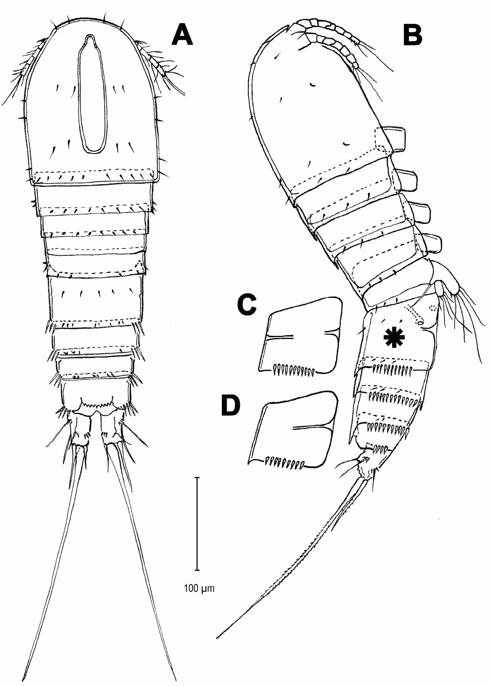
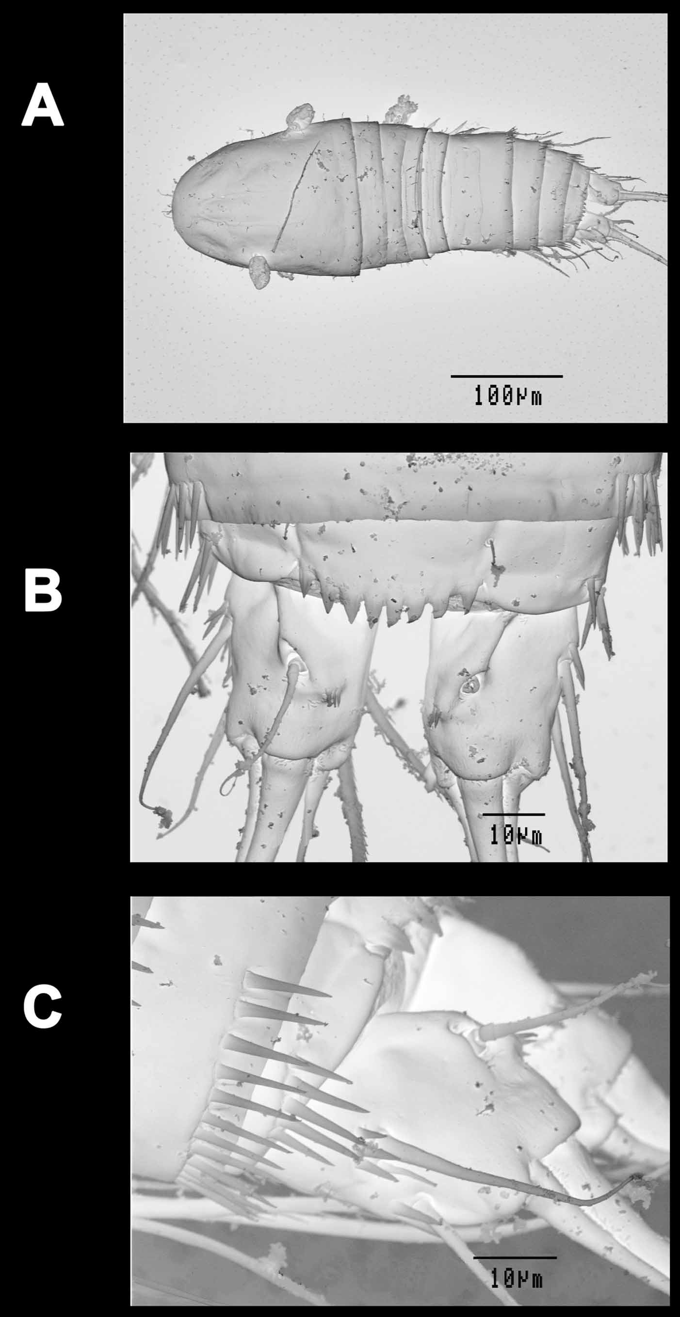

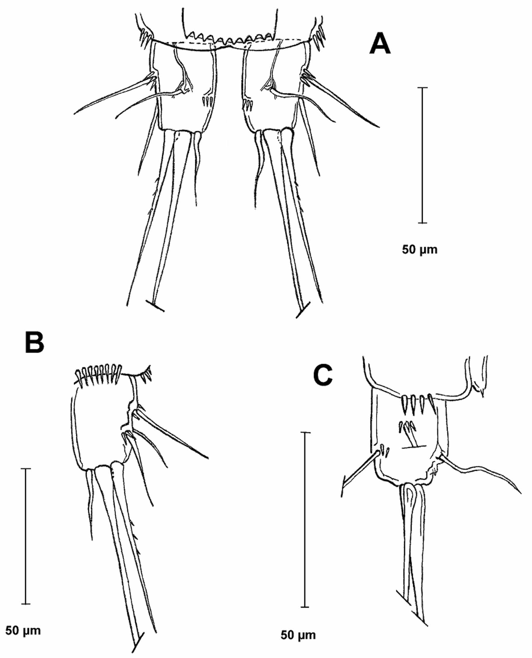
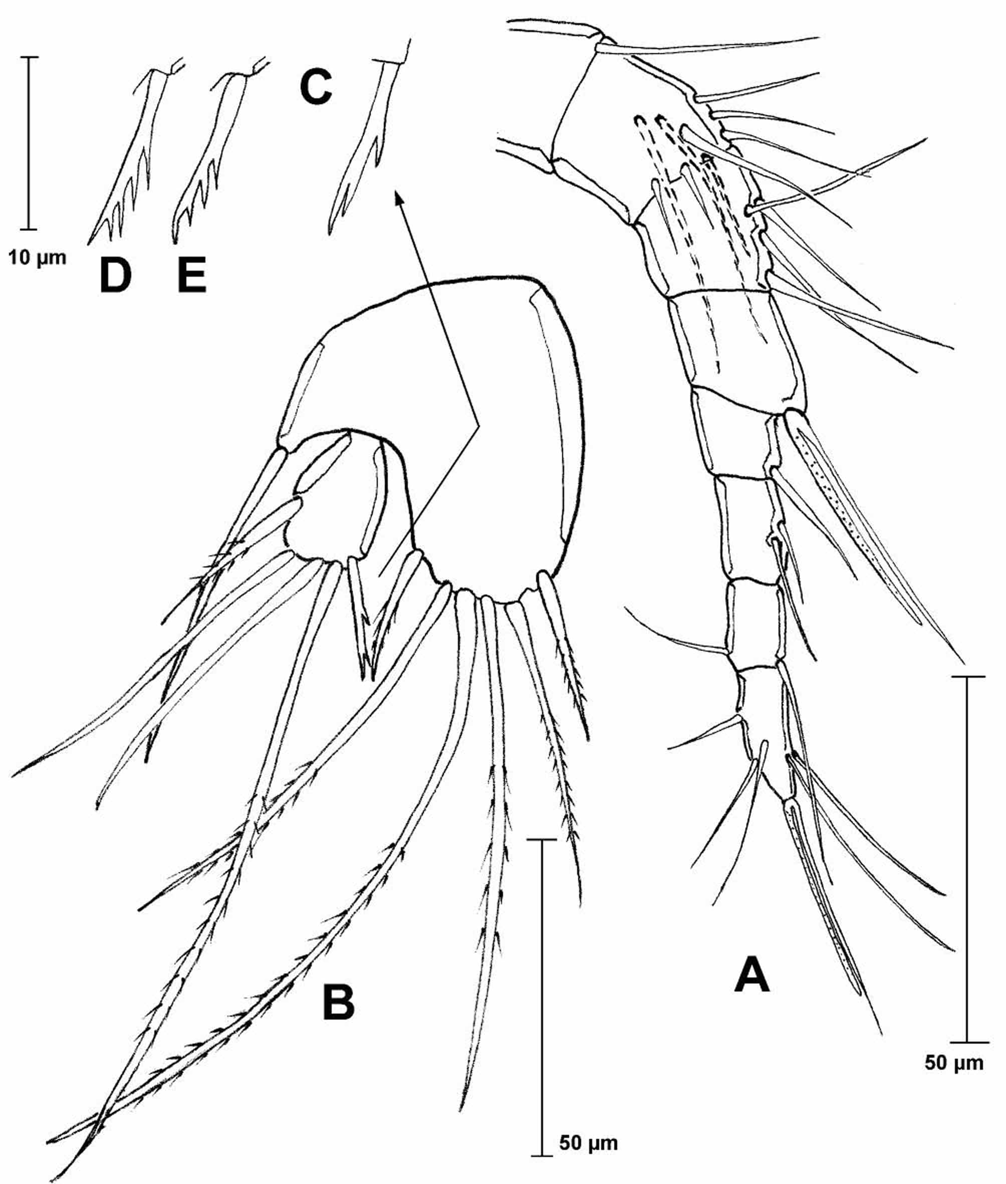
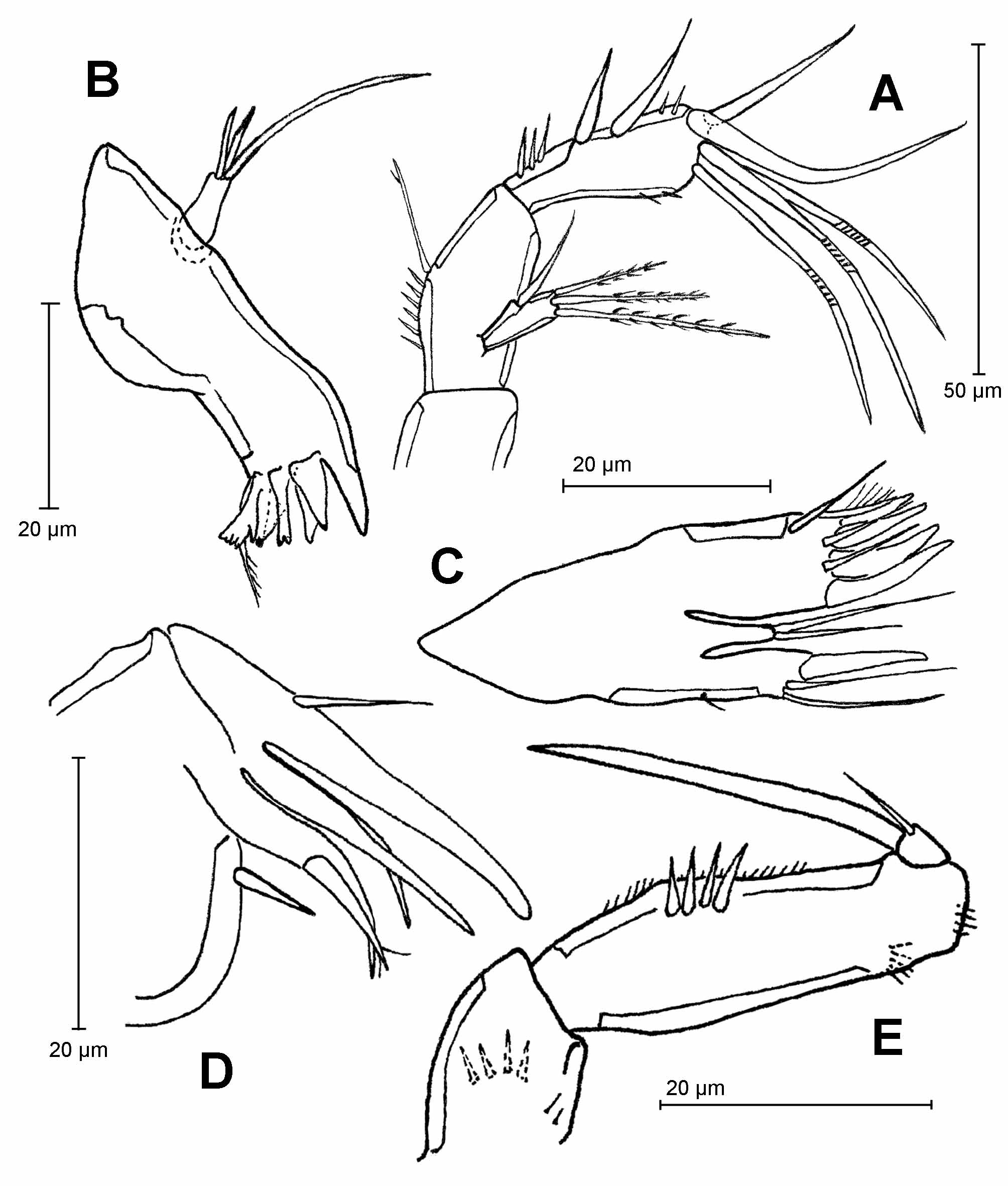
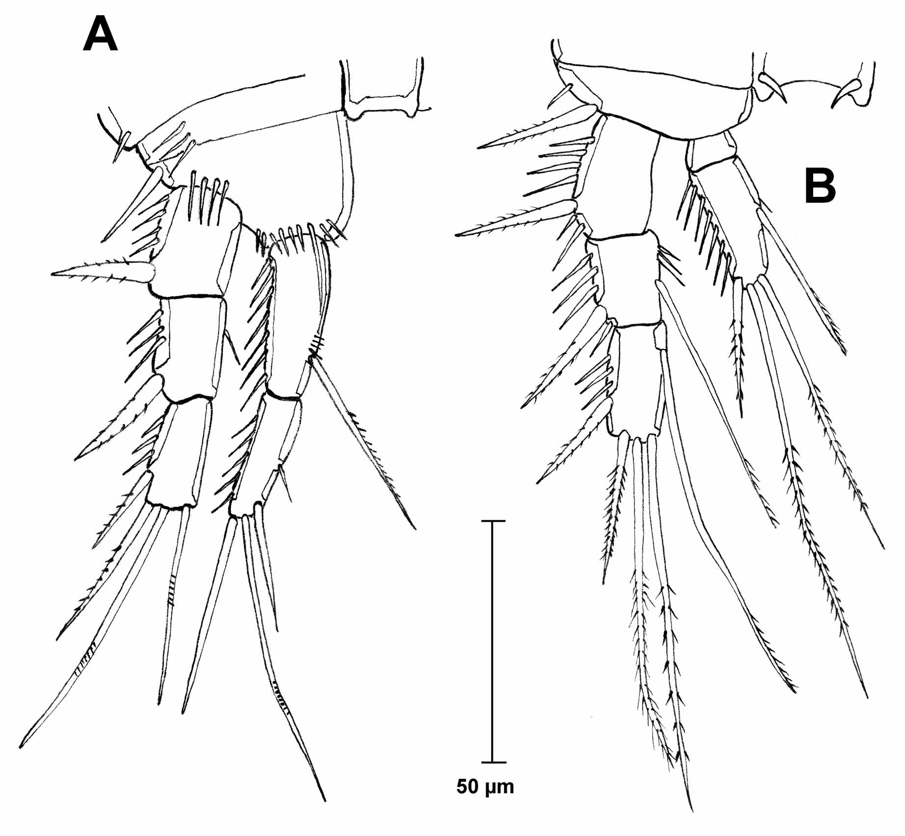
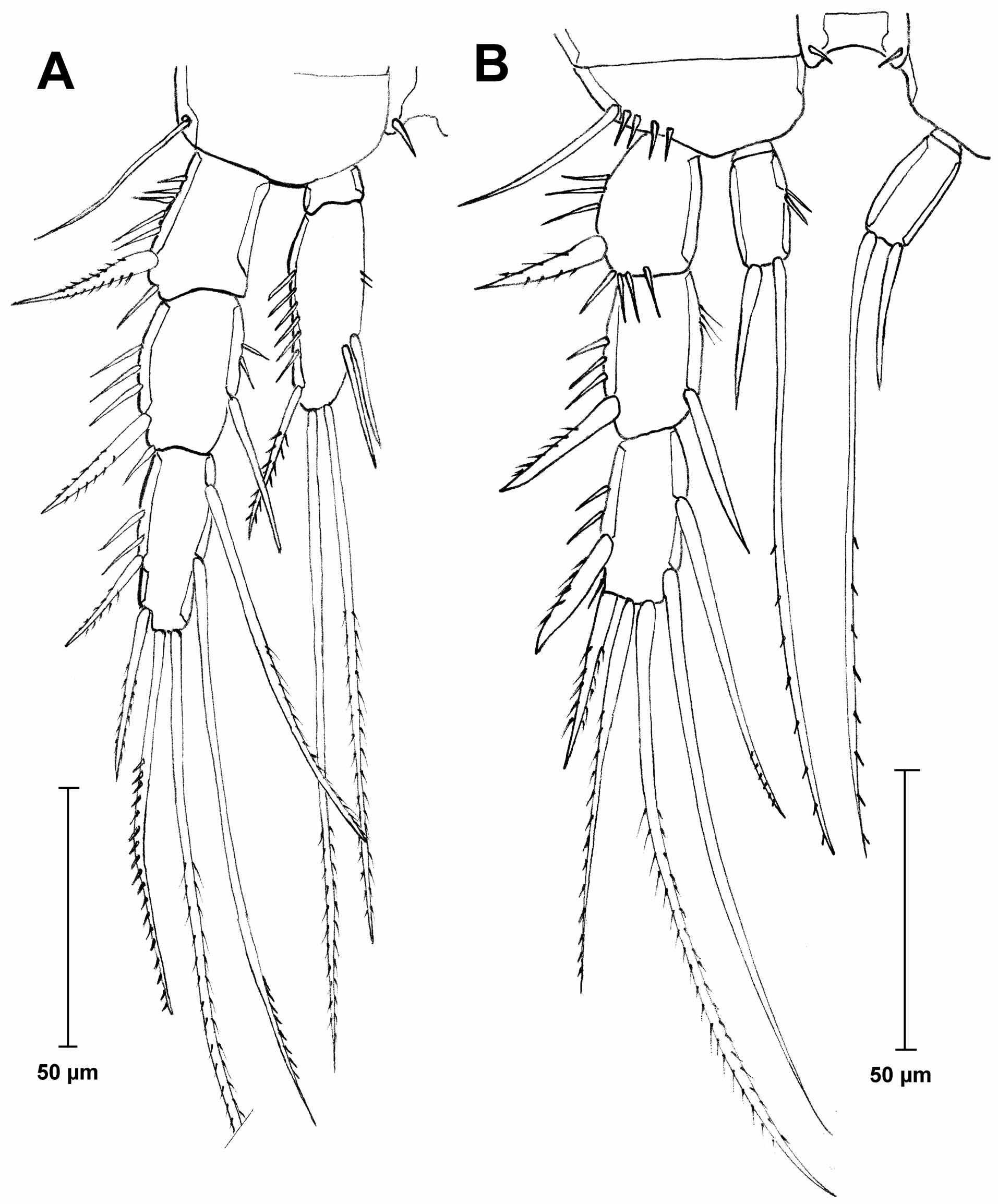
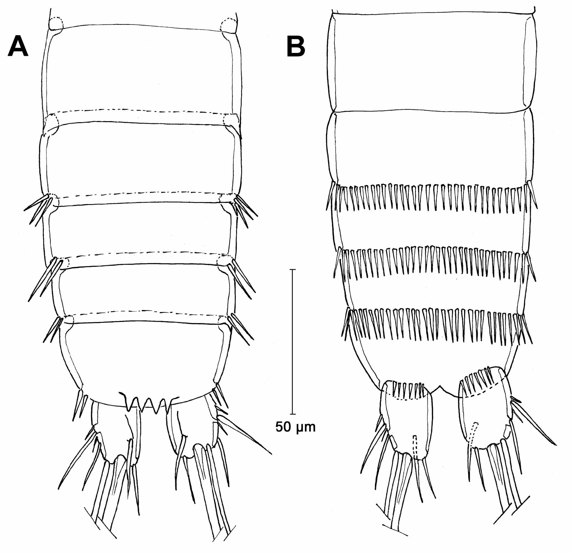
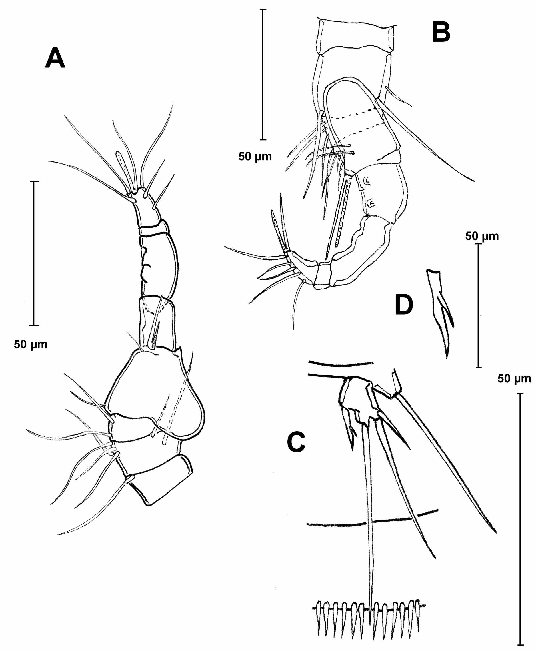
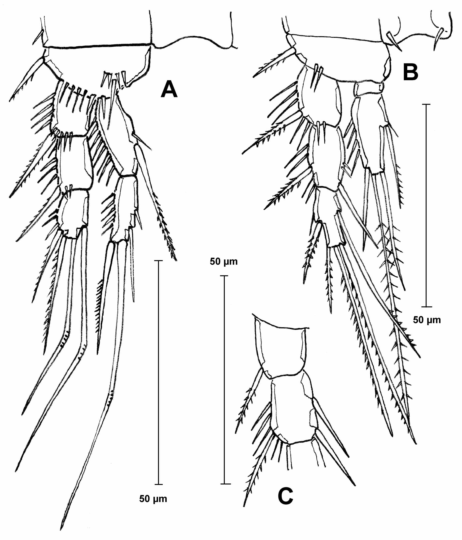

TABLE 1. List of species of the subgenus Canthosella, taxonomic status, known (+) and unknown (-) females and males, and geographic distribution. * species belonging to the group bearing a row of spines on the dorsal surface of the caudal ramus. ** species relocated by Janetzky et al. (1996)
| Nr. | Species | Status | Female | Male | Distribution |
|---|---|---|---|---|---|
| 1 | aliena Noodt, 1956 * | relocated ** | + | + | neotropical (Brazil), introduced into Germany |
| 2 | antillica (Petkovski, 1973) | + | + | neotropical (Cuba) | |
| 3 | bromelicola Ebert, 1976 | not published | - | + | neotropical (Brazil) |
| 4 | chocoensis n. sp. * | + | + | neotropical (Colombia) | |
| 5 | fluviatilis Chappuis, 1931 | + | + | oriental (Indonesia, Sumatra) | |
| 6 | goeldii Ebert, 1976 * | not published | + | - | neotropical (Brazil) |
| 7 8 | kalima (Delachaux, 1924) * lacustris Chappuis, 1931 | relocated ** | + + | - - | neotropical (Surinam) oriental (Indonesia, Sumatra) |
| 9 | mervini Janetzky, Martinez Arbizu & Reid, 1996 | + | + | neotropical (Jamaica) | |
| 10 | montana Ebert, 1976 | not published | + | + | neotropical (Peru) |
| 11 | muscicola (Chappuis, 1928) | relocated | + | + | oriental (Indonesia, Java) |
| NHMW |
Naturhistorisches Museum, Wien |
No known copyright restrictions apply. See Agosti, D., Egloff, W., 2009. Taxonomic information exchange and copyright: the Plazi approach. BMC Research Notes 2009, 2:53 for further explanation.
|
Kingdom |
|
|
Phylum |
|
|
Class |
|
|
Order |
|
|
Family |
|
|
Genus |
Attheyella (Canthosella) chocoensis
| Gaviria, Santiago & Defaye, Danielle 2012 |
pilagaensis
| Janetzky, Martinez Arbizu & Reid 1996 |
Attheyella (C.) mervini
| Janetzky et al. 1996 |
striblingi
| Reid 1990 |
vera
| Por & Hadel 1986 |
Attheyella (Canthosella) vera
| Por and Hadel 1986 |
silvicola Löffler, 1973
| Loffler 1973 |
Attheyella (Canthosella) silvicola Löffler, 1973
| Loffler 1973 |
Attheyella (Canthosella) antillica
| Petkovski 1973 |
siolii
| Kiefer 1967 |
vietnamica
| Borutzki 1967 |
Attheyella (Canthosella) vietnamica
| Borutzky 1967 |
Canthosella
| Chappuis 1931 |
Attheyella (Canthosella) fluviatilis
| Chappuis 1931 |
Attheyella (Canthosella) lacustris
| Chappuis 1931 |
Attheyella (Canthosella) muscicola (
| Chappuis 1928 |