
Philmontoides hageni ( Dohrn, 1905 ) Ingrisch, 2022
|
publication ID |
https://doi.org/ 10.11646/zootaxa.5182.2.1 |
|
publication LSID |
lsid:zoobank.org:pub:8920DE84-2BE6-4A68-A7F7-AC987F1F894E |
|
DOI |
https://doi.org/10.5281/zenodo.7053841 |
|
persistent identifier |
https://treatment.plazi.org/id/DB181868-FF85-FFD2-FF67-D62C2949F362 |
|
treatment provided by |
Plazi |
|
scientific name |
Philmontoides hageni ( Dohrn, 1905 ) |
| status |
comb. nov. |
Philmontoides hageni ( Dohrn, 1905) comb. nov.
Figs. 9A View FIGURE 9 , 10K–L View FIGURE 10 , 11A View FIGURE 11 , 12A View FIGURE 12 , 13A–D View FIGURE 13 , 16E–F View FIGURE 16 , 17A–B View FIGURE 17
Nicsara hageni Kirby, 1906 View in CoL
Salomona vittata Kuthy, 1910 View in CoL syn. nov.
Philmontis hageni Ingrisch, 2015 View in CoL
Types: Syntypes of L. hageni View in CoL (1 male, 1 female): New Guinea— 1 male, 1 female, leg. v. Hagen, Rohde—depository: Polish Academy of Science , Museum of the Institute of Zoology Warszawa, Poland ( MZPW) .
Holotype of S. vittata (female): Madang , Astrolabe-Bay , New Guinea, Montes Oertzen (5°28’S, 145°34’E), 1.i.- 31.xii.1897, leg. Biro —depository: Muséum d’Histoire Naturelle, Geneva ( MHNG). GoogleMaps
Remark on types (all examined): In the original publication, Dohrn (1905) describes male and female and gives measurements for two specimens, one male and one female. They were originally deposited in “Museum Stettin” and had been later transferred to MZPW Warszawa. In MZPW there are three specimens of this species identified by Dohrn, of which the male and one female are labeled as “ Type ” and the second female as “Cotype”. Another female identified by Dohrn as L. hageni was found in ZMB Berlin ”; this female was not labeled as a type.
(2) From the descriptions in Kuthy (1910) it is certain that he saw only a single female. For all species described in this publication, he did not give a note on the depository. But from his description, collector, and locality it is certain that the female specimen in MHNG must be regarded as the holotype of S. vittata Kuthy, 1910 . The more as Kuthys’ description “ovipositor basi subcompressus, in parte tertia latissimus, apice acuminatus” leaves no doubt that Salomona vittata belongs to Philmontoides . Moreover, there is no difference between this female and the females of Dohrn’s series of L. hageni .
Other specimens studied: Papua New Guinea: “ Neu-Guinea parte germanica”, leg. v. Hagen, Rohde 1 female cotype ( IZWP) ; “ New Guinea ”, leg. H. Dohrn — 1 female ( ZMB) ; New Guinea, leg. H. Fruhstorfer — 1 female ( MNHN) , 1 male ( ZMB) ; Papua New Guinea: Gulf, near Kerema, Murua Agricultural Station , (7°55’S, 145°47’E), 1–31.vii.1959, leg. F.X. Ryan — 1 female ( NBC) GoogleMaps ; Madang, Adelberg Mts [= Adelbert Range], Wanuma , elev. 800– 1000 m (4°35’S, 145°10’E), 25.x.1958, leg. J.L. Gressitt — 2 females, 1 male ( BPBM) GoogleMaps ; same locality, 26.x.1958, leg. J.L. Gressitt — 1 female, 2 males ( BPBM) GoogleMaps ; Morobe, 25 km South of Salamaua , elev. 1–80 m (7°13’S, 147°7’E), 25–26.i.1969, leg. J. Sedlacek — 1 male ( BPBM) GoogleMaps ; Busu River 32–48 km east of Lae , elev. 100 m (6°42’49.35’’S, 147°2’21.48’’E), 22.iii.1963, leg. J. Sedlacek — 1 female ( BPBM) GoogleMaps ; Huon Peninsula, Finschhafen (6°36’S, 147°51’E), 1952, leg. E. Wagner (Mission Ulap)— 1 female ( ZSM) GoogleMaps ; same locality, 10.iv.1963, leg. J. Sedlacek — 1 female ( BPBM) GoogleMaps ; same locality, 13.iv.1963, leg. J. Sedlacek — 2 females, 1 male ( BPBM) GoogleMaps ; same locality, 16.iv.1963, leg. J. Sedlacek — 2 females ( BPBM) GoogleMaps ; Laleng , elev. 1300–2000 m (6°31’S, 147°49’E), 23.iv.1963, leg. J. Sedlacek — 1 female ( BPBM) GoogleMaps ; New Guinea (NE), Western Highlands, Korn Farm , elev. 1560 m (5°50’S, 144°18’E), 19.x.1958, leg. J.L. Gressitt — 3 males ( BPBM) GoogleMaps .
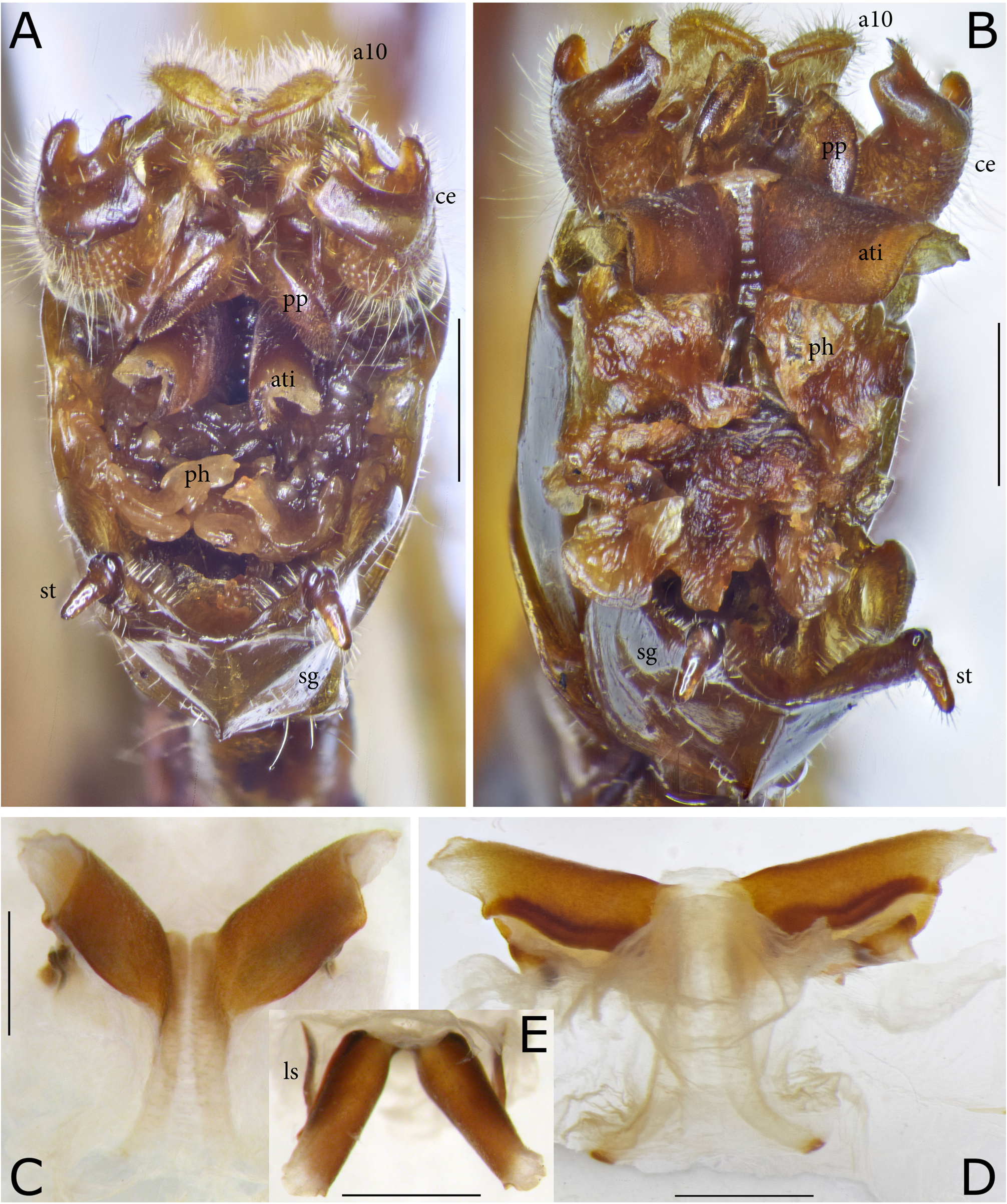
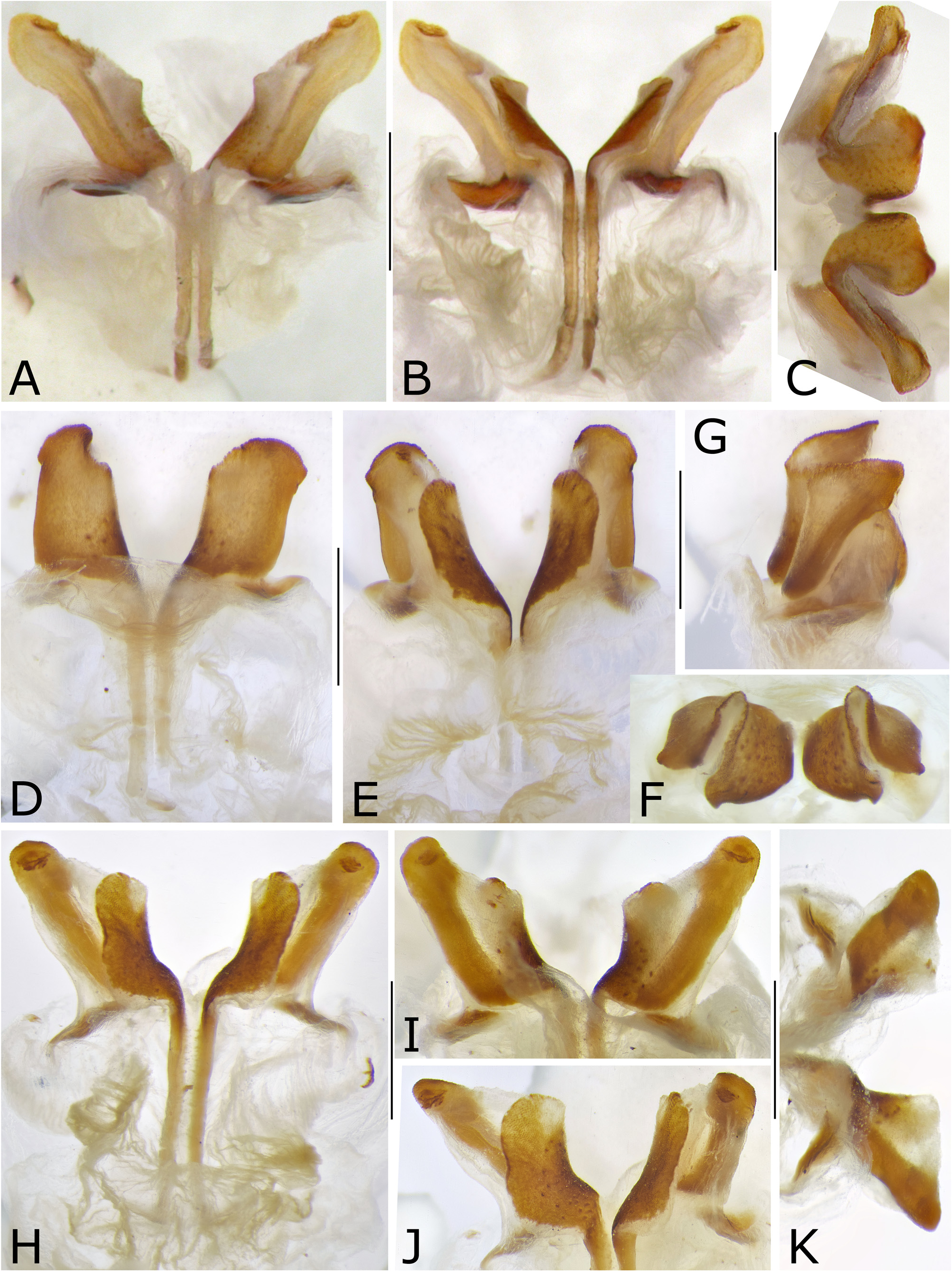
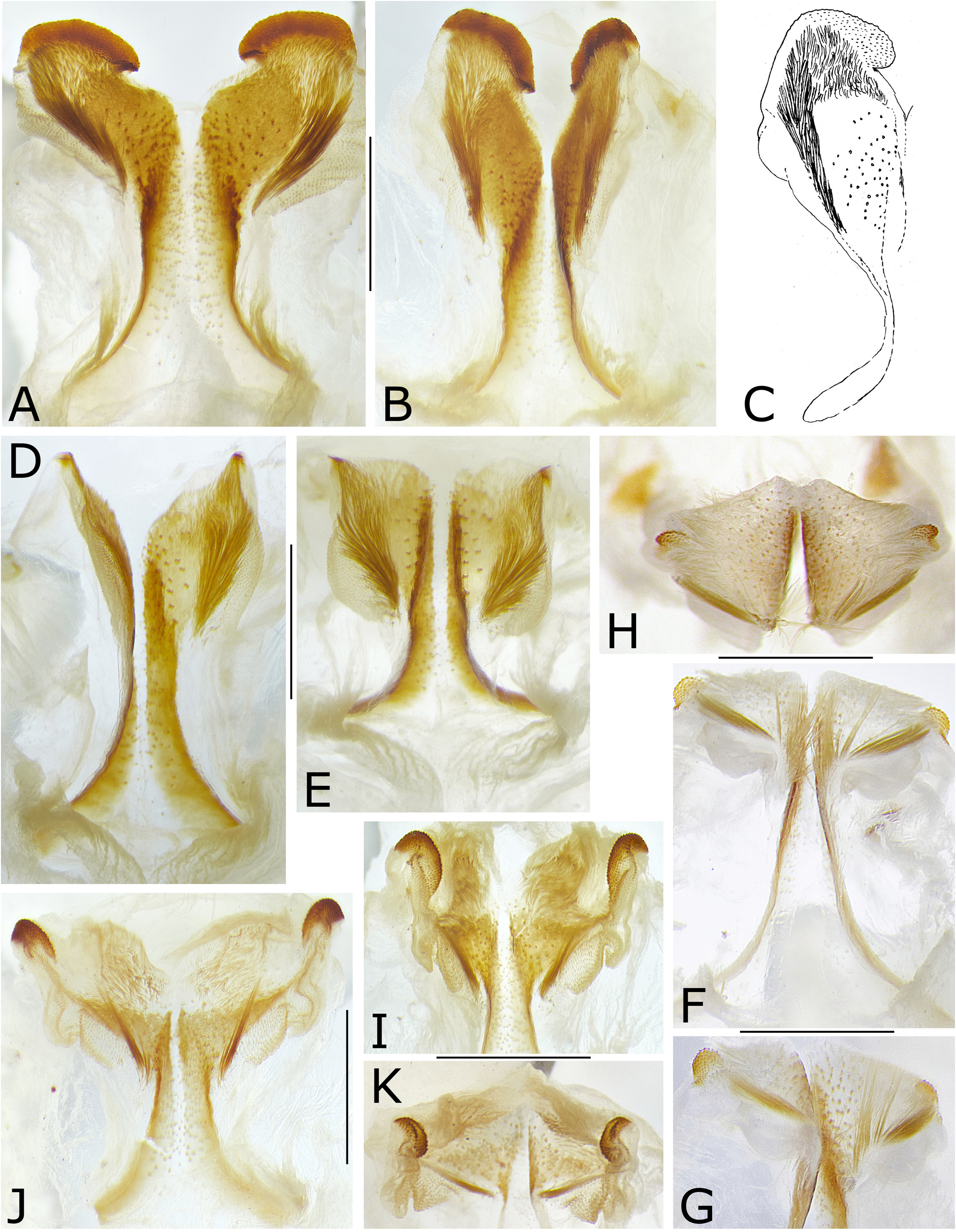
Diagnosis and discussion. P. hageni is unique for the shape of the male titillators, which consist of a pair of transparent and narrow basic stems that carry at end large vaulted plates with rough surface. These plates are folded around midline when stuck inside body ( Figs. 13A, E View FIGURE 13 ) but open to a longitudinally vaulted surface when free ( Figs. 13B–D View FIGURE 13 ). This structure of the titillators may have evolved from widened hyaline structures standing vertically to the main titillator axis as occur e.g., in P. striatus sp. nov. and P. wau sp. nov. ( Figs. 15F–K View FIGURE 15 ). On the other hand, a further development of the “umbrella-like” titillators led to more complex structures, in which both halves of the vaulted surface became different in size and shape as e.g., in P. lobatus ( Naskrecki & Rentz, 2010) or P. globosus sp. nov., or they became almost fully separated branches as in P. disjunctus sp. nov. ( Fig. 14 View FIGURE 14 ).
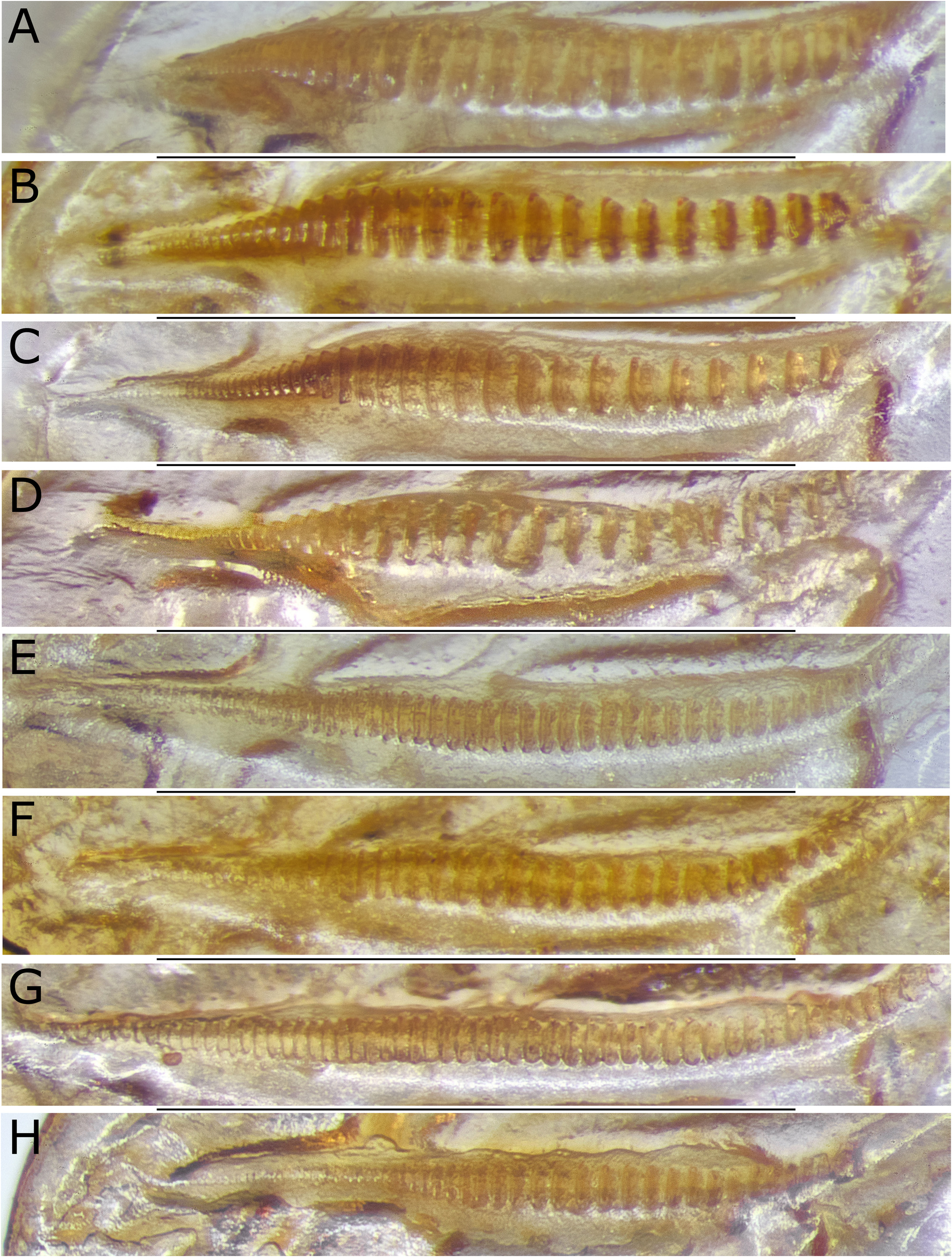
Regarding the male stridulatory file on the underside of the left tegmen, P. hageni , P. lobatus , P. globosus and P. disjunctus have files with large, stout, and spaced teeth in the main part of the file, becoming smaller and narrow in apical area, while the other species of the genus have smaller and more densely arranged teeth. Also, the wide and large appendages of the male tenth abdominal tergite are similar between these four species and differ from the shapes in the other species of the genus. The wide male cerci of P. hageni , with concave internal surface with two spines, one on dorsal end and one on apical rim, differ however from those of P. lobatus , P. globosus and P. disjunctus , instead they resemble more the cercus shapes in P. affinis , P. commodus sp. nov., P. wau sp. nov. and P. striatus sp. nov. From the latter species, males of P. hageni differ however in details of the cerci and in the shape of the appendages of the tenth abdominal tergite. Females differ from other species of the genus by the shape of the subgenital plate.
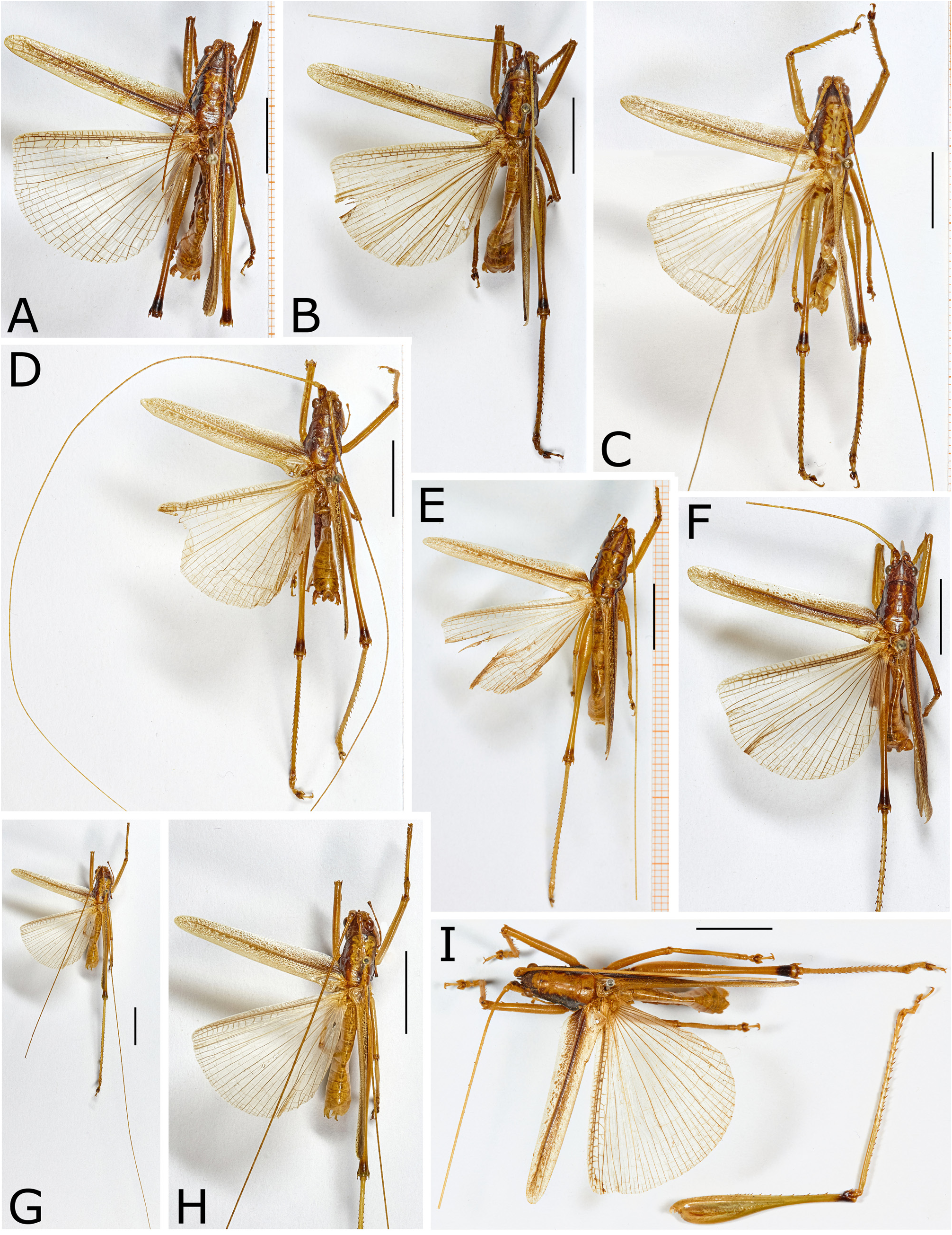
Description. Elongate species with tegmen reaching about end of hind femur ( Fig. 9A View FIGURE 9 ). Pronotum elongate, disc with anterior and posterior margins convex, the latter covering only very base of stridulatory area of tegmen; transverse furrows interrupted in midline; lateral lobes not very deep with ventral margin slightly concave, at end forming a rounded angle with the lateral area of the hind margin that is projecting laterad and clearly visible from above. Prosternal spines long, in two females of medium length; mesosternal lobes obtuse; metasternal lobes rounded and with an angle or fold. Femora with the following number of spines on ventral margins: (1) 5-8 / 3-10; (2) 6-9 / 1-3; (3) 9-15 / 9-18 (n = 18); hind knee lobes bispinose. In 4 males and 7 females also knee lobes of fore femur bi-spinose on both sides with second spines smaller than first spines; one female had a second spine at knee lobe of mid femur.
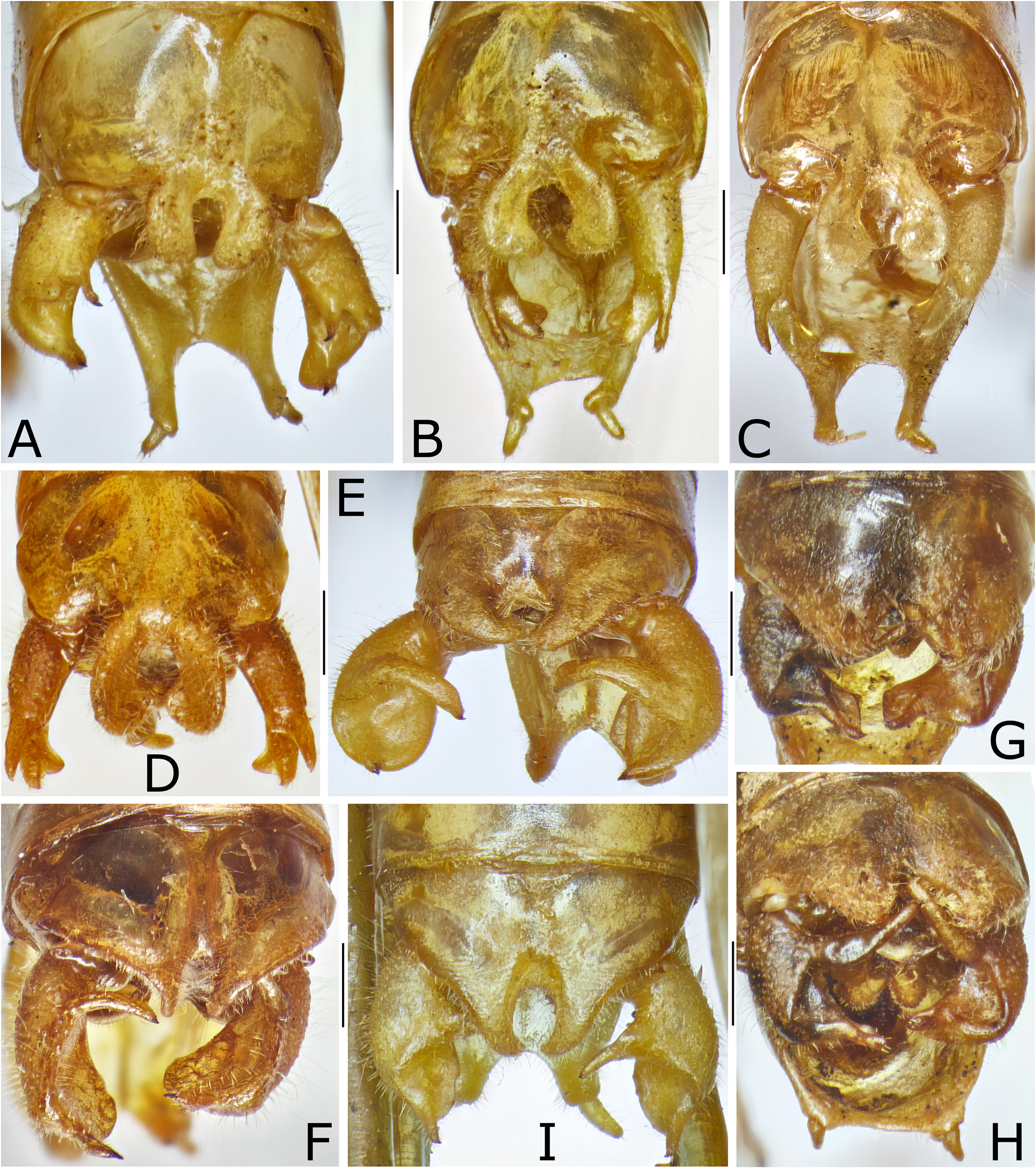
Male. Stridulatory file in about basal two thirds with large and spaced teeth, after a sinusoidal step, teeth abruptly narrowing and becoming denser and hardly perceptible toward end; Stridulatory file 1.27–1.34 mm long with 33–42 teeth, area with large teeth before step 0.92–1.05 mm with 16–20 teeth (n = 7 including type; Fig. 12A View FIGURE 12 ); some of the files appeared to be longer than the measurements given, but without countable teeth in that prolonged area. Tenth abdominal tergite little prolonged; apical margin with two compressed projections, together almost forming a circle with a long excision in midline, lateral areas of projections setose ( Fig. 11A View FIGURE 11 ). Epiproct with concave lateral margins, swollen, diverging posteriorly and their apical angles projecting; apical margin convex. Paraprocts bowl-shaped with strongly concave dorsal and convex ventral surface. Cerci short, conical, apico-internal surface concavely excavated; dorsal margin terminating into two short lobes: internal lobe with a spine at tip, external lobe obtuse; ventral margin curved mediad, terminating into a rounded ventral and a longer narrow dorsal lobe with a spine at tip ( Figs 10K–L View FIGURE 10 , 11A View FIGURE 11 ). Subgenital plate deeply excised from base with excision terminating into a narrow furrow that little surpasses mid-length of plate, afterward with a faint medial carina; lateral areas ascending and along rim little curved laterad again; the subgenital plate terminates into a pair of narrow, in apical half cylindrical projections that carry at end on ventral surface the styli ( Fig. 11A View FIGURE 11 ). Titillators in basal area whitish hyaline, simple, forming narrow, semi-transparent bands with out-curved base, in apical area extended laterally into large vaulted expansions with rough, brown surface; lateral end whitish hyaline and with wavy margin; on internal side covered by whitish membrane that also embeds the dark brown, narrow elongate and sinusoidally curved lateral sclerites ( Fig. 13C–E View FIGURE 13 ).
Female. Subgenital plate: ventral disc with convex, converging and upcurved lateral areas narrowing posteriorly and at end divided into a pair of in ventral view triangular, in lateral view upcurved, rounded lobes, triangularly excised in between; central area flat or faintly concave, with a strong medial carina along mid-length of disc; on both sides at very base with a roughly oval baso-lateral sclerite arising from a membranous latero-proximal expansion of the plate.
Coloration. Face of general color, antennal scrobae with internal surface darkened, scapus with internal surface dark brown; vertex with dark brown bands behind compounds eyes, or with a third brown band in middle, or vertex completely brown with a light band between eyes; pronotum with disc of general color, lateral lobes dark brown with one, two or three light spots; disc light with small brown spots at anterior margin or at anterior and posterior margins, and eventually a short or long brown stroke in middle split by a light line; tegmina along anterior and posterior margins of light color, partly with dark spots, in central area dark brown with light veinlets, radius and cubitus brown, media thickened and black; hind femur with black pre-genicular ring.
Measurements (10 males, 16 females).—Body w/wings: male 35–43, female 36–49; body w/o wings: male 27.5–37.0, female 25–45; pronotum: male 7.0–8.2, female 7.2–9.5; tegmen: male 22–30, female 20–30; hind femur: male 22.5–27.0, female 22.5–27.0; longest antenna: male>60 broken, female>70–220; ovipositor: female 12.5– 16.0 mm.


No known copyright restrictions apply. See Agosti, D., Egloff, W., 2009. Taxonomic information exchange and copyright: the Plazi approach. BMC Research Notes 2009, 2:53 for further explanation.
|
Kingdom |
|
|
Phylum |
|
|
Class |
|
|
Order |
|
|
Family |
|
|
Genus |
Philmontoides hageni ( Dohrn, 1905 )
| Ingrisch, Sigfrid 2022 |
Philmontis hageni
| Ingrisch 2015 |
L. hageni
| Ingrisch 2015 |
Salomona vittata
| Kuthy 1910 |
Nicsara hageni
| Kirby 1906 |
Lobaspis
| Hageni Dohrn 1905 |