
Philmontis C. Willemse, 1966
|
publication ID |
https://doi.org/ 10.11646/zootaxa.5182.2.1 |
|
publication LSID |
lsid:zoobank.org:pub:8920DE84-2BE6-4A68-A7F7-AC987F1F894E |
|
DOI |
https://doi.org/10.5281/zenodo.7053813 |
|
persistent identifier |
https://treatment.plazi.org/id/DB181868-FF9A-FFC8-FF67-D42A2DD7F770 |
|
treatment provided by |
Plazi |
|
scientific name |
Philmontis C. Willemse, 1966 |
| status |
|
Philmontis C. Willemse, 1966 View in CoL
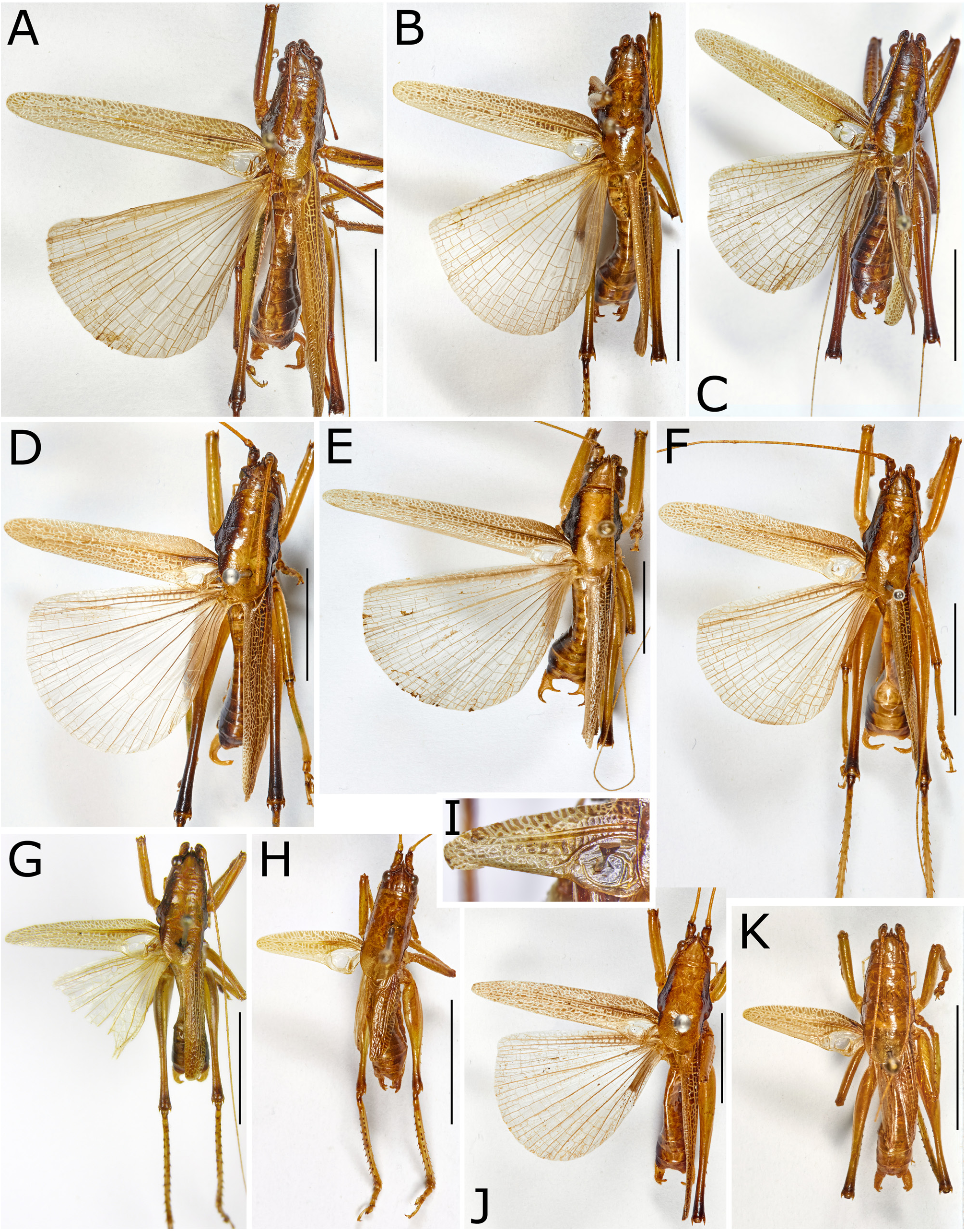
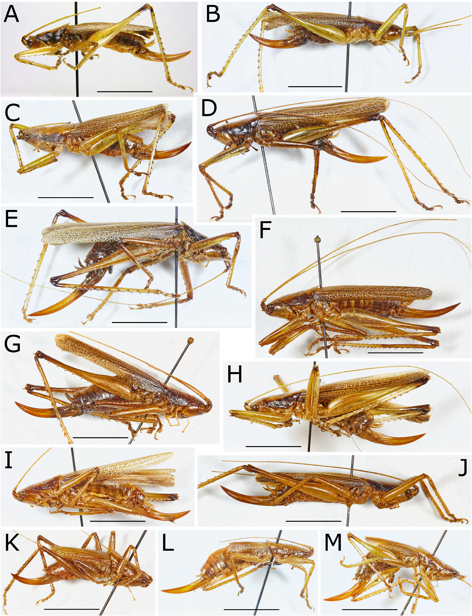
Figures 1–8 View FIGURE 1 View FIGURE 2 View FIGURE 3 View FIGURE 4 View FIGURE 5 View FIGURE 6 View FIGURE 7 View FIGURE 8 , map 1 View MAP 1
Typus generis: Philmontis nigrofasciatus C. Willemse, 1966 View in CoL by original designation.
Generic diagnosis. Mainly medium sized, but also small and few moderately large species. Pronotum prolonged, in males covering stridulatory area when at rest, in females little more than anterior margin of tegmen; lateral lobes very narrow. Prosternal spines short, rarely absent. Hind femur with spines only on anterior (external) margin and hind knee lobes with only a single spine. Male last abdominal tergite with a more or less pronounced incision from apical margin; male cerci in most species with partly excavated internal surface and often with incurved apical area; male subgenital plate terminating into a pair of long and narrow apical projections with a stylus at end; male titillators terminating into a small disc-shaped plate. Stridulatory file on underside of left male tegmen in most species with a prolonged apical area of indistinct minute teeth. Female ovipositor curved and gradually narrowing to acute tip; female subgenital plate with distinct lateral extensions at base, and with the plate terminating into a pair of long apical lobes varying from narrow with acute tip to wide with obtuse end. The differences compared to Philmontoides gen. nov. described below are already specified in introduction.
Description. Small, medium sized or moderately large species. Antennae long, reaching far behind body. Fastigium verticis conical, in anterior area slightly compressed, shorter than scapus; ventral margin separated by a shallow sinuosity from fastigium frontis. Frons shining, subsmooth, little grooved along clypeo-frontal suture, with or without few very shallowly impressed dots. Pronotum strongly prolonged behind in male, thus that the ventroposterior angle lies before middle of pronotum length; in female less prolonged; posterior margin of pronotum rounded in both sexes, in males covering stridulatory area of tegmen; pronotum surface shining, subsmooth, only in apical area and along margins little subrugose; disc slightly convex with lateral angles broadly rounded, apical area faintly raised and indistinctly shouldered, transverse sulci and secondary furrows very weak, lateral lobes longer or much longer than high, auditory swelling small. Most species fully winged but wings only little surpassing abdomen, small species often brachypterous; tegmen of fully winged species varying from almost covering abdomen to reaching beyond hind knees, gradually narrowed toward rounded tip. Prosternum provided with two moderately long, medium sized or short spines or only with tubercles, in few small species or only in some specimens of them fully missing. Mesosternal lobes rounded or angularly rounded, metasternal lobes rounded; medial plate with or without a short process or obtuse tubercle at posterior angles. Anterior tibia in cross-section quadrangular with dorsal angles rounded. Fore and mid femora with spines on both ventral margins, on hind femur only on anterior (external) margin, in few specimens with single spinules also on internal margin. Knee lobes of pro- and mesofemur spinose on both sides; on posterior side of profemur spine often short or obtuse; knee lobes of hind femur unispinose with rare individual exceptions.
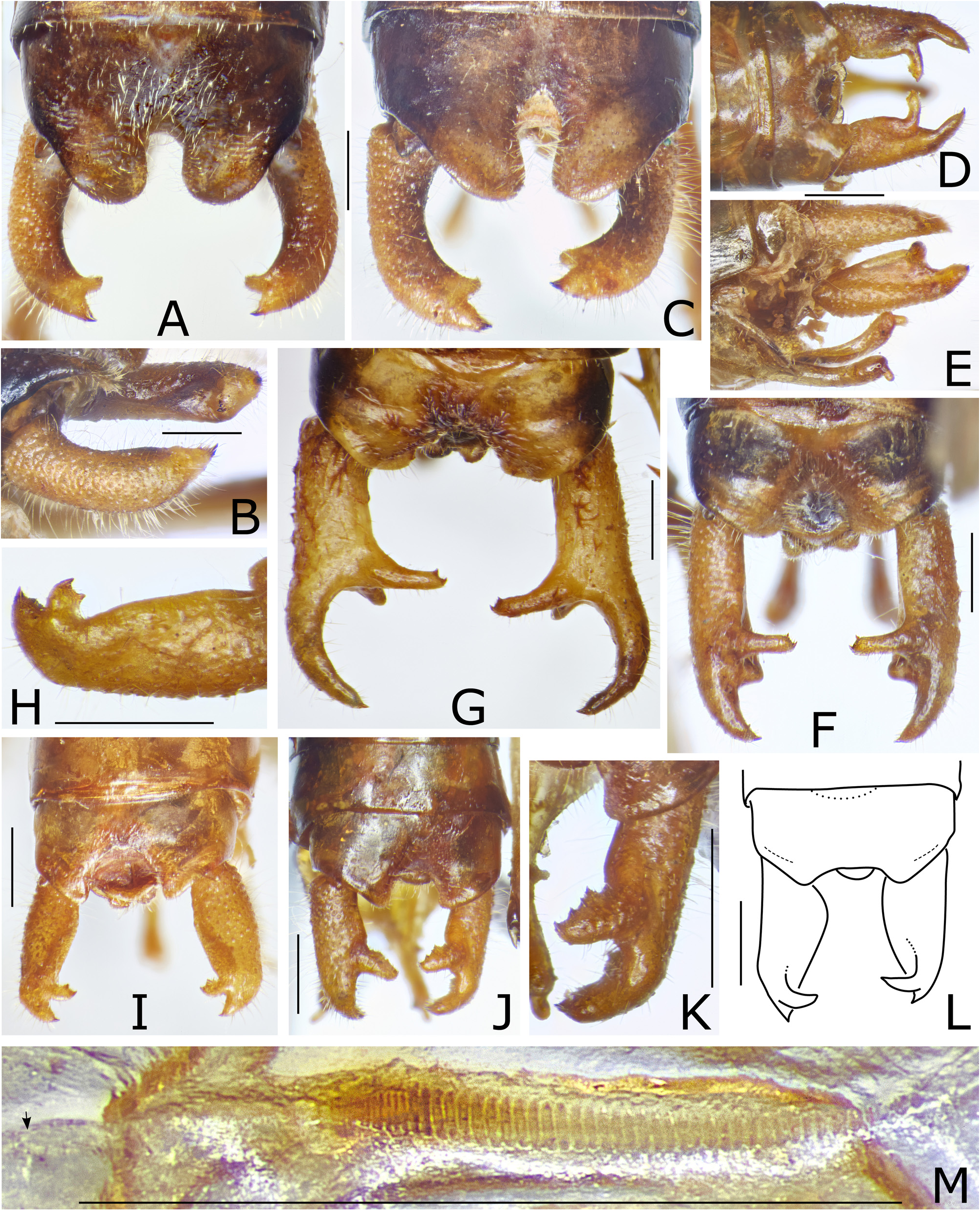
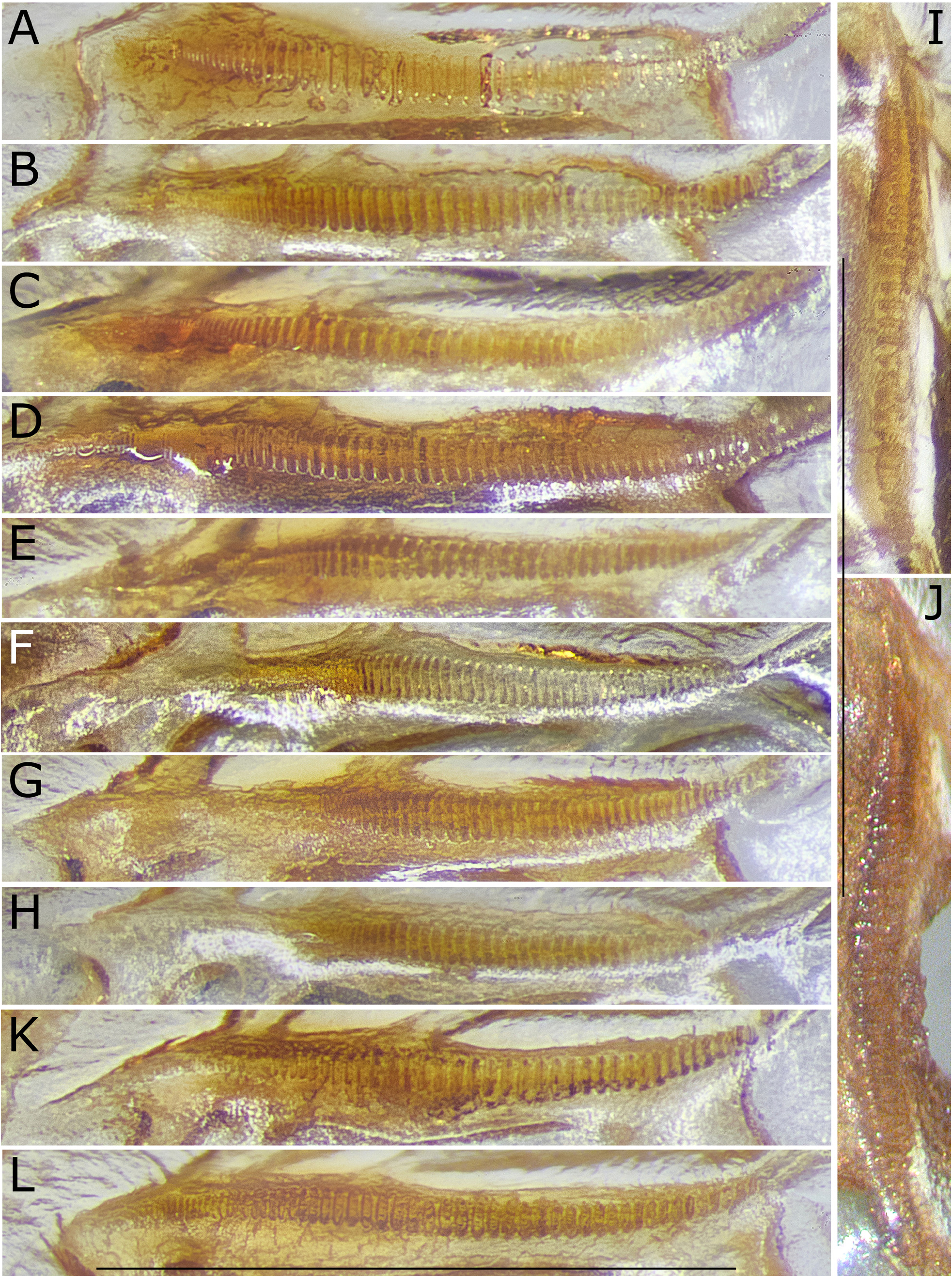
Male. Stridulatory file with teeth in apical area largely reduced and often indistinct; area with reasonably large teeth varies between species and is often separated by a step from apical area. Tenth abdominal tergite with apical margin angularly projecting at both sides, wide-roundly excised in middle. Epiproct oriented vertically at base, later often curved apicad. Paraprocts compressed, auricularly projecting at both sides of epiproct and sometimes even surpassing dorsal surface of epiproct; with short, obtuse apical projections. Cerci with ventro-internal margin often roundly expanded. Subgenital plate with obtuse lateral carinae; lateral areas ascending and then curved laterad again; lateral margins convex, approaching each other posteriorly; with or without long apical projections; styli small or very small. Titillators rather uniform between species; separate, central areas running parallel, basal and apical areas curved laterad; basal areas usually short and broad; apical areas flattened, in some species widened, in others as narrow as in basal area, always narrowing toward end; apex forming a small, sclerotized disc twisted in a 90° angle against preceding area.
Female. Pronotum less strongly prolonged than in male, covering only bases of tegmina; lateral lobes as narrow as in male; auditory swelling distinct but without humeral sinus. Seventh abdominal tergite with lateral areas shortened or excised to provide room for extensions of baso-lateral projections of subgenital plate; rarely unmodified. Epiproct triangularly rounded. Cerci long, conical, slightly curved, apex pointing. Ovipositor moderately long, in little more than basal third stout, afterward regularly upcurved and narrowed to acute tip. Subgenital plate with dorsal expansions from sub-basal area ascending on both sides of ovipositor.
Coloration. Rather uniform between species. Partly green when alive, specimens studied often discolored and yellowish brown or ochreous. Frons concolorous, antennal scrobae dark brown or with brown spots, scapus often with brown spots; vertex including fastigium verticis and dorso-posterior part of genae with or without dark elements. Pronotum with paranota blackish brown, often including light spots; disc green or yellowish brown. Tegminal pattern variable. Legs green, hind knees brown or with a brown spot at top. Anterior tibiae often with a dark spot below tympana.
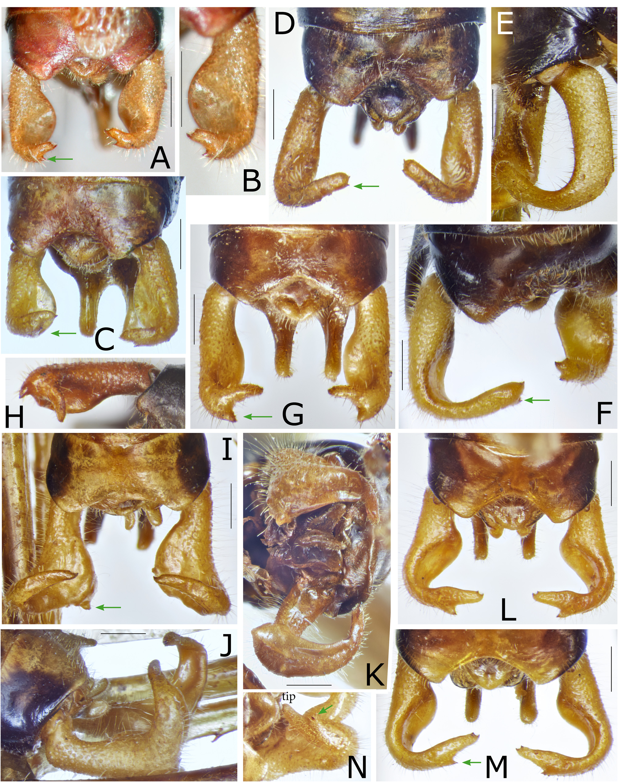
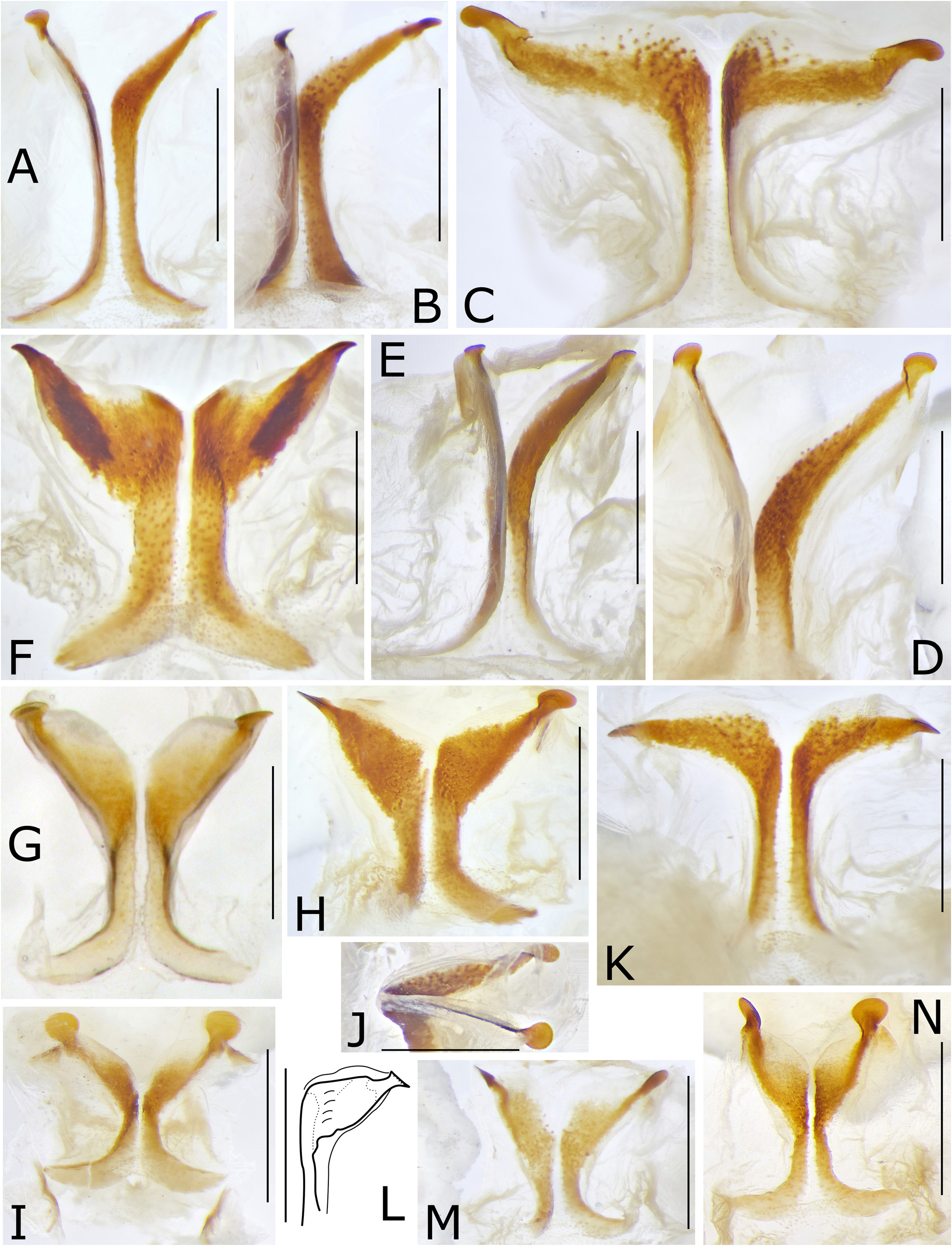
Discussion. The species of Philmontis can be assigned to three groups: (1) mainly medium large species similar to P. nigrofasciatus with in-curved male cerci having part of the internal surface concavely deepened and the apical area almost rectangularly bent against the basal area, while the female subgenital plate terminates into a pair of long projections that are widely separated from each other at base; (2) medium sized to large species with unique modification of the male cerci, and the female subgenital plate having the apical lobes either strongly widened or divided into narrow lobes from central area; (3) small brachypterous species with simplified abdominal appendages and more uniform coloration. Comparing the male cerci of group (2) that have straight or moderately curved stems and one or two internal projections from little behind mid-length or in subapical area of the cercus stem with species of group (1) with incurved male cerci that carry a small cone or spine in sub-apical or more basal area (marked in Fig. 2 View FIGURE 2 by an arrow), it is not unlikely that species of group (2) like P. pandus sp. nov. and P. profusus sp. nov. ( Figs 3F–G View FIGURE 3 ) represent a more basic condition of the male cercus, while in species related to P. nigrofasciatus (group 1, Fig. 2 View FIGURE 2 ) what appears as a large in-curved apical area of the cercus could be instead the internal projection while the apical area of the cercus is reduced to a small cone or spine. When we assume that the male cerci help opening the female subgenital plate during copulation, the huge modification of the male cerci in P. forcipatus becomes understandable as an adaptation to the female subgenital plate, which in this species terminates into pair of wide apical lobes while in most other species of the genus the female subgenital plate terminates into pair of narrow or very narrow apical lobes. The male of Philmontis extensus sp. nov. is still unknown. But we may expect that it also has strongly modified cerci as P. forcipatus . In P. spinosus sp. nov. the apical area of the cercus is shortened but the internal projection is short too ( Figs 3A–C View FIGURE 3 ). Females of that species have subgenital plates that terminate into long and narrow apical projections that are however not separated at base as in the other species of the genus but divide from the central area ( Fig. 8F View FIGURE 8 ).
Tentative key to species of the genus Philmontis Willemse, 1966
Key to known species—Males
1. Medium sized or moderately large, slightly brachypterous to fully winged species with tegmina reaching or surpassing tip of abdomen ( Figs 1A–G, J View FIGURE 1 )................................................................................2.
- Small, markedly brachypterous species with tegmina covering little more than half of abdomen ( Figs 1H–I, K View FIGURE 1 )...........9.
2. Male cerci in at least basal half cylindrical, terminating either into two short projections of subequal size or into three projections of varying size: a curved apical projection, a cylindrical dorso-internal projection with two apical teeth, and a cylindrical or compressed angular ventro-internal projection ( Figs 3A–C, 3F–G View FIGURE 3 )..............................................3.
- Male cerci on internal surface with a concave excavation of variable size and with ventral margin expanded ventrad; apical area of cercus always strongly curved or bent mediad; or cercus more strongly modified ( Fig. 2 View FIGURE 2 )..........................5.
3. Male tenth abdominal tergite terminating into a pair of extended apical lobes with rounded end and narrow interspace between lobes. Cerci nearly cylindrical but moderately curved; at end divided into two short conical lobes; distal lobe with a spinule at tip ( Figs 3A–C View FIGURE 3 )................................................................. Philmontis spinosus sp. nov.
- Male tenth abdominal tergite terminating into a pair of shorter apical lobes with wider interspace. Cerci in about basal half straight, afterward dividing into three lobes of unequal shape ( Figs 3F–G View FIGURE 3 )........................................4.
4. Male cerci terminating into a long, curved apical projection with one spinule at tip, a dorso-internal projection with converging margins in basal area, becoming cylindrical toward end that carries two spinules, and a ventro-internal projection with obtuse, cylindrical end ( Fig. 3G View FIGURE 3 )................................................................ P. profusus sp. nov.
- Male cerci terminating into a less long, curved and conical apical projection with stout base and one spinule at end, a straight, narrow cylindrical, dorso-internal projection with two spinules at tip, and a ventral projection forming a compressed, triangular plate with obtuse ventro-apical angle ( Fig. 3F View FIGURE 3 ).............................................. P. angulatus sp. nov.
5. Male cerci running for a short distance ventro-laterad at base, then bent, running posteriorly with the internal margin strongly expanded; in about mid-length cercus bent dorsad, is narrowing and with concave proximal surface; apical area of cercus curved mediad, at tip provided with a short spine ( Figs 2I–K View FIGURE 2 ).......................... P. forcipatus ( Willemse, 1966)
- Male cerci less strongly modified; curved mediad behind mid-length or in more apical area......................... 6.
6. Male cerci rather narrow, concave impression on internal surface not very strong and ventral margin of cercus hardly expanded; in-curved apical area straight and almost parallel-sided, with rounded end carrying a minute spinule at tip and another spinule in sub-apical area on ventral margin ( Figs 2D–F View FIGURE 2 )............................................. P. angustus sp. nov.
- Male cerci with ventro-internal margin conspicuously expanded with a large grooved area on internal surface; incurved apical area carrying a distinct spine or tooth on ventral margin at beginning or in mid-length of incurved apical area and one or two spinules at tip........................................................................................7.
7. In-curved apical area of cercus almost as long as straight basal area, with a spine around mid-length on ventral margin and with two spinules at tip; excavation of internal surface in basal area very strongly expressed ( Figs 2L–N View FIGURE 2 )....... P. flexus sp. nov.
- In-curved apical area of cercus distinctly shorter than straight basal area; ventral expansion on internal side with surface concave but less strongly grooved........................................................................8.
8. Cerci with in-curved apical area carrying a spine with widened base from ventral margin of about apical third of in-curved apical area and another spine from apical angle of truncate tip of cercus and a minute spinule from proximal angle of tip of cercus ( Figs 2A–C View FIGURE 2 ).................................................... Philmontis nigrofasciatus Willemse, 1966 View in CoL
- Cerci carrying a distinct subacute cone at very beginning of in-curved and markedly down-curved apical area, and a minute spinule at end of obtuse tip ( Figs 2G–H View FIGURE 2 )..................................................... P. murmur sp. nov.
9. Cerci with internal projection arising just before end of cercus.................................................10.
- Cerci with internal projection arising markedly before end of cercus............................................11.
10. Cerci with a curved internal projection arising just before end of cercus and terminating into a single acute tip; internal margin of cercus in about basal half with oval extension. Tenth abdominal tergite with apical margin only shallowly excised in middle ( Fig. 3L View FIGURE 3 )....................................................................... P. minimus Willemse, 1966
- Cerci with a curved internal projection arising from internal margin in subapical area and terminating into two acute spines; internal surface of cercus behind basal third with shallow oval impression. Tenth abdominal tergite with apical margin rather deeply excised in middle ( Figs 3H–I View FIGURE 3 )....................................................... P. pandus sp. nov.
11. Cerci rather stout, in basal area about cylindrical; behind mid-length with a wide, compressed internal projection with short anterior and long posterior margin connected by a wide, oblique apical margin carrying four small, acute teeth; cercus behind internal projection with narrowing margins, then curved mediad with stout, rounded end that carries a spine at tip ( Figs 3J– K View FIGURE 3 ).................................................................................. P. robustus sp. nov.
- Cerci conical with deeply concave internal surface; in subapical area, from ventral margin with a narrow and little curved internal projection terminating into two acute spines; apical area of cercus narrow and compressed; behind internal projection strongly narrowed toward dorso-lateral margin that carries at tip an acute spine ( Figs 3D–E View FIGURE 3 )............ P. pumilus sp. nov.
Key to known species—Females
1. Subgenital plate terminating into a pair of broad apical lobes with obtuse end ( Figs 8A–E View FIGURE 8 ).......................... 2.
- Subgenital plate terminating into a pair of narrow, band-shaped or spine-like apical lobes............................3.
2. Subgenital plate in general outline oval, widest in about mid-length, apical lobes regularly curved and narrowed to broadly rounded end ( Figs 8A–C View FIGURE 8 )................................................................ P. extensus sp. nov.
- Subgenital plate with a step in sub-basal area, followed by concave and then convex lateral margins that become obliquely sub-truncate at end ( Figs 8D–E View FIGURE 8 )................................................... P. forcipatus ( Willemse, 1966)
3. Subgenital plate widened at base, afterward with converging lateral margins, apical lobes long, dividing in an acute angle from each other and narrowing to acute tips ( Figs 8F–G View FIGURE 8 )........................................... P. spinosus sp. nov.
- Subgenital plate of different shape, apical projections arising from lateral or more central area of plate, but always separated at base by a wide interspace...............................................................................4.
4. Subgenital plate with apical projections robust, about as long as or longer than the entire basal area and often compressed ( Figs 7A–L View FIGURE 7 )..............................................................................................5.
- Subgenital plate with apical projections delicate, shorter than the basal plate ( Figs 7M–O View FIGURE 7 , 8H–M View FIGURE 8 ).....................9.
5. Subgenital plate with apical projections substraight and markedly approaching each other toward subacute tip, subbasal area with a transverse bulge ( Figs. 7C–E View FIGURE 7 )....................................................... P. angustus sp. nov.
- Subgenital plate with apical projections somewhat sinuate, only little or not at all approaching each other posteriorly, tip subacute, obtuse, or truncate............................................................................6.
6. Subgenital plate with baso-lateral extensions more strongly projecting laterad; central disc with a pair of impressions behind insertion of lateral extensions ( Figs 7K–L View FIGURE 7 )..................................................... P. flexus sp. nov.
- Subgenital plate with baso-lateral extensions oriented dorsad, hardly seen from below. Females of the following three species are difficult to identify without corresponding males..........................................................7.
7. Subgenital plate with baso-lateral extensions long, with narrow proximal and widened distal area; apical projections of subgenital plate at base strongly bent ( Figs 7I–J View FIGURE 7 ). Ovipositor about 12.5 mm long....................... P. banz sp. nov.
- Subgenital plate with baso-lateral extensions less prolonged and of different shape. Ovipositor about 10.0– 11.5 mm long...8.
8. Wings distinctly surpassing end of abdomen. Subgenital plate wider with baso-lateral extensions strongly curved dorsad; apical projections markedly bent dorsad at base, in further curse sinuate with truncate end ( Figs. 7F–H View FIGURE 7 )........ P. murmur sp. nov.
- Wings reaching about end of abdomen. Subgenital plate less wide with baso-lateral extensions less strongly curved dorsad; apical projections less strongly and more regularly curved to subacute tip ( Fig. 7A–B View FIGURE 7 )... .. P. nigrofasciatus Willemse, 1966 View in CoL
9. Larger species with tegmen surpassing abdomen. Female subgenital plate short, in lateral view rather regularly curved; at end with a pair of thin and short apical spines separated from each other ( Figs 7M–O View FIGURE 7 )................... P. angulatus sp. nov.
- Small, usually brachypterous species with shortened wings that do not reach the end of abdomen.................... 10.
10. Subgenital plate with baso-lateral extensions mainly laterally extended but at end with a narrow up-curved projection toward body ( Fig. 8H–I View FIGURE 8 )....................................................................... P. profusus sp. nov.
- Subgenital plate with baso-lateral extensions abruptly bent dorsad and widening toward end; in lateral view appearing nearly U-turned............................................................................................11.
11. Subgenital plate with a fine medial furrow extended into a triangular membranous area at end, sclerotized disc terminates into a pair of spine-like, curved, apical projections; baso-lateral extensions with shallowly grooved surface ( Figs 8J–K View FIGURE 8 ).............................................................................................. P. pandus sp. nov.
- Subgenital plate with medial seam not extended apically; hind margin subtruncate with narrow, spine-like lateral projections; baso-lateral extensions up-curved, twisted and provided with a pit ( Figs. 8L–M View FIGURE 8 )..................... P. pumilus sp. nov.


No known copyright restrictions apply. See Agosti, D., Egloff, W., 2009. Taxonomic information exchange and copyright: the Plazi approach. BMC Research Notes 2009, 2:53 for further explanation.