
Morochares xuzaifui Loktionov, Lelej & Liu, 2018
|
publication ID |
https://doi.org/ 10.11646/zootaxa.4462.4.3 |
|
publication LSID |
lsid:zoobank.org:pub:452FE48A-28D8-4991-B9D1-5C63B8050D9D |
|
DOI |
https://doi.org/10.5281/zenodo.5979883 |
|
persistent identifier |
https://treatment.plazi.org/id/DC394A30-FFA7-FFF3-74EA-3DC0FDB40C74 |
|
treatment provided by |
Plazi |
|
scientific name |
Morochares xuzaifui Loktionov, Lelej & Liu |
| status |
sp. nov. |
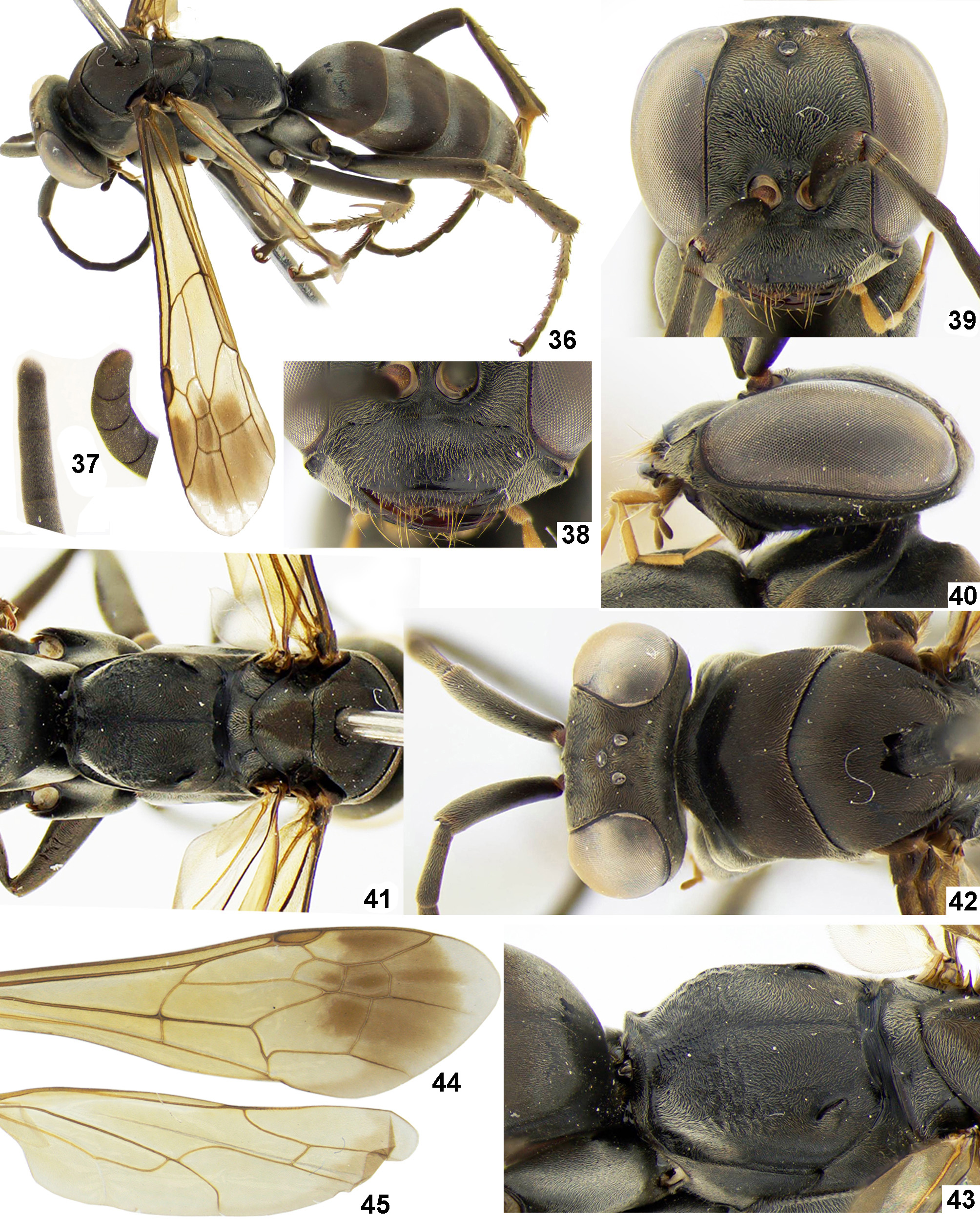
Morochares xuzaifui Loktionov, Lelej & Liu View in CoL , sp. nov.
( Figs 36–45 View FIGURES 36–45 )
Material examined. Holotype, ♀, China, Guangdong, Mt. Nan Kun Shan, 22.V.2010 ( Zaifu Xu ), No 2016000201 [ SCAU] . Paratypes: 1♀, China, Guangdong, Mt. Nan Kun Shan, 4–6.VI.2011 (Zaifu Xu), No 2016001301 [ IBSS] ; 1♀, China, Zhejiang, Wenzhou, Mt. Wuyanling, 28.VII–5.VIII.2005 ( Jingxian Liu ), No 2 0 16000679 [ SCAU] .
Diagnosis. Female. The female of Morochares xuzaifui sp. nov. can be easily separated from those of other congeners by having the forewing yellowish-brown, with a subapical brown fascia and a narrow whitened apical band ( Fig. 44 View FIGURES 36–45 ) and the head in frontal view with the vertex slightly produced beyond the eye top and slightly roundly convex medially ( Fig. 39 View FIGURES 36–45 ). Other characters of importance are: the clypeus with anterior margin straight ( Fig. 38 View FIGURES 36–45 ); the scape 1.5–1.6 times as long as F1; apical flagellomere roundly blunt apically; the propodeum with coarse and more or less straight transverse rugae on dorsum posteriorly and posterior face ( Fig. 43 View FIGURES 36–45 ); and maxillary palps from yellow to light brown ( Fig. 39 View FIGURES 36–45 ). Male. Unknown.
Description. FEMALE. Length: body 12.0– 14.5 mm; forewing 8.8–11.0 mm. Head 1.15–1.22 times as wide as height; MID 0.51–0.54 times as long as head width in frontal view, half of MID 1.03–1.20 times as long as eye width ( Fig. 39 View FIGURES 36–45 ). Ocelli large, not raised; ocellar triangle right-angled; POD: OOD = 1.0–1.2 ( Fig. 42 View FIGURES 36–45 ). Head in frontal view with vertex slightly produced beyond eye top and slightly roundly convex medially ( Fig. 39 View FIGURES 36–45 ). Posterior margin of vertex in dorsal view inconspicuously concave medially or straight ( Fig. 42 View FIGURES 36–45 ). Head in lateral view with frons slightly convex ( Fig. 40 View FIGURES 36–45 ). Temple in dorsal view slightly developed ( Fig. 42 View FIGURES 36–45 ). Gena in profile narrowing towards vertex ( Fig. 40 View FIGURES 36–45 ). Face on both sides of antennal sockets slightly concave. Malar space short ( Fig. 38 View FIGURES 36–45 ). Clypeus slightly convex, 1.08–1.09 times as wide as LID and 2.67–2.95 times as wide as height; anterior margin straight or inconspicuously concave; anterolateral corner rounded ( Fig. 38 View FIGURES 36–45 ). Mandible stout with small subapical tooth. Labrum anterior margin emarginate medially ( Fig. 38 View FIGURES 36–45 ). Maxillary palps 3–6 about same length, fourth segment slightly longer. Flagellum filiform; scape slightly bent; ratio of scape, pedicel and F1–F 4 in holotype 61: 10: 38: 33: 30: 27; scape 1.5–1.6 times as long as F1, and 0.83–0.85 times as long as F1 and F2 combined, and 1.27–1.36 times as long as UID; F1 3.64–3.80 times as long as maximum width and 0.81–0.84 times as long as UID; apical flagellomere roundly blunt apically ( Fig. 37 View FIGURES 36–45 ).
Mesosoma. Pronotum in dorsal view somewhat elongated, medially 0.38–0.42 times as long as maximum width ( Fig. 42 View FIGURES 36–45 ); anterior face low and noticeably inclined, roundly merging into dorsum ( Fig. 40 View FIGURES 36–45 ); posterior border subangulate ( Fig. 42 View FIGURES 36–45 ). Dorsum of mesoscutum convex in anterior half. Dorsum of mesoscutellum and metanotum slightly convex. Metapostnotum constricted medially and slightly concave posteromedially ( Figs 41, 43 View FIGURES 36–45 ). Propodeum in dorsal view 0.89–0.93 times as long as wide ( Fig. 41 View FIGURES 36–45 ); dorsum in lateral view barely convex; dorsum roundly merging with lateral and posterior faces, with longitudinal and shallow furrow medially, sometimes indistinct ( Fig. 43 View FIGURES 36–45 ).
Legs. Proleg without spines, except following: protibia outer face with 2–3 short spines, protibia with few different length spines apically, protarsomere 1 with two short spines on outer face and two longitudinal rows of very short spines ventrally, protarsomeres 2 and 3 with very short spines ventrally; protarsi shortened, protarsomere 1 1.27–1.33 times as long as protarsomere 2–4 combined. Meso- and metafemur without spines except two inconspicuous spines dorso- and lateroapically; meso- and metatibia, meso- and metatarsomere 1 with scattered long and suberect spines; meso- and metatarsomeres 2 and 3 ventral face with three longitudinal rows of short spines; meso- and metatarsomere 4 ventral face with median longitudinal row of short spines and 1–2 spines on each side of row; metatibia longer spur 0.57–0.71 times as long as metatarsomere 1. Tarsomere 5 without spines ventrally. Tarsal claws symmetrical and bifid, inner tooth broad and obliquely truncated.
Wings translucent, yellowish-brown ( Figs 44, 45 View FIGURES 36–45 ). Forewing ( Fig. 44 View FIGURES 36–45 ) with brown fascia on marginal cell (except basally), SMC2 (entirely or apical half), SMC3 (entirely) and 2M (apical part) across wing, and with short somewhat whitened apical band; pterostigma light brown; SMC2 1.50–1.84 times as long as high, narrowed on vein Rs by 0.65–0.70 times its own length on vein M, receiving crossvein 1m-cu at basal 0.65–0.69; SMC3 0.97– 1.09 times as long as SMC2 on vein M, 0.73–1.03 times as long as SMC2 on vein Rs, narrowed on vein Rs by 0.55– 0.71 times its own length on vein M, receiving crossvein 2m-cu at basal 0.61–0.71; crossvein 2rs-m straight or hardly arched towards wing apex; crossvein 3rs-m curved or arched towards wing apex; crossvein cu-a straight, originating right to or just beyond separation of vein M+CuA; vein M ending slightly before wing margin; vein Cu1 almost touching wing margin. Hind wing ( Fig. 45 View FIGURES 36–45 ) with barely brownish apical portion; crossvein cu-a interstitial or slightly anterofurcal, arched before confluence with vein A.
Metasoma slightly wider than mesosoma in dorsal view. Posterior margin of T1 usually straight, T2–T5 and S1–S5 slightly emarginate medially. S5 somewhat compressed laterally, forming indistinct longitudinal median carina in posterior half.
Sculpture. Body matt, except clypeus apically, mandible and metapostnotum posteromedially polished, metasoma sometimes somewhat polished. Body with inconspicuous micropunctures. Metapostnotum with fine transverse stria ( Fig. 43 View FIGURES 36–45 ). Propodeum with transverse coarse and more or less straight rugae on dorsum posteriorly and posterior face; dorsum microshagreened ( Fig. 43 View FIGURES 36–45 ). S6 with carina polished posteromedially. Antenna and legs matt.
Colour and pubescence. Body black ( Fig. 36 View FIGURES 36–45 ); top of apical flagellomere somewhat yellowish; mandible dark brown, sometimes light brown subapically; maxillary palps from yellow to yellowish-brown; claws and spurs brown; longitudinal brush on metatibia inner face golden-brown. Body without setae except following: upper frons and pronotum with scattered thin short erect setae; labrum and mandible with few thin and pale setae; S1–S4 with short scattered pale setae; S5 with short or long pale setae; T6 and S6 with long pale setae. Head, mesosoma and legs with iridescence micropubescence; frons with somewhat yellowish micropubescence; dorsum of pronotum, mesoscutum, mesoscutellum, meso- and metapleuron and sides of propodeum with somewhat coppery micropubescence; propodeum posterior face with silver micropubescence. Metasomal segments with iridescent micropubescence; T2–T4 anteriorly with band of silver micropubescence more distinct in anterolateral portion ( Fig. 36 View FIGURES 36–45 ).
MALE. Unknown.
Distribution. China: Zhejiang and Guangdong.
Etymology. This species is named after well-known Chinese hymenopterologist, Prof. Xu Zai-fu (1965–2017).
Remarks. The female of Morochares xuzaifui sp. nov. is similar with those of M. sinica sp. nov. and M. wolfi sp. nov. by having the forewing yellowish-brown, but it can be easily distinguished from them by the following characters: the scape 1.5–1.6 times as long as F1 and the F1 1.27–1.36 times as long as UID in M. xuzaifui sp. nov. vs the scape 1.24–1.28 times as long as F1 and the F1 1.05–1.09 times as long as UID in M. sinica sp. nov.; the head in frontal view with the vertex slightly produced beyond the eye top and slightly convex medially ( Fig. 39 View FIGURES 36–45 ) in M. xuzaifui sp. nov. vs the vertex not produced beyond the eye top and almost straight medially in M. sinica sp. nov. ( Fig. 7 View FIGURES 6–14 ); the temple in dorsal view slightly developed ( Fig. 42 View FIGURES 36–45 ) in M. xuzaifui sp. nov. vs not developed in M. sinica sp. nov. ( Fig. 9 View FIGURES 6–14 ); the clypeal anterior margin straight ( Fig. 38 View FIGURES 36–45 ) in M. xuzaifui sp. nov. vs distinctly roundly produced in M. wolfi sp. nov. ( Fig. 32 View FIGURES 27–35 ).
No known copyright restrictions apply. See Agosti, D., Egloff, W., 2009. Taxonomic information exchange and copyright: the Plazi approach. BMC Research Notes 2009, 2:53 for further explanation.