
Hydrolagus mirabilis, Collett, 1904
|
publication ID |
https://doi.org/ 10.1111/jfb.14302 |
|
DOI |
https://doi.org/10.5281/zenodo.10993028 |
|
persistent identifier |
https://treatment.plazi.org/id/DC4C87C8-AC61-BD4B-FCC2-F8EE44F2F954 |
|
treatment provided by |
Felipe |
|
scientific name |
Hydrolagus mirabilis |
| status |
|
3.2 | Hydrolagus mirabilis View in CoL
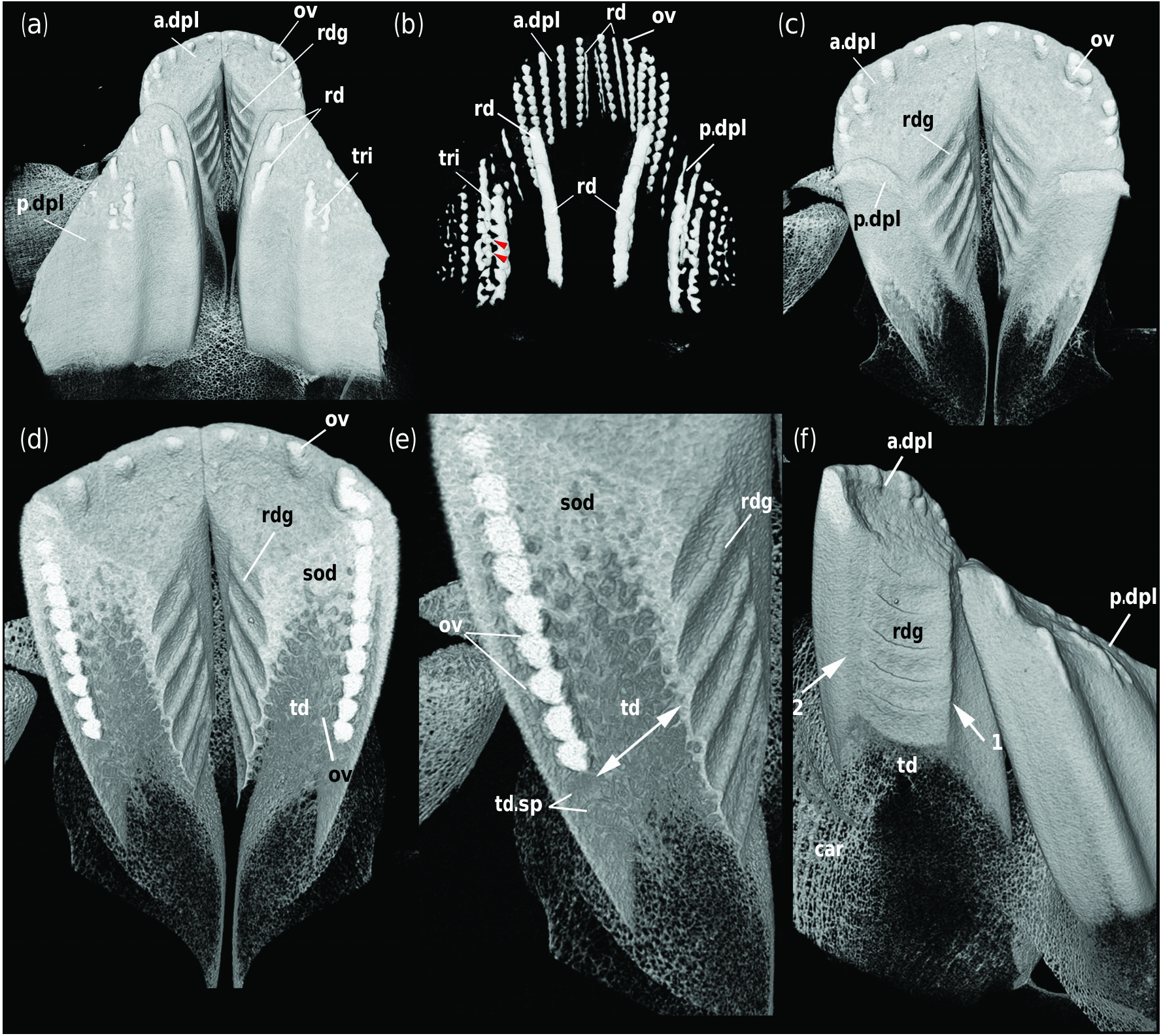
The anterior and posterior dental plates of a young juvenile and adult of Hydrolagus ( Figures 4 View FIGURE 4 and 5) are comparable to those of Chimaera , with an ovoid series of whitlockin in the anterior plate and a parasymphyseal rod in the adult (Figure 5b), and a small number of extra rods in the ovoid series in the juvenile ( Figure 4b View FIGURE 4 ). With respect to the posterior plate, in the earlier growth stage both an ovoid series and rods are present along the labial margin of the plate, with two rods present lingually and medially. In addition, a tritoral pad develops posterior to these two rods, appearing to form via the incorporation of individual ovoids ( Figure 4b View FIGURE 4 , arrowheads on left of image, Supporting Information Files S1–S3). In the adult, broad, well–developed tritoral pads are present in this position and with respect to the more medial rods, the more posterior has become intensely vascularized to resemble the more labial pad. However, the more anterior rod shows only a few openings that represent incorporation of blood vessels within the mineralized dentine. Along the labial margin, both series of ovoids and rods are present (Figure 5b).
In both growth stages, trabecular dentine below the oral surface has been infilled with sclerotic dentine ( Figures 4d,e View FIGURE 4 and 5d,e) and becomes deeply worn, exposing the ovoids and tritoral pads that resist deep wear ( Figures 4c,d,f View FIGURE 4 and 5c,d,f). Serial ridges are present on the lingual surface of the anterior dental plate, with five thick ridges in the juvenile, comparable in morphology to those in Chimaera ( Figure 4 View FIGURE 4 ). However, in the adult the ridges are more numerous but less distinct (Figure 5). Bulbous expansions at the end of these ridges are absent in both these growth stages ( Figures 4c,f View FIGURE 4 and 5c,f). As in Chimaera , there may be correspondence between the ridges and the forming ovoids ( Figures 4e View FIGURE 4 and 5e, double arrow). In addition, trabecular dentine forms in advance of these ridges forming in both growth stages ( Figures 4f View FIGURE 4 and 5f), but always inside a shell of outer dentine. The posterior furrow and oral-aboral ridge are present in both growth stages although the furrow appears to be deeper in the adults ( Figures 4f View FIGURE 4 and 5f, arrows 1 and 2).
No known copyright restrictions apply. See Agosti, D., Egloff, W., 2009. Taxonomic information exchange and copyright: the Plazi approach. BMC Research Notes 2009, 2:53 for further explanation.