
Anodontophora tuvensis, Pomorski, Romuald J., 2007
|
publication ID |
https://doi.org/ 10.5281/zenodo.177440 |
|
DOI |
https://doi.org/10.5281/zenodo.6241284 |
|
persistent identifier |
https://treatment.plazi.org/id/DC6DDF7F-FFF7-FFD1-FF21-2D72FA9B7839 |
|
treatment provided by |
Plazi |
|
scientific name |
Anodontophora tuvensis |
| status |
sp. nov. |
Anodontophora tuvensis sp. nov.
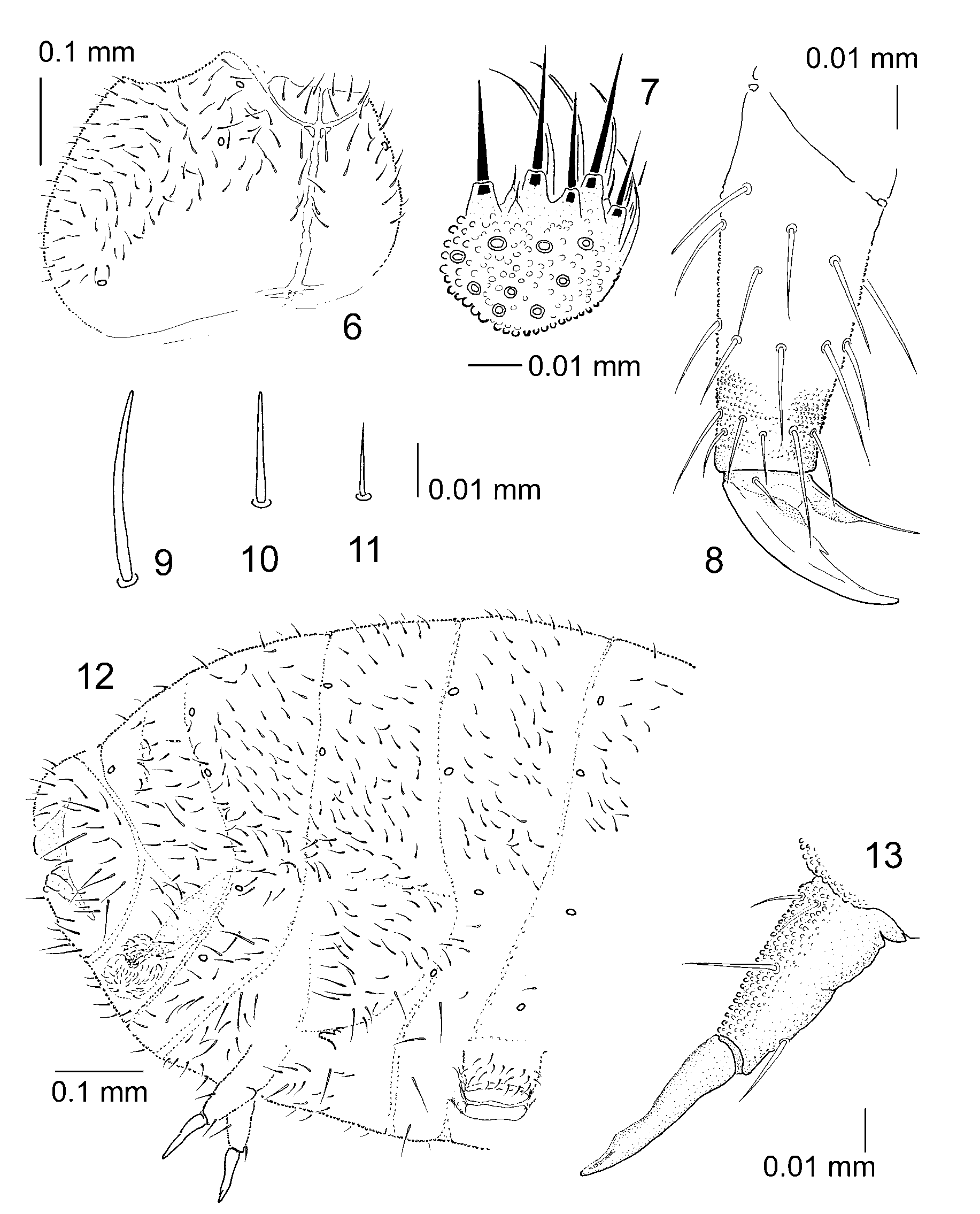
Figs 1-13 View FIGURES 1 – 5 View FIGURES 6 – 13
Description. Color uniform bluish-grey. Length of holotype (reproductive male) 1.5 mm. Body shape cylindrical, short, without anal spines ( Fig. 1 View FIGURES 1 – 5 ). Granulation of body surface fine and uniform, with distinctly marked antennal bases.
Antennae nearly as long as head. Antennal segment IV with subapical organite ( Fig. 3 View FIGURES 1 – 5 ). Microsensillum on antennal segment IV in latero-external position, above posterior setae ( Fig. 5 View FIGURES 1 – 5 ). Subapical organite present. Antennal III sense organ consisting of 4 guard setae, 5 papillae, 2 spherical, finely granulated sensory clubs, and 2 typical sensory rods ( Figs 4 and 5 View FIGURES 1 – 5 ). Antennal segment III with microsensillum slightly below antennal organ III.
A small area of finer granulation in the position of postantennal organ which is absent ( Fig. 2 View FIGURES 1 – 5 ). Labial palp of type O with 9 proximal setae ( Fig. 7 View FIGURES 6 – 13 ).
Pseudocellar formulae: dorsal 32/233/33332, ventral 3/000/2202; 2 pseudocelli on each of three subcoxae (arrangement of pseudocelli as in Figs 1 View FIGURES 1 – 5 , 6 and 12 View FIGURES 6 – 13 ). Pseudocelli well chitinised. First ventral cephalic pseudocellus located near mouth apparatus. Anterior cephalic pseudocelli arranged in a triangle, two located at antennal base, one somewhat behind submedially ( Figs 1 and 2 View FIGURES 1 – 5 ). Parapseudocelli absent.
Dorsal chaetotaxy as in Fig. 1 View FIGURES 1 – 5 , nearly symmetrical, poorly differentiated into apically rounded macro- and mesosetae and apically pointed microsetae ( Figs 9–11 View FIGURES 6 – 13 ). Body sensilla invisible. Thoracic terga II and III with microsensilla laterally.
Ventral tube with 19+19 setae and 1+1 setae at base. Retinaculum with 3+3 teeth and without setae at base. Manubrium with numerous setae posteriorly. Dens with 1 anterior and 3 posterior setae, mucro as in Fig. 13 View FIGURES 6 – 13 . Claw with inner denticle only. Empodial appendage somewhat shorter than claw, with small basal lamella ( Fig. 8 View FIGURES 6 – 13 ). Tibiotarsal distal whorl with 11 setae. Male ventral organ absent.
Type material. Holotype, reproductive male (mounted on slide); Russia, southern Krasnoyarskiy Kray, Republic of Tuva, western Sayan Mts., northern slope of Oiskiy Ridge, ca 1800 m a.s.l., near Olenya Rechka, mountain tundra with Salix and Betula rotundifolia , in moss; 27 VI 1990; leg. S. Stebaeva. The holotype is in the collection of the Department of Systematic Zoology and Zoogeography, University of Wrocław.
Etymology. The species has been named after the Republic of Tuva, where the specimen was found.
Acknowledgments. I am most grateful to Sofiya K. Stebaeva (Moscow) for the specimens and my departmental colleague Andrzej ElŻanowski for editorial help. This work was supported by intramural grant 1018/S/IZ/2005.

No known copyright restrictions apply. See Agosti, D., Egloff, W., 2009. Taxonomic information exchange and copyright: the Plazi approach. BMC Research Notes 2009, 2:53 for further explanation.