
Cerogobius petrophilus, Kovačić & Bogorodsky & Troyer & Tornabene, 2019
|
publication ID |
https://doi.org/ 10.11646/zootaxa.4565.2.2 |
|
publication LSID |
lsid:zoobank.org:pub:1E01FCF7-0D6D-4E8C-BDF2-E2D8068934B5 |
|
DOI |
https://doi.org/10.5281/zenodo.5928731 |
|
persistent identifier |
https://treatment.plazi.org/id/DE205C17-FF86-FFB1-FF3A-FFD94A77DDC3 |
|
treatment provided by |
Plazi |
|
scientific name |
Cerogobius petrophilus |
| status |
sp. nov. |
Cerogobius petrophilus sp. nov.
Horned goby
( Figures 1 View FIGURE 1 –12, Table 1)
Holotype. SMF 35961, female, 16.32 + 3.54 mm, Red Sea , Saudi Arabia, Thuwal, Al Fahal reef, N 22°13.656' E 38°58.185', rubble-sand patch at base of reef, 8 m, coll. E.M. Troyer, 22 October 2017. GoogleMaps
Paratypes (collected with holotype) GoogleMaps . PMR VP4501 View Materials , female, 15.36 + 3.21 mm; SMF 35962, female, 16.72 + 3.47 mm; SMF 35963, female, 14.81 + 2.97 mm; SMF 35964, female, 14.09 + 2.80 mm ; SMF 3 About SMF 5965, female, 12.97 + 2.84 mm; UW 158279, 3 , 14.1 + 3.0 mm (cleared and stained), 12.6 + 2.0 mm (cleared and stained), 12.4 + 2.8 mm (preserved in 95% ethanol), all tissue sampled .
Diagnosis. As for genus.
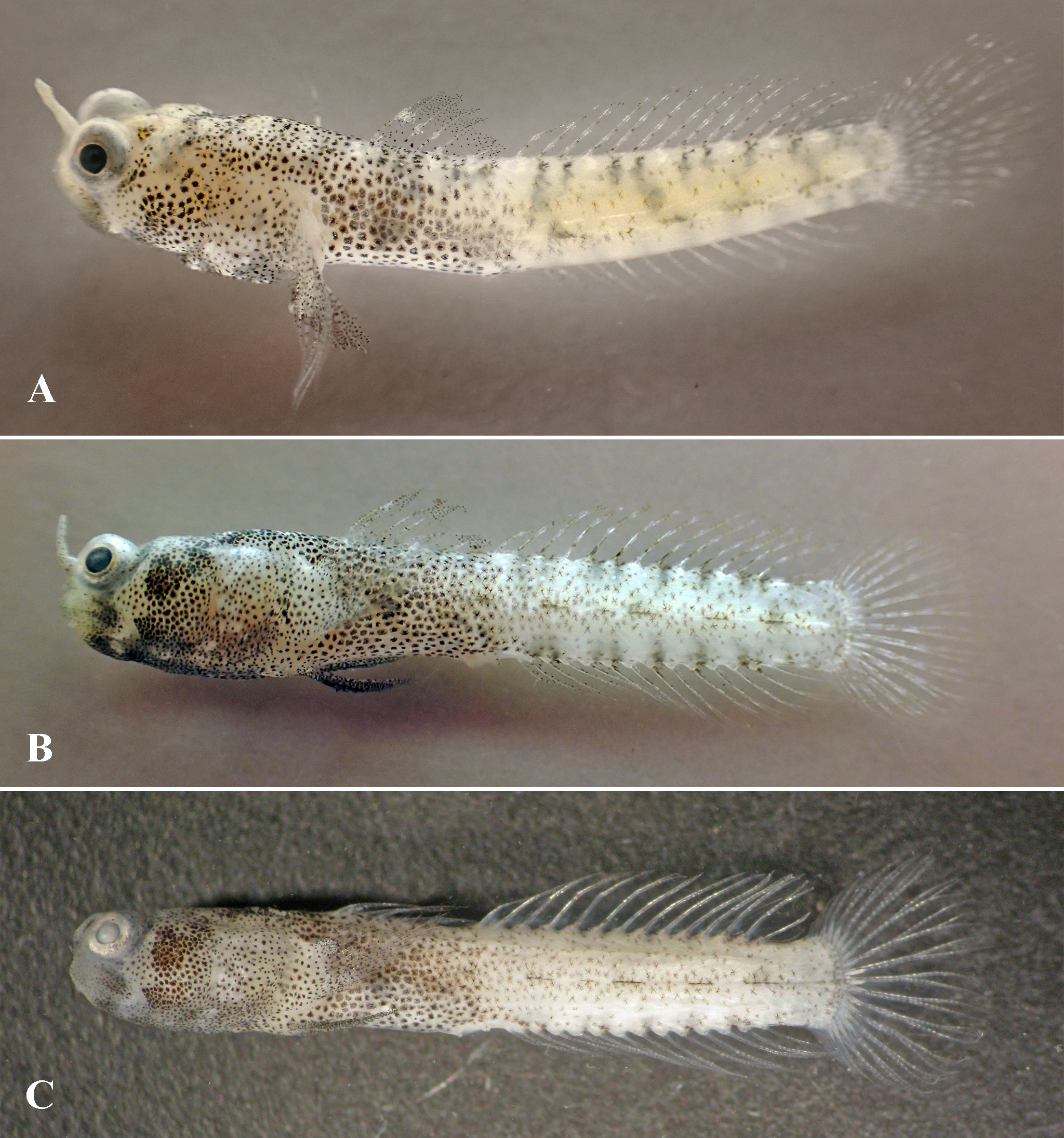
Description (morphometric values in the text are presented first for the holotype followed by ranges for paratypes; meristic values, if variable, the same). Body elongate, its depth at anal-fin origin 8.2 (7.2–7.9) in SL, laterally compressed posteriorly, with caudal peduncle deep and short, caudal-peduncle depth 0.9 (0.8–0.9) of body depth at anal-fin origin, caudal peduncle depth 1.12 (1.04–1.17) of caudal peduncle length ( Fig. 3 View FIGURE 3 ). Head of moderate size, the length 3.0 (3.1–3.3) in SL, width 5.4 (5.3–5.8) in SL, depth 6.1 (5.8–6.3) in SL, and slightly depressed, its depth 0.9 (0.9–1.0) of width. Snout very steep in profile from lateral view, and short, its length 0.5 (0.4–0.5) of eye diameter, 9.4 (8.1–10.4) in head length, with unpaired horn-like tentacle at the level of nostrils in snout midline, slightly shorter in height measured from the anterior side than the eye diameter ( Fig. 1A View FIGURE 1 ). Anterior nostril a tube without process from rim; posterior nostril a short tube ( Fig. 1A View FIGURE 1 ). Eyes dorsolateral, moderately large, eye diameter is 4.5 (4.0–4.5) in head length, orbit projecting far forward and distinctly elevated above dorsal profile. Interorbital narrow, 4.4 (4.5–5.6) of eye diameter. No tentacle above eye. Mouth terminal, oblique, jaws ending anteriorly about equally, lower jaw not projecting anteriorly. Mouth moderately large, posterior angle of jaws behind vertical through posterior edge of eye. Upper lip wider than cheek, covered with a thin dermal flap, extending upwards over cheek and nearly reaching eye ( Fig. 1B View FIGURE 1 ) (only in paratype SMF 35963 dermal flap shorter, Fig. 1D View FIGURE 1 ). Cheek very narrow, depthless than upper lip width. Teeth in both jaws in a single row posteriorly, becoming 3-4 rows anteriorly, with outer row as enlarged recurved canines, inner rows smaller, pointed, and irregularly spaced. Small mental frenum at mid-ventral behind lower lip. Branchiostegal membranes fused to isthmus, gill openings restricted to pectoral-fin base. Lower limb of first gill arch joined to gill cover by membrane. No spines on preopercle.
Fins. First dorsal fin VI, second dorsal fin I,12–13 (I,12:2, I,13:5); anal fin I,12–13 (I,12:2, I,13:5); branched caudal-fin rays 13, segmented 17. Pectoral-fin rays 13–14 (13:8, 14:3, both sides, right side cut in paratype SMF 35964), all rays branched except lowermost and uppermost upper rays not free at tips. Pectoral girdle without flaps on anterior edge. Pelvic fins I,5 + 5,I, left and right fin completely separated, distant and without frenum, rays branched (except fifth ray), fourth ray the longest, rays progressively shorter towards the first ray and pelvic spine ( Fig. 1C View FIGURE 1 ). Spines of first dorsal fin not elongate or filamentous, first to fifth spine of first dorsal fin subequal in length, third and fourth only slightly longer, sixth spine short; fourth or fifth to sixth spine of first dorsal fin nearly reaching to origin of second dorsal fin when folded down. Origin of first dorsal fin behind vertical at pectoral-fin base. Origin of anal fin posterior to vertical at origin of second dorsal fin i.e. opposite about first segmented ray of second dorsal fin or slightly behind. Pectoral fins not reaching posteriorly to below origin of second dorsal fin. Pelvic fins short, ending distantly in front of anus, 0.7 (0.6–0.8) of distance between origin of fins and anus, shorter than pectoral fins, 0.8 (0.8–0.9) of pectoral-fin length. Caudal fin rounded, shorter than head, 1.5 (1.5) in head length.
Squamation. Head and body completely scaleless. Cyanine Blue stain confirmed lack of any scales or scale pouches on the body.
Cephalic sensory systems ( Fig. 2 View FIGURE 2 ). No head canals. Rows of head sensory papillae were counted on the left side of all type material, except paratype SMF 35962 in which papillae were hard to notice on the damaged skin surface. In addition, some rows were not visible in every specimen. Rows of head sensory papillae were heavily reduced, with rows missing or reduced to one to few papillae and additional individual papillae present on the position of absent head canals. Preorbital rows: snout with three median preorbital series, each as single papilla, upper row r (1) at middorsal between eyes and posterior nostrils, and row s (1) at anterior nostril, and with s 3 (1) above upper lip. Lateral series c on snout in three parts: superior horizontal row c 2 between anterior and posterior nostril (2); inferior rows: upper horizontal c 2 (2) and lower horizontal c 1 (2) above upper lip. Suborbital rows: rows a and b absent; four very short transverse suborbital rows of sensory papillae (1–4) present, row 1 oblique, reaching upper lip, row 2 short and distant from eye, close to row d, row 3 and 4 short, close to posteroventral edge of eye and distant from row d (1: 4–5, 2: 2, 3: 2–3, 4: 3); row d (3–4 + 2–3) short, above posterior part of upper lip, and continuing shortly on cheek. Preoperculo - mandibular rows: external row e (8–9 + 2–6) longitudinal and uniserial, divided into anterior and posterior sections; internal row i (4–6 + 2–4) longitudinal and uniserial, divided into anterior and posterior sections, mental row f (2–3) behind frenum. Oculoscapular rows: anterior upper longitudinal row x 1 divided (3 + 1–2); posterior longitudinal row x 2 (2); posterior part of row x 1 and row x 2 not visible in every specimen; anterior lower transverse row z as single papilla, not visible in every specimen; rows q and y not present; transverse axillary rows as 1 and as 2 as single papilla, not visible in every specimen; as 3, la 1 and la 2 not present. Opercular rows: two papillae at position of the missing preopercular canal (marked PC on Fig. 2 View FIGURE 2 ); transverse row ot (2–4); superior longitudinal row os (1–2); and inferior longitudinal row oi (1) not visible in every specimen. Anterior dorsal rows: two papillae behind posterior upper edge of eye on the position of the missing anterior oculoscapular canal (marked OC on Fig. 2 View FIGURE 2 ); transverse row n (2–4) behind eye, rows o and m not present, row g longitudinal (3–5); row h (1–2) not visible in every specimen. Interorbital rows: two pairs of larger papillae are present in interorbital area anteriorly and posteriorly on the place of the missing anterior oculoscapular head canal (marked OC on Fig. 2 View FIGURE 2 ).
Color of live and fresh female ( Fig. 3A and 3B View FIGURE 3 ). Head and body translucent i.e. showing limited transparence. Cheek, postorbital head, prepectoral and prepelvic areas, and anterior body to below origin of the second dorsal fin densely covered with variable-sized melanophores. Remaining body covered with few blackish speckles; row of eight, small, irregular black spots along base of second dorsal fin. Snout, tentacle, and orbits pale cream. First dorsal fin with translucent membranes, outer half of fin with many black dots, basal half with few dots on spines and membranes, about middle of first four spines and first interspinous membrane with whitish speckles. Second dorsal and caudal fins with translucent membranes and a row of alternating white and blackish speckles along spine and soft rays. Anal fin with whitish rays and translucent membranes. Pelvic fins densely dotted with numerous melanophores.
Color of preserved female ( Fig. 3C View FIGURE 3 ). Coloration of preserved female similar to fresh specimen. Body opaque and whitish with dark brown pigmentation with large melanophores mostly on head and in anterior part of body and no other pigments visible. Head and body to below origin of the second dorsal fin densely pigmented with variable-sized melanophores with very poorly recognizable pattern. Snout weakly pigmented, dark vertical bar around posterior edge of eye and pigmented bar on check from eye to lips, followed by a pale band, containing scattered melanophores, that extends from nape over posterior cheek and angle of jaws to below it. Preopercle darker and less densely pigmented opercle and pectoral-fin base. Ventral side of head heavily pigmented with tiny melanophores except for pale area below each side angle of jaw. Breast in front of pelvic fin pale, belly pigmented. Eyes with bright whitish iris and blurred whitish pupil due to the alcohol fixation. The melanophores on the body behind the vertical through origin of the second dorsal are scattered, smaller and more obscure than anteriorly. Only visible patterns are dashed narrow line along lateral midline and dark spots at the origin of each dorsal and anal fin ray. First dorsal fin dotted with dark brown melanophores and with white dots restricted to anterior origin and at spines as oblique pattern of dots going downwards from spines I to spine VI; second dorsal fin with white spine and rays, and mostly translucent interradial membranes, melanophores, if present, rare at spine and some ray origins and also rarely scattered on rays; caudal and anal fin with transparent membranes. Pectoral and pelvic fins intensively dotted with dark brown melanophores, matching anterior body pigmentation ( Fig. 1C View FIGURE 1 ).
Alive coloration of non-collected material of unidentified sex ( Figs. 5 View FIGURE 5 A-C). Coloration of head blackish ( Figs. 5A, B View FIGURE 5 ) or pale cream ( Fig. 5C View FIGURE 5 ), covered with many irregular melanophores. In the specimen with blackish head ( Figs. 5A, B View FIGURE 5 ), snout, tentacle, orbit and jaws pale gray suffused with pale orange; postorbital head mainly black except for few small white spots on opercle and for an irregular, oblique, white band, containing scattered melanophores, that extends from nape to posterior chin. Individuals with blackish head are possibly males, considering their dark coloration compared to the type specimens which all are females.
Etymology. The genus name Cerogobius is formed from the Greek word keras (Latin = ceros; horn) and the Latin word gobio (small fish or gudgeon), and is in reference to the horn-like tentacle on the head. The specific name petrophilus is formed from the Latin words petra (rock) and philus (having an affinity for), and is in reference to the rocky habitat where the species was found.
Distribution. At present only known from the northern part of the Red Sea, from collection taken at Thuwal in Saudi Arabia, and SCUBA observations at the southern tip of Ras Mohammed national park and in Marsa Shagra, Marsa Alam, in Egypt.
Habitat. Collected on rubble-sand patches at the base of isolated coral blocks near the margin of Al Fahal reef in the central Red Sea, near Thuwal, Saudi Arabia ( Fig. 5D View FIGURE 5 ). The holotype and paratypes were found living inside small holes in large pieces of dead coral rock covered with short algae at a depth of 8 m. Two other specimens, not examined in this study, were collected at a depth of 15 m in May 2017 at the same locality. Observations in other localities were made at depths of about 5 m.
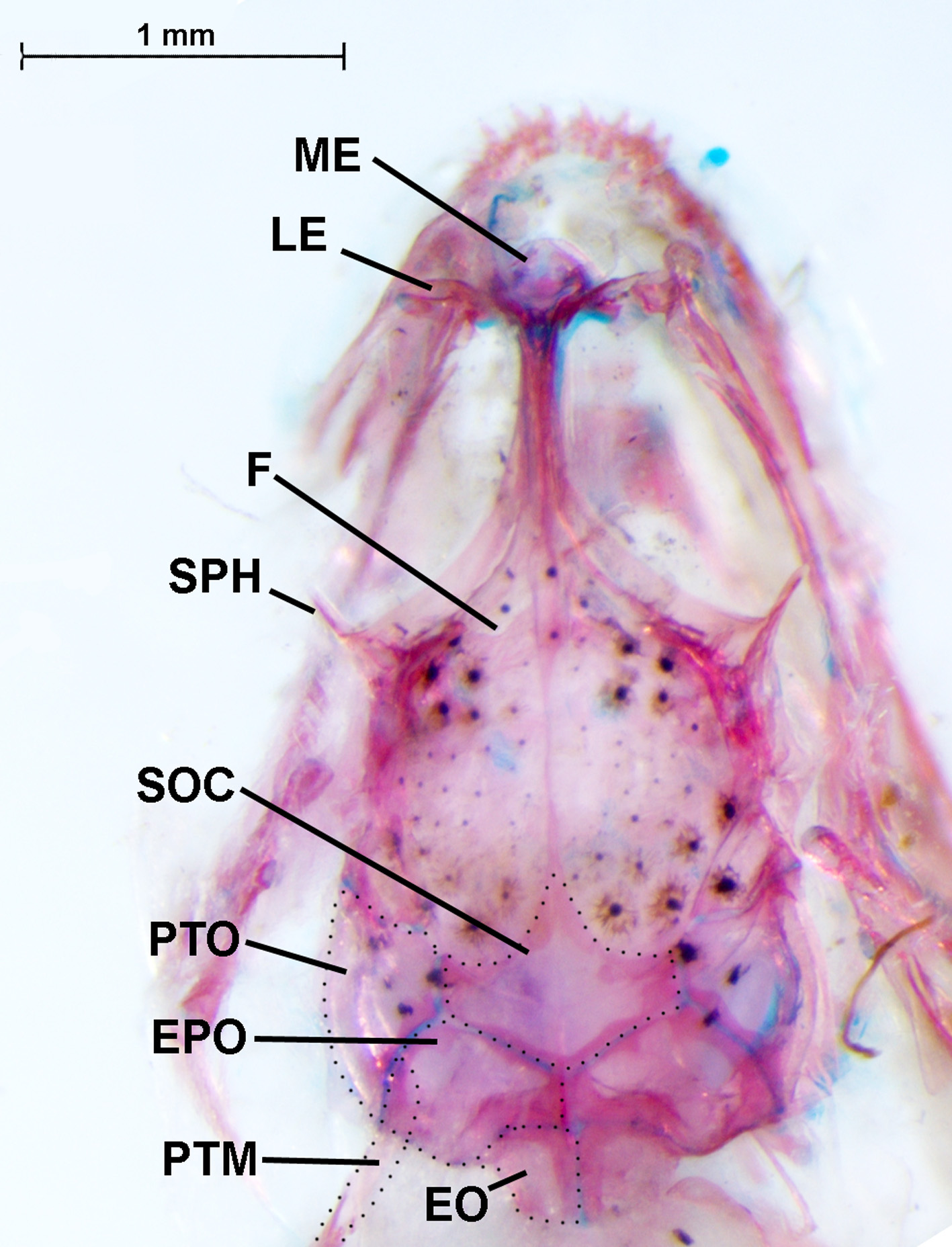
Osteology. In general, the osteology of Cerogobius petrophilus is remarkably conservative in comparison to other gobies. The overall osteology shows a broad suite of characters that are shared across most generalized of species of the Gobiidae , especially species with a 32211-0 dorsal pterygiophore pattern and six spines in the first dorsal fin. Images of the main skeletal elements ( Figs. 6 View FIGURE 6 –12) and generalized descriptions are provided here to serve as the basis of future comparative osteological studies.
Cranium ( Fig. 6 View FIGURE 6 ). The roof of the cranium in C. petrophilus is primarily comprised of the frontals and sphenotics anteriorly, and the supraoccipital, pterotic, epiotics posteriorly. The sphenotics have a prominent lateral flange that forms the posterolateral margin of the orbit, with the anterior orbit being formed from the lateral ethmoids. The supraoccipital bears a low dorsal crest on its posterior half, and is enclosed posteriorly by the epiotics, laterally by the pterotics, and anteriorly by the frontals. The pectoral girdle connects to the cranium dorsally via the posttemporal, which is attached to the epiotic.
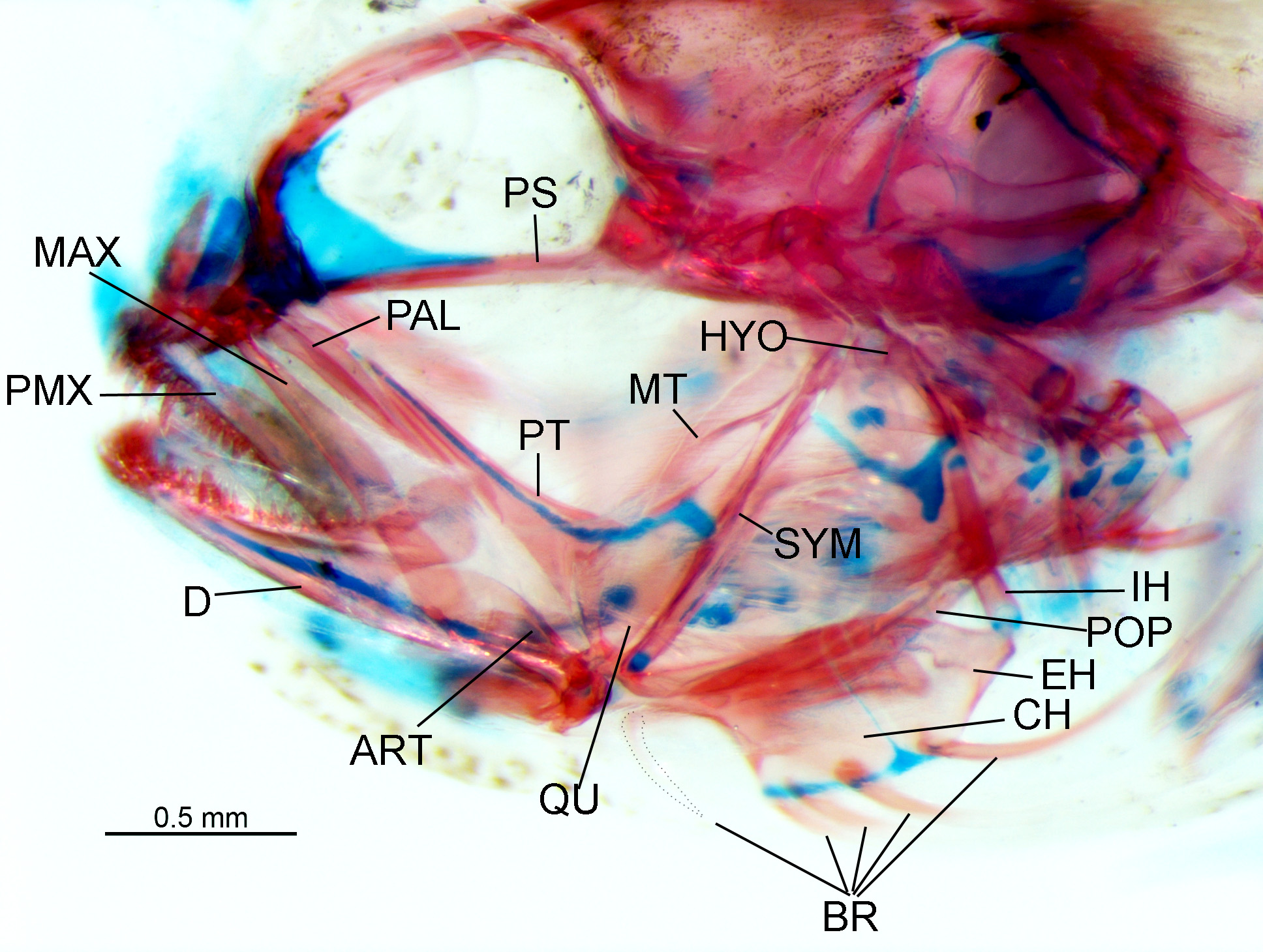
Jaws and suspensorium ( Fig. 7 View FIGURE 7 ). The premaxilla is nearly as long as the maxilla, and bears a moderately long ascending process that is approximately as tall as half the horizontal length of the premaxilla. The dentary is stout and robust with a large coronoid process. A thin Meckel’s cartilage runs along the medial face of the dentary and continues posteriorly onto the articular. The anterior arm of the suspensorium is formed from the descending process of the palatine and ectopterygoid, the latter overlapping the former along approximately one-half of its length. The palatine is roughly T-shaped, with the anterior heads articulating with the lateral ethmoid and a lateral process on the anterior head of the maxilla. A wide metapterygoid abuts the symplectic along its posteroventral surface, and joins the quadrate via a cartilaginous connection anteroventrally. The symplectic connects to the hyomandibular via a thin layer of cartilage, unlike the connections between the metapterygoid and the hyomandibular, and between the hyomandibular and the preopercle, which are both to bone.
Hyoid arch ( Fig. 7 View FIGURE 7 ). A relatively elongate, narrow interhyal serves as the main connection between the epihyal+ceratohyal complex and the hyomandibular. The branchiostegal rays are poorly ossified in the specimens examined here. The first branchiostegal ray originates from the thin, elongate anterior arm of the ceratohyal. The next three branchiostegal rays originate from the stout, deep, trapezoid-shaped posterior end of the ceratohyal, which itself is joined synchondrally do the epihyal. The last branchiostegal ray originates from the epihyal, which is triangular in shape.
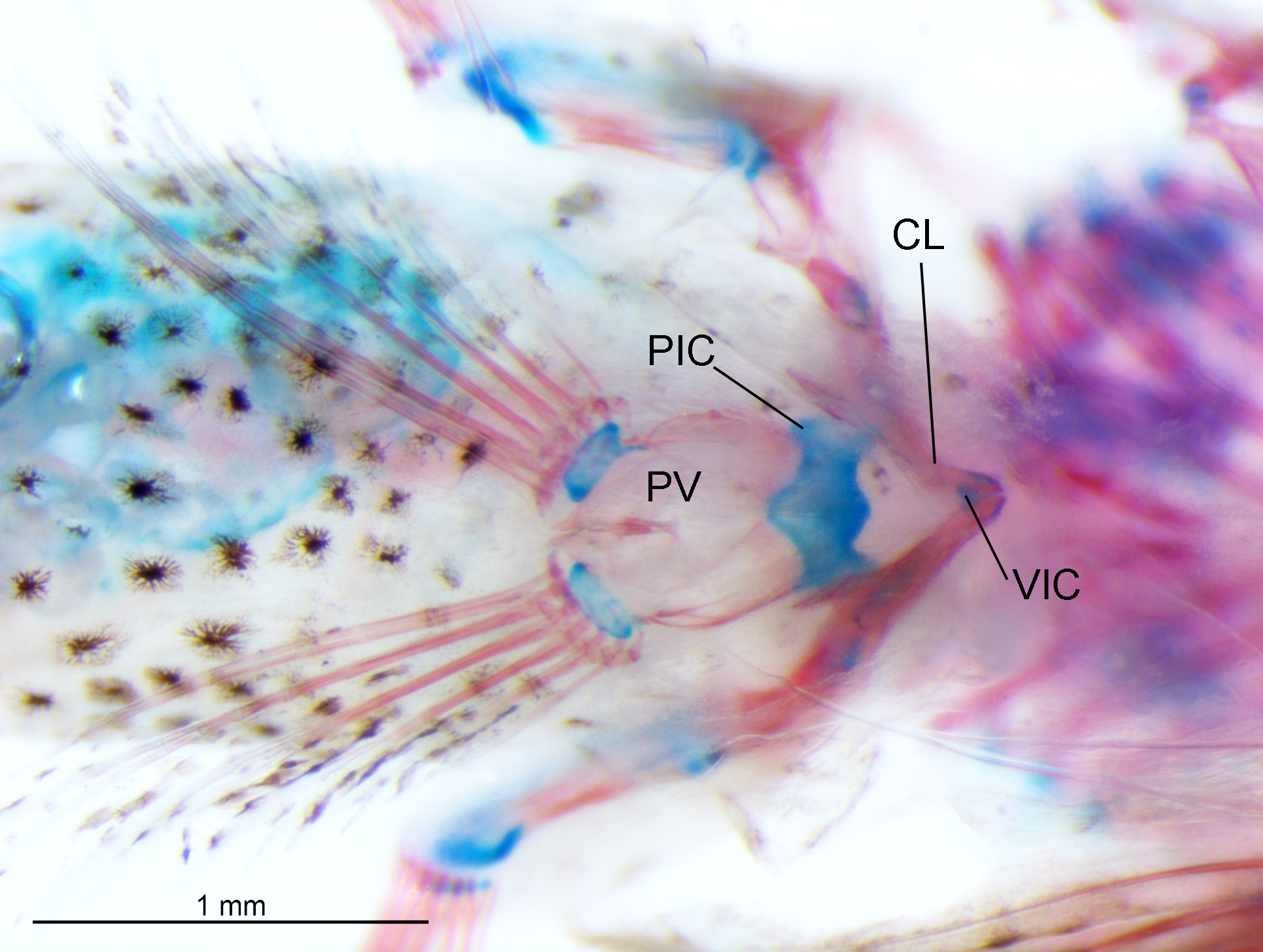
Pectoral and pelvic girdle ( Figs. 8–9 View FIGURE 8 View FIGURE 9 ). The posttemporal has two well-developed anterior limbs that connect the pectoral girdle to the neurocranium via attachments to the epiotic (dorsal limb) and the intercalar (ventral limb). There are four pectoral radials that are poorly ossified and surrounded by a thin layer of cartilage. The dorsalmost radial is small and triangular, with the remaining radials being larger and square to dumbbell shaped. The scapula is small, unossified, and has a large foramen dorsally. The scapula synchondrally connects the cleithrum anteriorly, the coracoid ventrally, and the pectoral radials laterally. The cleithrum has a deeply forked dorsal surface, and is attached ligamentously to the supracleithrum. Ventrally, the cleithrum has two pointed processes. The elongate anterior process connects to the corresponding process on the cleithrum of the opposing side via the ventral intercleithral cartilage. The shorter posterior process on the ventral surface of the cleithrum connects to the pelvic bone via the pelvic intercleithral cartilage.
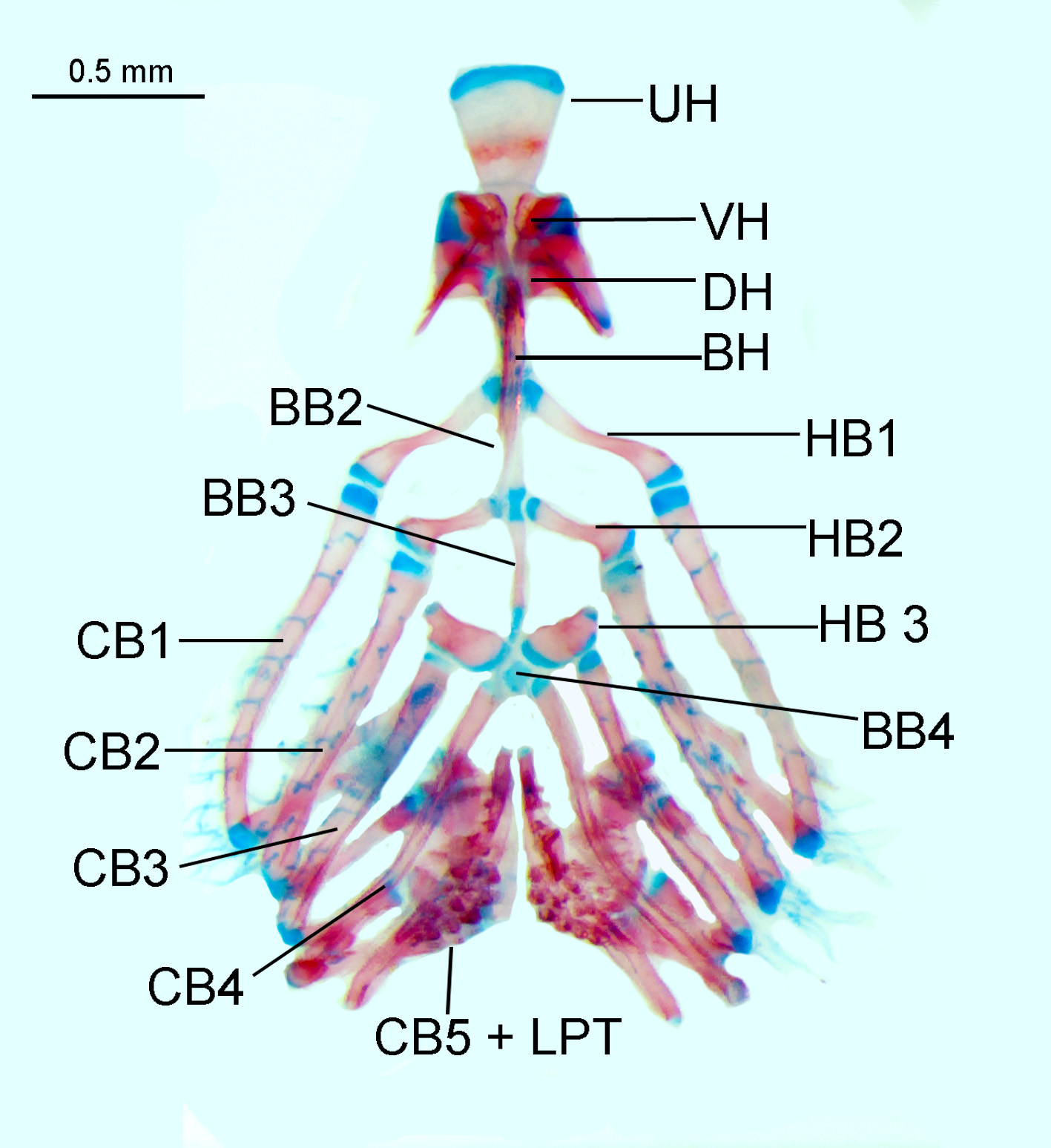
Gill arches ( Figs. 10 View FIGURE 10 –11). The primary elements of the ventral gill arches consist of a series of five pairs of ceratobranchials. Ceratobranchials 1–4 are slender and elongate, with ceratobranchials 1–3 each being bound to hypobranchials 1–3, respectively. Hypobranchials 1 and 2 are slender and elongate. Hypobranchial 3 is short and stout. Ceratobranchial 4 connects directly to basibranchial 4, which is small and unossified. Basibranchials 1–3 are elongate and well ossified. Ceratobranchial 5 forms a slender lower pharyngeal tooth plate, which is completely separate from the tooth plate on the opposing side. The urohyal is broadly spatulate with a cartilaginous anterior margin. The dorsal gill arches are composed primarily of four pairs of epibranchials. Epibranchial 1 has a deeply forked dorsal head, with the posterior fork connecting to the pharyngobranchial toothplate 2 via an elongate interarcual cartilage. Epibranchials 2 and 3 articulate with pharyngobranchial toothplates 2 and 3 respectively. Epibranchial 4 articulates with pharyngobranchial toothplate 4, which is largely fused to pharyngobranchial toothplate 3 to form a single toothed surface.
Axial and caudal skeleton (Fig. 12). There are 10 precaudal vertebrae and 17 caudal vertebrae. The dorsal-fin pterygiophore pattern is 3-22110. Two anal-fin pterygiophores insert anterior to the first haemal arch. There are ossified epineurals visible in association with vertebrae 1–8, with unossified elements visible on at least the next two vertebrae. There are no dorsal or ventral postcleithra visible. Several elements in the axial and caudal skeleton are not well ossified or did not take up alizarin red stain, but are nevertheless still present as hard, translucent elements. Most second dorsal-fin pterygiophores are unstained, as are the epural, hypural 5, parhypural, and the ventral half of the last haemal spine. The urostyle is fused to hypurals 3 + 4 (fused), which are separated by gap from hypurals 1 + 2 (fused). The dorsal and ventral procurrent cartilage each support three unsegmented procurrent caudal-fin rays. Single segmented fin-rays are associated with the epural, hypural 5, and the parahypural. Hypurals 1 + 2 and 3 + 4 are each associated with seven branched fin-rays.
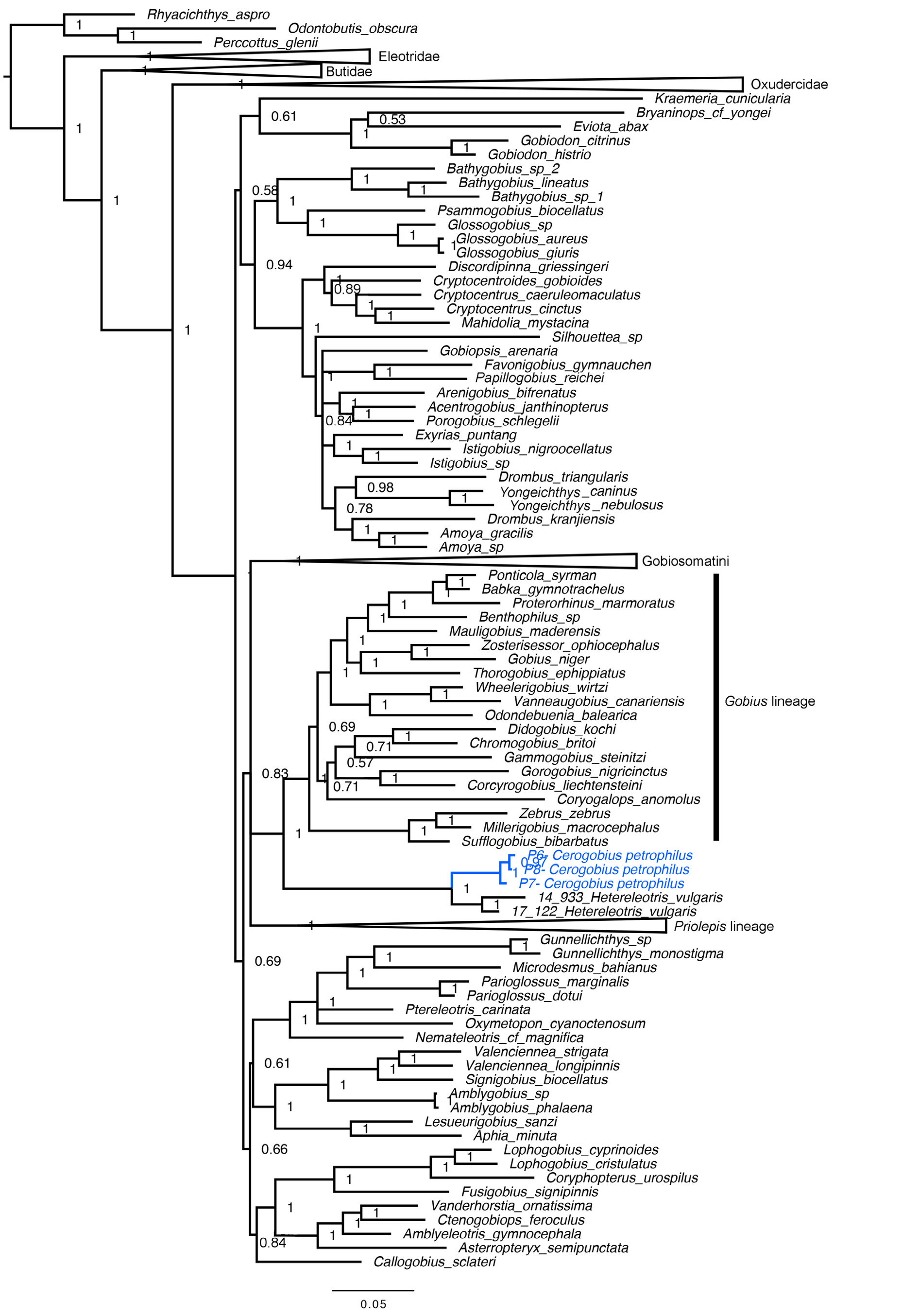
Genetics and phylogeny. The molecular phylogenetic analysis supports a close relationship between Hetereleotris vulgaris and Cerogobius petrophilus ( Fig. 4 View FIGURE 4 ), matching the evidences from morphology (see Remarks for the genus and Discussion).


No known copyright restrictions apply. See Agosti, D., Egloff, W., 2009. Taxonomic information exchange and copyright: the Plazi approach. BMC Research Notes 2009, 2:53 for further explanation.
|
Kingdom |
|
|
Phylum |
|
|
Class |
|
|
Order |
|
|
Family |
|
|
Genus |