
Anovia circumclusa (Gorham)
|
publication ID |
https://doi.org/ 10.5281/zenodo.187887 |
|
DOI |
https://doi.org/10.5281/zenodo.5658816 |
|
persistent identifier |
https://treatment.plazi.org/id/DE378783-FFD8-D767-FF1E-FA73FDA2FA2A |
|
treatment provided by |
Plazi |
|
scientific name |
Anovia circumclusa (Gorham) |
| status |
|
( Figs. 1–41)
Zenoria circumclusa Gorham, 1899: 262 . Korschefsky, 1931: 108. Blackwelder, 1945: 443. Anovia circumclusa Gordon, 1971 : p.1; Gordon, 1972: 27 –29. (Type depository: BMNH).
Diagnosis: The larva of this species resembles all other known noviine larvae, but is distinguishable by the presence of many chalazae on the lateral strumae of the abdominal segments ( R. cardinalis has 2, and R. koebelei has 4). Anovia circumclusa adults are best recognized by the structure of the male genitalia. In A. circumclusa , the basal lobe is slender and does not extend laterally beyond the internal paramere margin, while in all other Anovia species the basal lobe is quite broad distally, and overlaps the medial paramere margin. Also, the basal piece is widest basally in A. circumclusa , not distally as in A. virginalis .
Egg. Length 0.5 mm, width 0.25 mm. Elongate-oval, color bright magenta. Surface granular, often covered with waxy exudate ( Fig. 1). Eggs typically oriented horizontally, not placed on end; laid singly or in small clusters on exposed leaf surfaces; often laid on or under prey ( Majerus 1994, JAF pers. obs.)
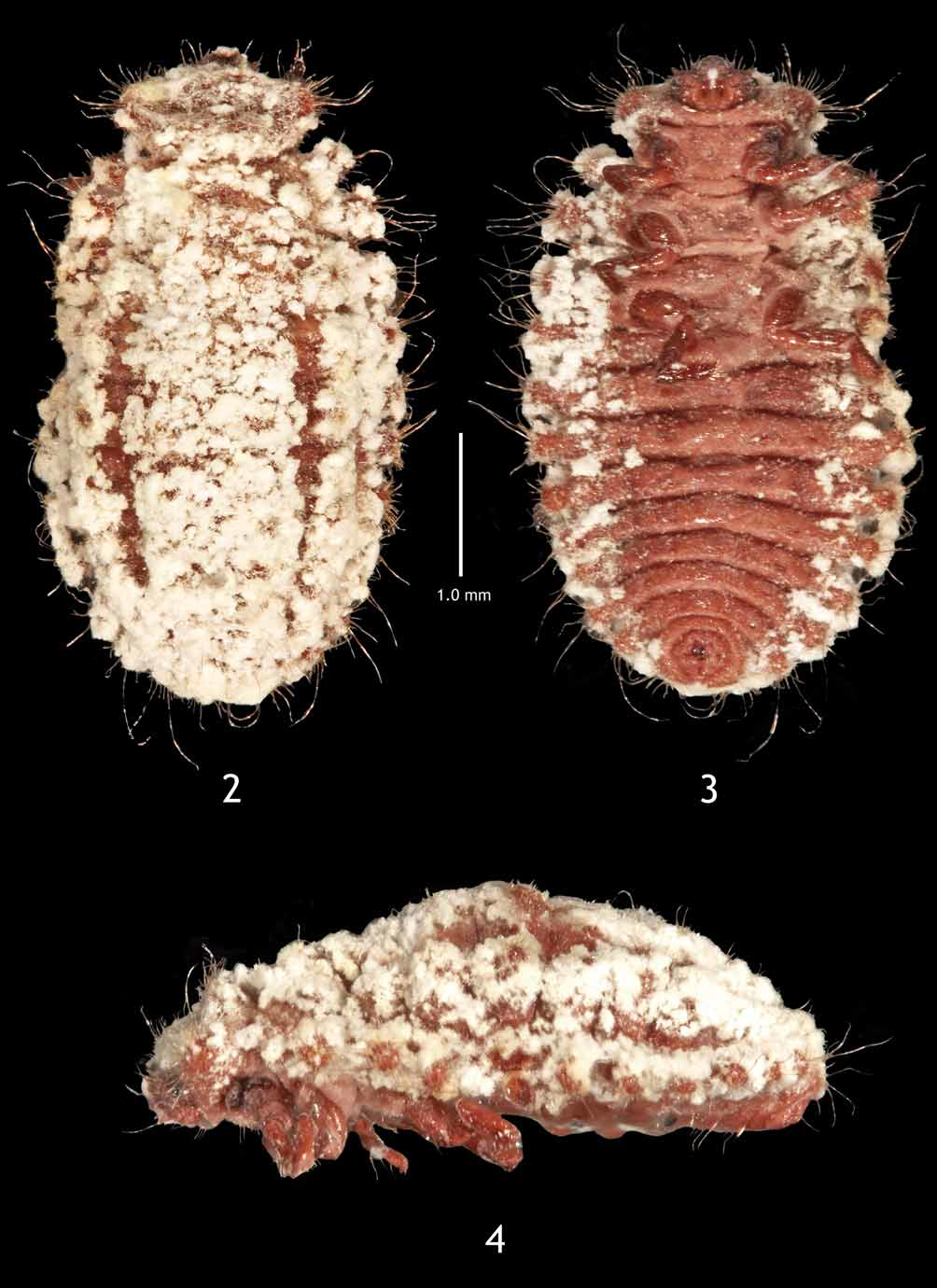
Mature Larva. Length 5–7 mm, ( Figs. 2–4 View FIGURES 2 – 4 ). Body ovoid, convex, widest at midpoint, laterally arcuate. Color bright magenta with waxy, white exudate. Dorsal surface moderately setose, finely granulate, covered with waxy exudate ( Figs. 2–4 View FIGURES 2 – 4 ). Setae pale, erect, simple, length variable.
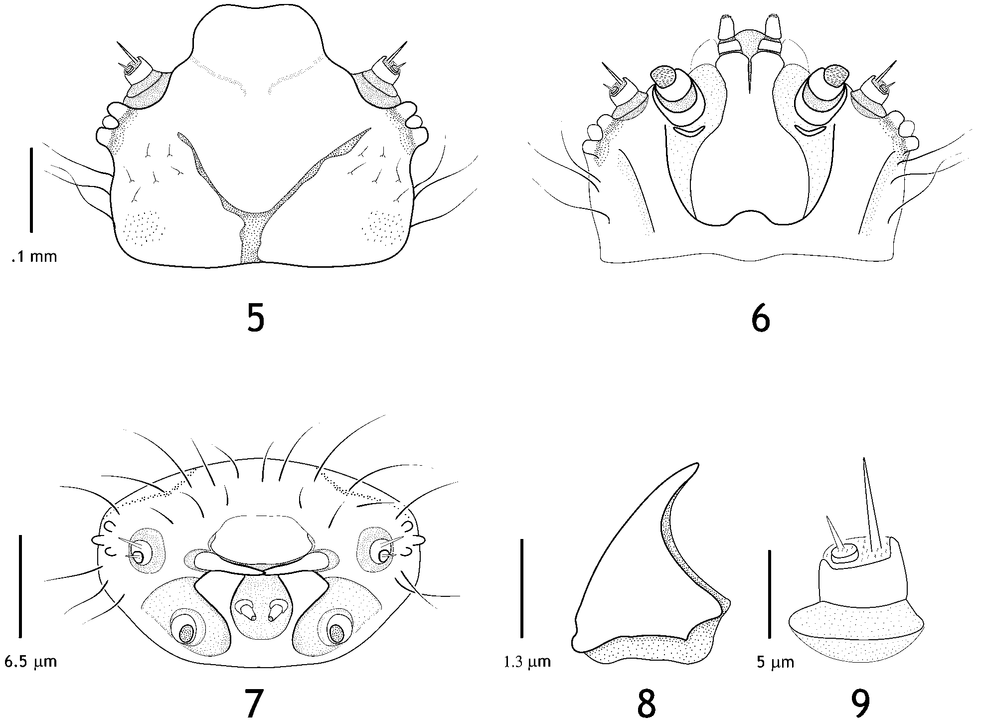
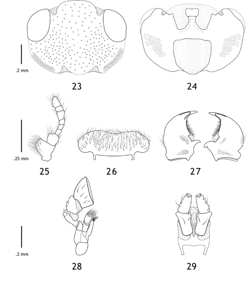
Head ( Figs. 5–7 View FIGURES 5 – 9 ) prognathous, darkly pigmented, subquadrate, at least twice as long as wide; dorsal and lateral surfaces with several chalazae; seta-like asperities lateral to frontal arms ( Fig. 5 View FIGURES 5 – 9 ). Frontal arms vshaped; epicranial stem short, about as wide as long; median endocarina absent. Stemmata arranged in triangular pattern, three on each side. Antenna inserted anteromesad to stemmata, 2-segmented ( Fig. 9 View FIGURES 5 – 9 ). Antennomere I robust, length ~ 1/3 width; II small, length subequal to width, sensorium longer than antennomere I. Labrum distinct, subrectangular, weakly bilobed apically ( Figs. 5, 7 View FIGURES 5 – 9 ). Mandible triangular, enlarged basally, falcate apically ( Figs. 7, 8 View FIGURES 5 – 9 ). Maxillolabial complex retracted ( Figs. 6, 7 View FIGURES 5 – 9 ). Maxilla with cardo and stipes fused to form solid, sclerotized structure with slender, arm-like extensions passing anteriorly and laterally around labial palpi; maxillary palpomere 2-segmented; I much broader than long; II about as broad as long ( Figs. 6, 7 View FIGURES 5 – 9 ). Mala membranous, transverse. Hypopharyngeal bracon present, well-developed.
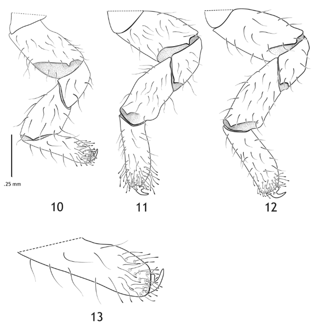
Thoracic segments each with a pair of sclerotized plates; meso-and metathorax each with a pair of lateral strumae; struma bearing many chalazae ( Figs. 2–4 View FIGURES 2 – 4 ). Legs long, robust, strongly sclerotized dorsally, semimembranous and unpigmented ventrally ( Figs. 10–12 View FIGURES 10 – 13 ). Coxa transverse ( Fig. 3 View FIGURES 2 – 4 ). Femur robust, almost as broad as long ( Figs. 3 View FIGURES 2 – 4 , 10–12 View FIGURES 10 – 13 ). Tibia elongate, ventral surface distally setose; distal setae flat, clavate ( Figs. 10–13 View FIGURES 10 – 13 ). Tarsungulus strongly curved, basal tooth well-developed. Abdomen 10-segmented; segments I–IX with 2 pairs of sclerotized tubercles, 1 pair of chalazate strumae, and 1 pair of annular spiracles; X bearing pygopod ( Fig. 3 View FIGURES 2 – 4 ).
Pupa. Length 4.5–5.5 mm, width 2.5 – 3.5 mm, exarate ( Figs. 14–16 View FIGURES 14 – 16 ). Dorsal habitus elliptical, convex, partially covered in last larval exuvium, attached by cauda to substrate. Color (excluding exuvium) magenta with pale setae ( Fig. 15, 16 View FIGURES 14 – 16 ). Dark, stout, bristle-like setae present on dorsal surface of head, pronotum, and humeral angles ( Fig. 15 View FIGURES 14 – 16 ).
Head length subequal to width. Antenna short, not extending beyond outer margin of eye, club indistinguishable from flagellum. Apical maxillary palpomere strongly securiform ( Fig. 16 View FIGURES 14 – 16 ).
Abdomen with 9 ventrites, I and II reduced and hidden beneath metacoxae; dorsal surface of abdomen with paired transverse tubercles on segments I–VIII; anterolateral angles with annular spiracles; IX with bipartite urogomphi.
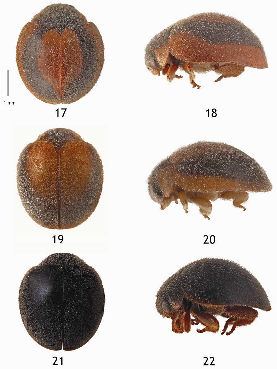
Adult. Length 4–4.5 mm. Dorsal habitus hemispherical, laterally arcuate, convex; head strongly deflexed, not visible from above; color variable ( Figs. 17–22 View FIGURES 17 – 22 ). Vestiture pale, short, moderately dense, posteriorlydirected.
Head width about twice head length; dorsal surface with evenly spaced, small, shallow punctures; ventral surface narrower; postoccipital margin sinuate ( Figs. 23, 24 View FIGURES 23 – 29 ). Eyes large, covered entirely by pale, suberect setae. Antennal insertion exposed, anteromesad to inner eye margin. Antenna with 8 articles; antennomere I asymmetrical, laterally expanded; II subglobose; III–V subequal in length and width; VI–VIII forming loose club, VI–VII asymmetrical, expanded medially; VI about as long as IV + V, VII shorter, VIII broadly tapered apically ( Fig. 25 View FIGURES 23 – 29 ). Clypeus small, fused to frons ( Figs. 23, 24 View FIGURES 23 – 29 ). Frontoclypeal suture absent. Labrum ( Fig. 26 View FIGURES 23 – 29 ) emarginate medially, expanded beyond clypeus laterally. Mandible apically bidentate, teeth sickle-shaped, not in same plane, ventral tooth longer than dorsal one; prosthecal fringe well-developed ( Fig. 27 View FIGURES 23 – 29 ). Lacinia slender, elongate, apically setose ( Fig. 28 View FIGURES 23 – 29 ). Galea broad, elongate, truncate and apically setose. Maxillary palp 3-segmented, palpifer well-developed; palpomere I elongate, about three times as long as basal width, broadest apically, membranous surface exposed; II apically divergent, mesal edge short, membranous surface exposed; III with distal edge almost twice the length of proximal edge, lateral edge twice the length of mesal one ( Fig. 28 View FIGURES 23 – 29 ). Labium narrow, labial palp 2-segmented; palpomeres I and II subequal in size, palpomere II gradually narrowed distally to apical sensory area ( Fig. 29 View FIGURES 23 – 29 ).
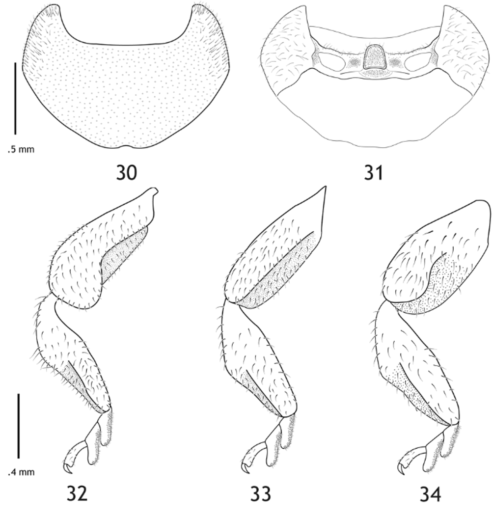
Pronotum with dorsal surface punctate, moderately setose; anterior angles extending forward just beyond lower margin of eye ( Figs. 18, 20, 22 View FIGURES 17 – 22 ); anterior edge horizontal just behind head capsule; posterior edge markedly sinuate, slightly notched at scutellum ( Fig. 30 View FIGURES 30 – 34 ). Prosternum narrow; prosternal process abruptly raised, rectangular with margins entire; procoxal cavities slightly transverse, closed internally ( Fig. 31 View FIGURES 30 – 34 ).
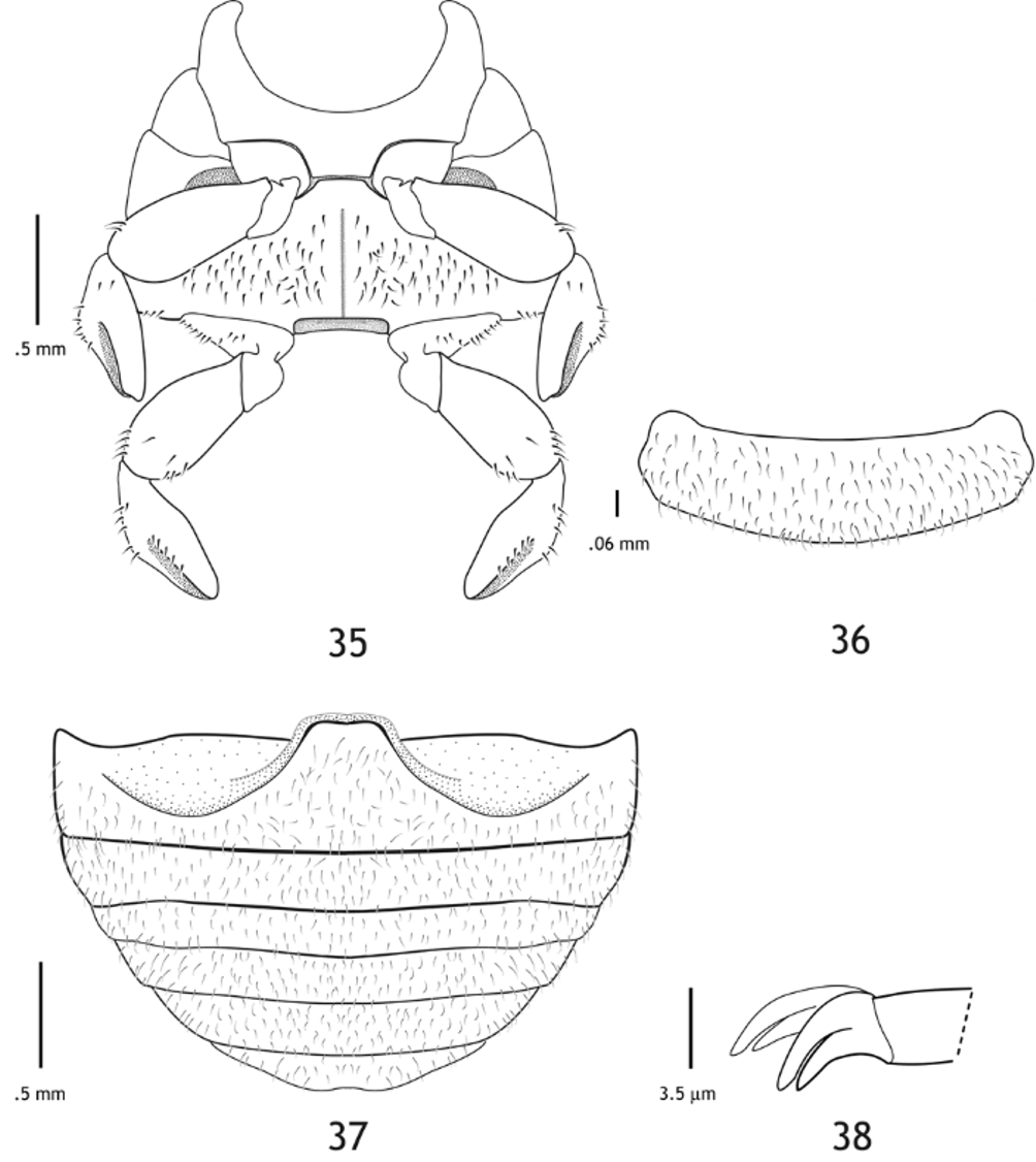
Scutellum triangular. Meso- and metathorax ventrally flattened, pubescent ( Fig. 35 View FIGURES 35 – 38 ). Mesoventrite short, narrowest posteriorly. Metaventrite wider than long, finely punctate. Legs ( Figs. 32–34 View FIGURES 30 – 34 ) flattened, broad and stout. Femur deeply grooved ventrally for reception of tibia; groove bicarinate, sharply defined, extending almost entire length of femur. Profemur with anterior groove expanded prior to apex. Tibia slightly widened at mid-length, ventral surface broader than dorsal, deeply grooved for reception of tarsus; groove bicarinate. Tarsal formula 3-3-3; tarsomeres I and II elongate, lobed ventrally with spongy pubescence; III elongate, cylindrical; male tarsal claw bifid; female tarsal claw with long triangular tooth ( Fig. 38 View FIGURES 35 – 38 ).
Elytron subhemispherical to hemispherical, laterally arcuate, finely punctate, non-striate; epipleuron complete to posterior margin, ventral surface moderately rugose. Wing with reduced cantharoid venation, absent in distal half, with strong medial and cubital veins, one anal vein, jugal lobe present. Abdomen with broad, slightly cleft intercoxal process; postcoxal line incomplete to lateral margin; 6 ventrites; I–V rectangular, progressively narrower in width posteriorly; VI narrower, tapering slightly to rounded apex; male with emarginate apex ( Fig. 37 View FIGURES 35 – 38 ), female with apex entire. Pygidium subrectangular, setose, broadly rounded apically ( Fig. 36 View FIGURES 35 – 38 ).
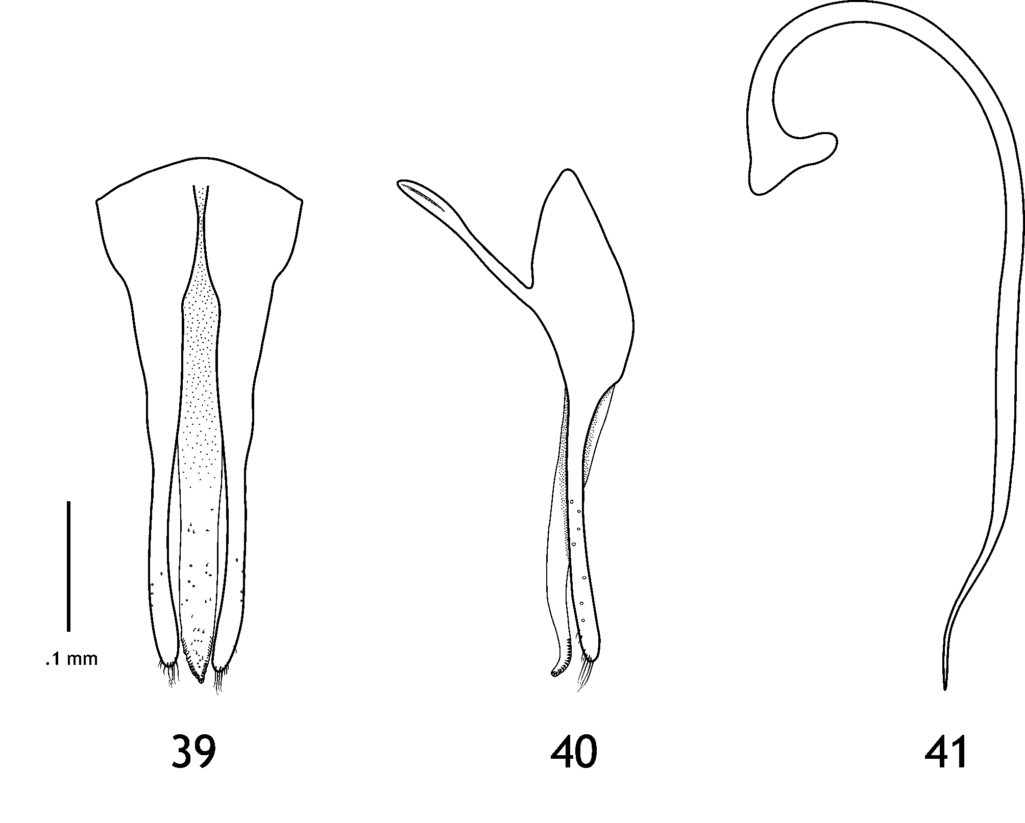
Male genitalia with phallobase widest anteriorly; basal lobe slender, not extended laterally beyond internal margin of parameres ( Figs. 39–41 View FIGURES 39 – 41 ). Sipho as in Fig. 41 View FIGURES 39 – 41 .
Material examined: see Table 2.

No known copyright restrictions apply. See Agosti, D., Egloff, W., 2009. Taxonomic information exchange and copyright: the Plazi approach. BMC Research Notes 2009, 2:53 for further explanation.
|
Kingdom |
|
|
Phylum |
|
|
Class |
|
|
Order |
|
|
Family |
|
|
Genus |
Anovia circumclusa (Gorham)
| Forrester, Juanita A., Vandenberg, Natalia J. & Mchugh, Joseph V. 2009 |
Zenoria circumclusa
| Gordon 1972: 27 |
| Blackwelder 1945: 443 |
| Korschefsky 1931: 108 |
| Gorham 1899: 262 |