
Suffrica, Henrard, Arnaud & Jocqué, Rudy, 2015
|
publication ID |
https://doi.org/ 10.11646/zootaxa.3972.1.1 |
|
publication LSID |
lsid:zoobank.org:pub:256EC29D-3CD6-499F-969A-C54B98DB041B |
|
DOI |
https://doi.org/10.5281/zenodo.6103030 |
|
persistent identifier |
https://treatment.plazi.org/id/DF1187D9-6D60-FFEB-FF1F-748FFF13FAFC |
|
treatment provided by |
Plazi |
|
scientific name |
Suffrica |
| status |
gen. nov. |
Suffrica View in CoL View at ENA gen. nov.
Figs 1 – 96 View FIGURES 1 – 5 View FIGURES 6 – 9 View FIGURES 10 – 14 View FIGURES 15 – 20 View FIGURES 21 – 24 View FIGURES 25 – 27 View FIGURES 28 – 31 View FIGURES 32 – 34 View FIGURES 35 – 41 View FIGURES 42 – 45 View FIGURES 46 – 53 View FIGURES 54 – 59 View FIGURES 60 – 63 View FIGURES 64 – 66 View FIGURES 67 – 69 View FIGURES 70 – 73 View FIGURES 74 – 81 View FIGURES 82 – 85 View FIGURES 86 – 89 View FIGURES 90 – 95 View FIGURE 96
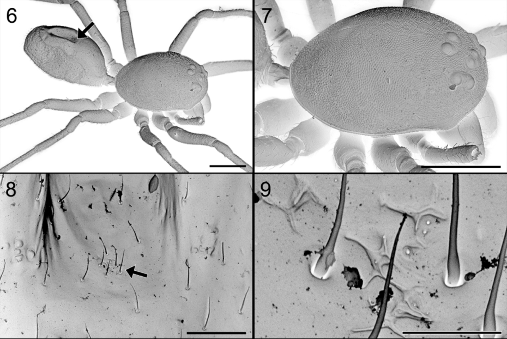
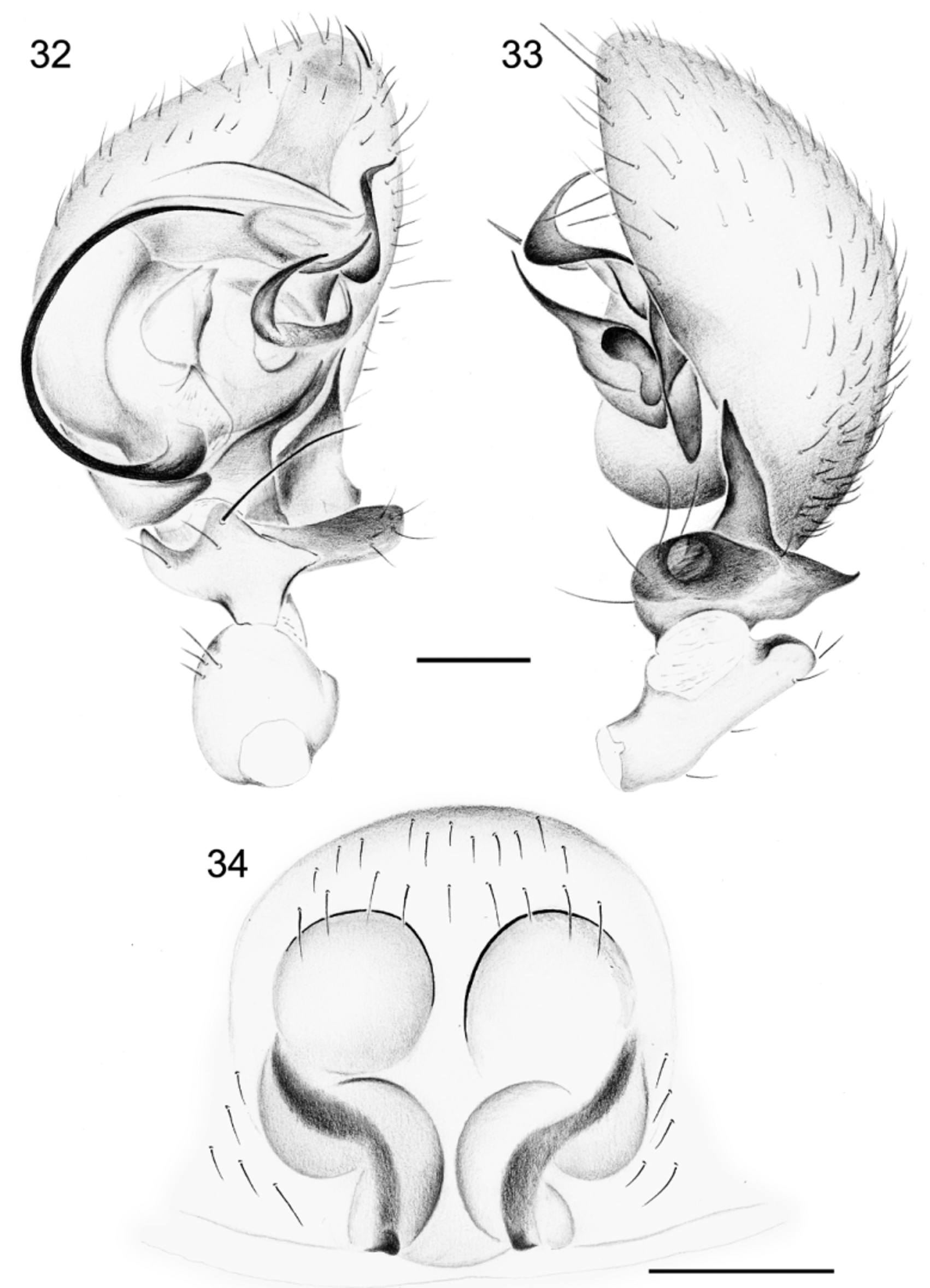
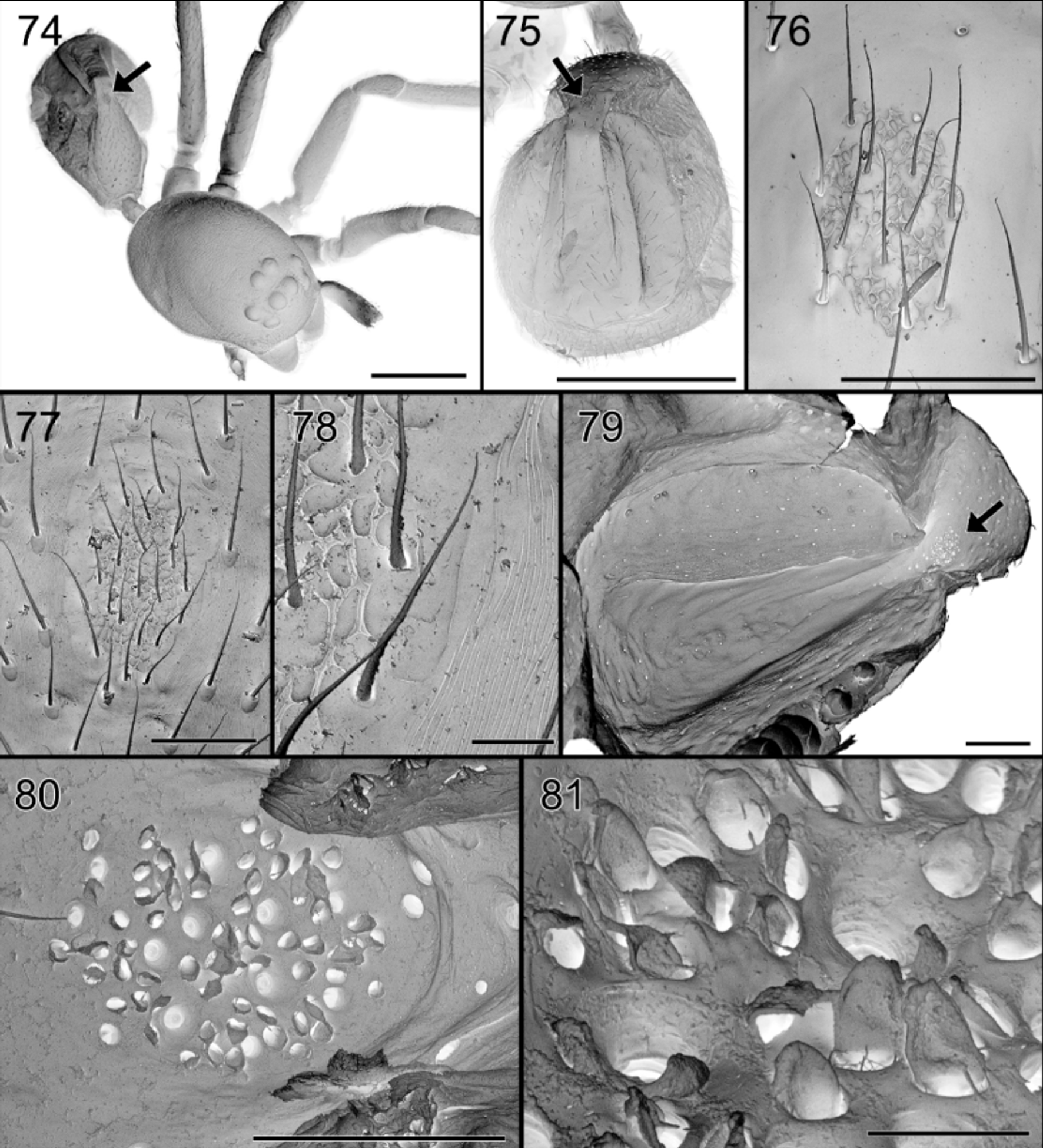
Diagnosis. Members of the genus Suffrica resemble Suffasia and Asceua in having a pair of femoral organs without barbed setae on each leg ( Figs 17–20 View FIGURES 15 – 20 ), a similar eye disposition and teeth on both cheliceral margins. They are distinguished by the narrow, domed cephalothorax ( Figs 2 View FIGURES 1 – 5 , 7 View FIGURES 6 – 9 ), the cheliceral promargin with two well separated slanting teeth, the proximal one larger ( Fig. 41 View FIGURES 35 – 41 ), a dorsal abdominal gland ( Figs 8, 9 View FIGURES 6 – 9 , 37 View FIGURES 35 – 41 , 47 View FIGURES 46 – 53 , 76–81 View FIGURES 74 – 81 ) and the dorsum of the abdomen with a groove in males ( Figs 6 View FIGURES 6 – 9 , 26 View FIGURES 25 – 27 , 65 View FIGURES 64 – 66 ).
Etymology. Suffrica is a contraction of Suffasia and Africa. The gender is feminine.
Type species. Suffrica exotica sp. nov.
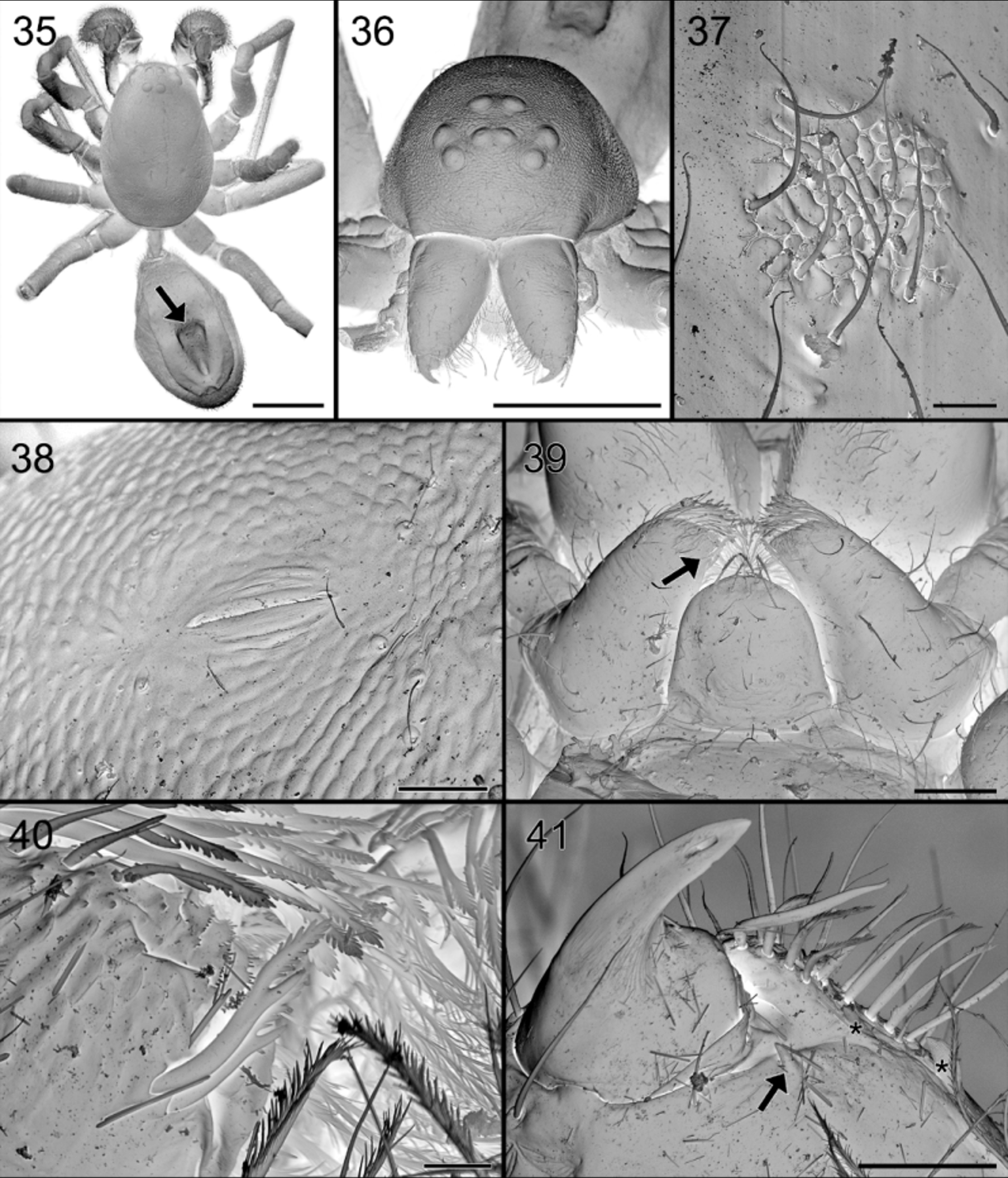
Description. Small spiders (1.9–2.9 mm). Carapace ( Figs 3 View FIGURES 1 – 5 , 6, 7 View FIGURES 6 – 9 , 36, 38 View FIGURES 35 – 41 ) narrow, oval (L/W = 1.5–1.7) and domed (L/H = 1.92–2.24), with smooth to slightly reticulated teguments, almost devoid of hairs; widest at level of coxae II–III, narrowed anteriorly to about 0.70 times maximum width in males and 0.75 times maximum width in females (cephalic width measured at level of PLE centers). Cervical grooves not marked.
Colour ( Figs 1–3 View FIGURES 1 – 5 , 25–27 View FIGURES 25 – 27 , 64–66 View FIGURES 64 – 66 ): carapace medium to dark brown; chelicerae and mouthparts dark brown; sternum medium brown; abdomen dorsum uniform dark in females, with two longitudinal lighter stripes in male, anteriorly with small, central lighter brown spot at level of abdominal gland, venter uniform grey, darker towards sides.
Eyes in two rows: anterior row straight, procurved as seen from in front ( Figs 3 View FIGURES 1 – 5 , 36 View FIGURES 35 – 41 ), posterior row strongly procurved. All eyes rounded and subequal. MOQ longer than wide, wider at the back than in front. Clypeus straight, height about 5 times diameter of ALE, with dispersed short setae.
Chilum single, superior margin wider than inferior one, slightly protruding in the center ( Fig. 3 View FIGURES 1 – 5 ). Chelicerae not fused ( Fig. 36 View FIGURES 35 – 41 ); intercheliceral triangle absent; promargin with two slanting teeth, the proximal one larger and retromargin with one small, sharp tooth ( Fig. 41 View FIGURES 35 – 41 ); fangs short and sharp, slightly curved. Labium roughly quadrangular with rounded distal margin, hardly narrowed at base ( Fig. 39 View FIGURES 35 – 41 ). Endites roughly triangular, strongly converging, distally with special asymmetric setae ( Fig. 40 View FIGURES 35 – 41 ); palps directed straight forward. Sternum slightly longer than wide, shield-shaped with slightly rounded sides, provided with triangular extensions corresponding with coxal concavities and with intercoxae.
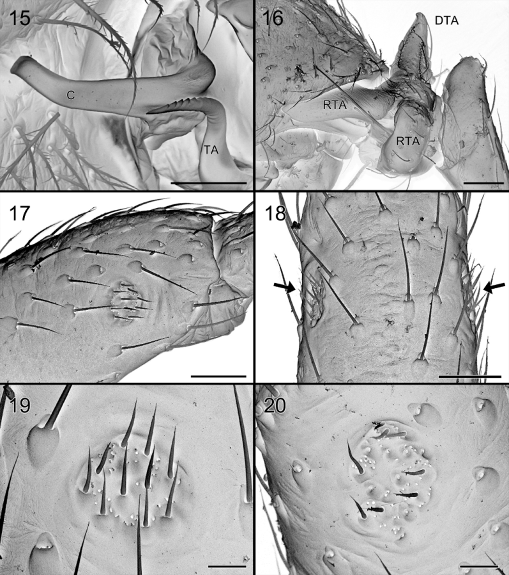
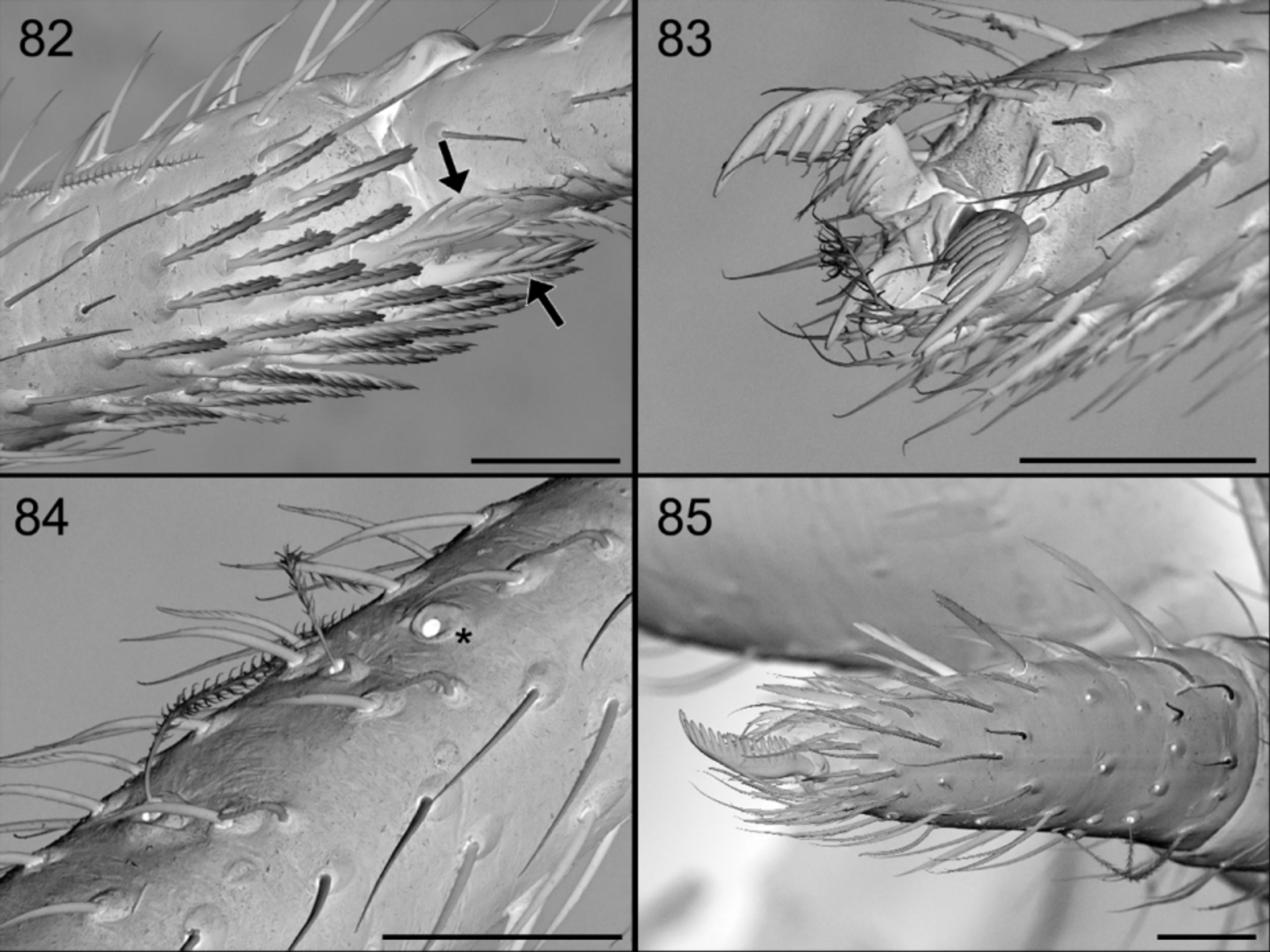
Legs slender. Formula 4123 or 4132. Three-clawed ( Fig. 83 View FIGURES 82 – 85 ), median one smooth, lateral ones medially pectinated as in most Zodariids. Spination reduced but a few dorsal spines on femora and some small ventral spines on tibiae and metatarsi; distal ones on metatarsi ramified ( Fig. 82 View FIGURES 82 – 85 ). Preening brush with short chisel-shaped setae on metatarsi II and III ( Fig. 82 View FIGURES 82 – 85 ). Femoral organ double, on prolateral and retrolateral face of all legs, with smooth conical hairs and numerous small pores in a shallow circular depression ( Figs 17–20 View FIGURES 15 – 20 ). Tarsal organ simple hole on distal edge of elevated plate ( Fig. 84 View FIGURES 82 – 85 ), located on proximal part of tarsi.
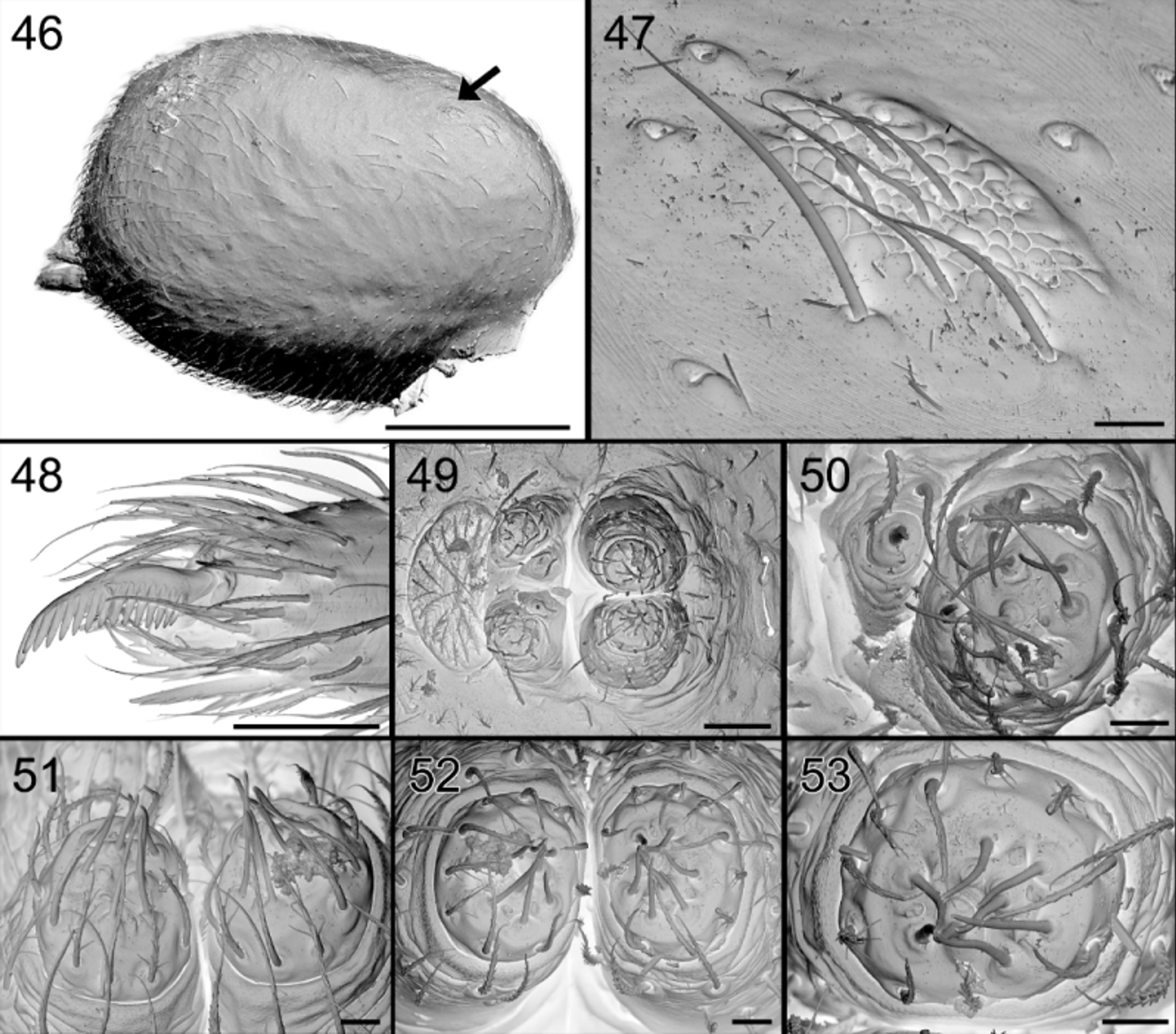
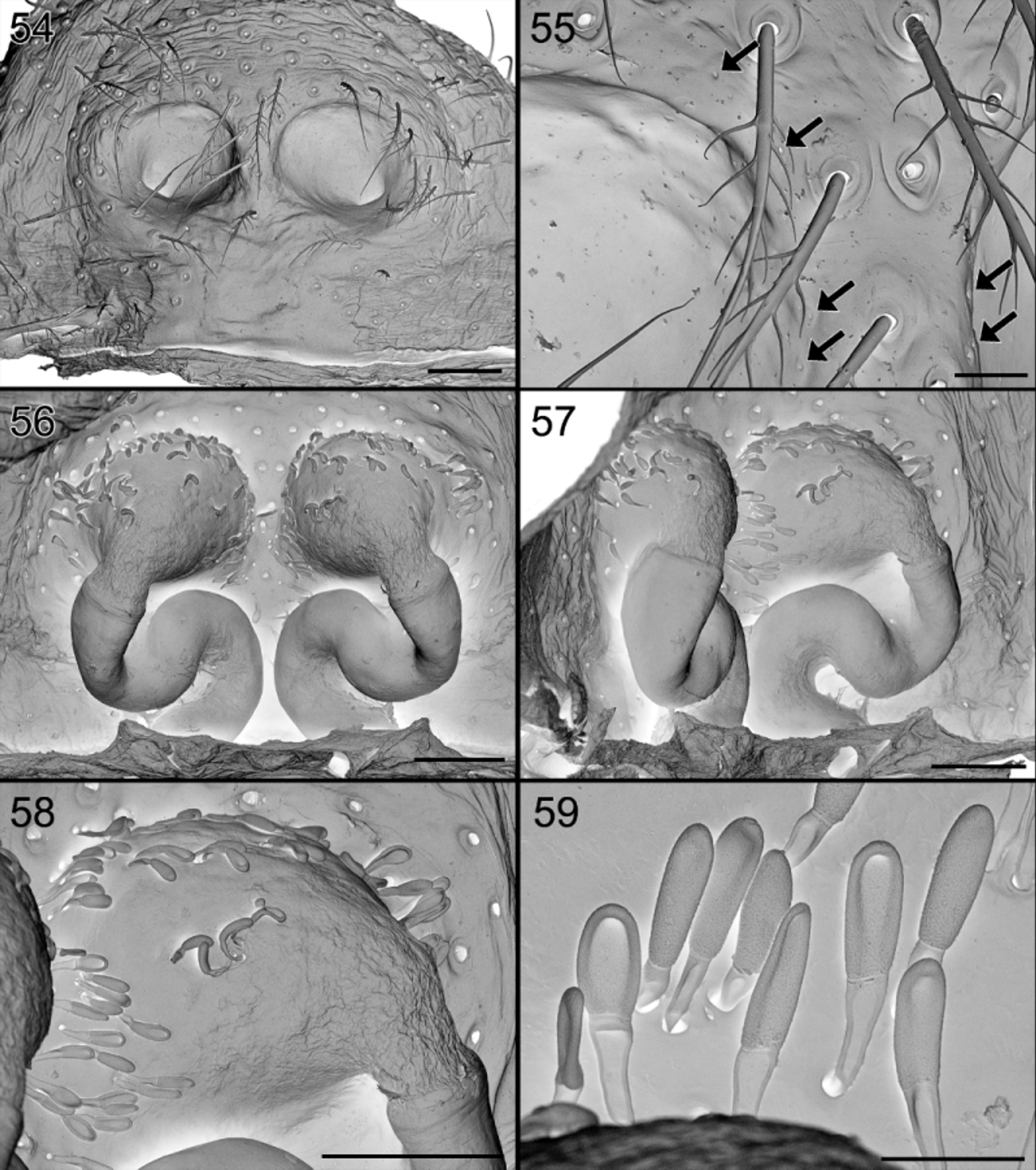
Abdomen oval. Dorsum in both sexes anteriorly with oval gland outlet with coarsely granulated tegument, tiny gland openings and smooth setae ( Figs 8, 9 View FIGURES 6 – 9 , 37 View FIGURES 35 – 41 , 47 View FIGURES 46 – 53 , 76 – 81 View FIGURES 74 – 81 ); internal view shows perforated oval area, with numerous pits, larger ones corresponding to the base of smooth setae, smaller ones accommodating the outlets of flat, oval glands ( Figs 80, 81 View FIGURES 74 – 81 ); abdomen provided in males with scutum covering ¾ of surface, with central longitudinal groove delimited by shallow ridges ( Figs 2 View FIGURES 1 – 5 , 6 View FIGURES 6 – 9 , 26 View FIGURES 25 – 27 , 35 View FIGURES 35 – 41 , 65 View FIGURES 64 – 66 , 75 View FIGURES 74 – 81 ). Tracheal spiracle procurved, widened to rounded opening at extremities ( Fig. 22 View FIGURES 21 – 24 ); just in front of spinnerets; fairly wide, around 0.25 times maximum width of abdomen. Males with four spinnerets ( Fig. 21 View FIGURES 21 – 24 ), females with six ( Fig. 49 View FIGURES 46 – 53 ). ALS ( Figs 22, 23 View FIGURES 21 – 24 , 51 – 53 View FIGURES 46 – 53 ), conical, biarticulate, with two major ampullate gland spigots in the center and five (in males) or six (in females) aciniform gland spigots around the former. PLS small, with eight aciniform gland spigots in female ( Fig. 50 View FIGURES 46 – 53 ), two in male ( Fig. 24 View FIGURES 21 – 24 ). MS absent in males, reduced in females with one aciniform gland spigot ( Fig. 50 View FIGURES 46 – 53 ). Colulus represented by a few setae. Sperm pore a wide slightly procurved slit.
Female palp with some strong prolateral spines on conical tarsus, with pectinated claw turned inwards over nearly 45°, without patch of chemosensitive setae ( Figs 48 View FIGURES 46 – 53 , 85 View FIGURES 82 – 85 ).
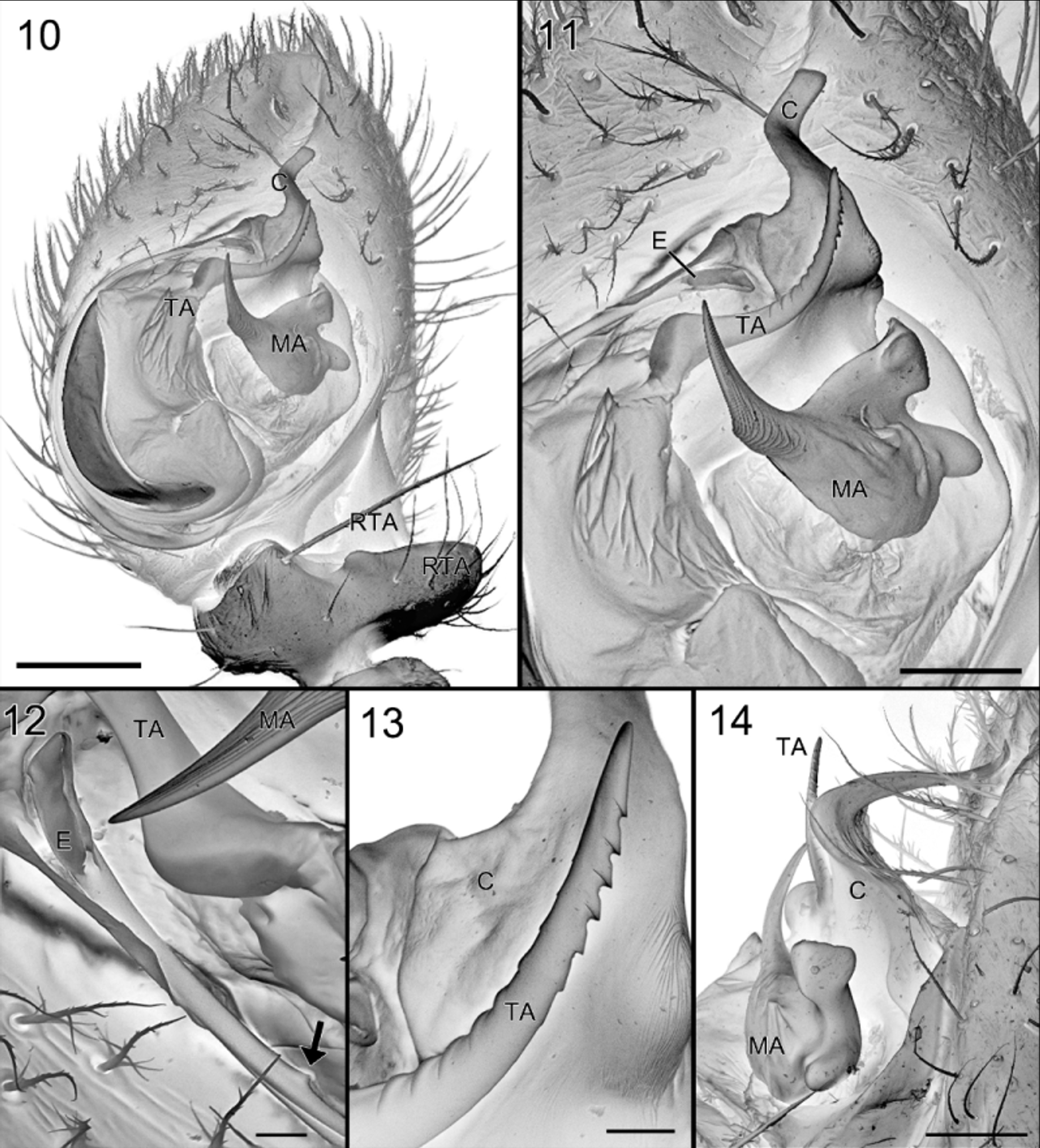
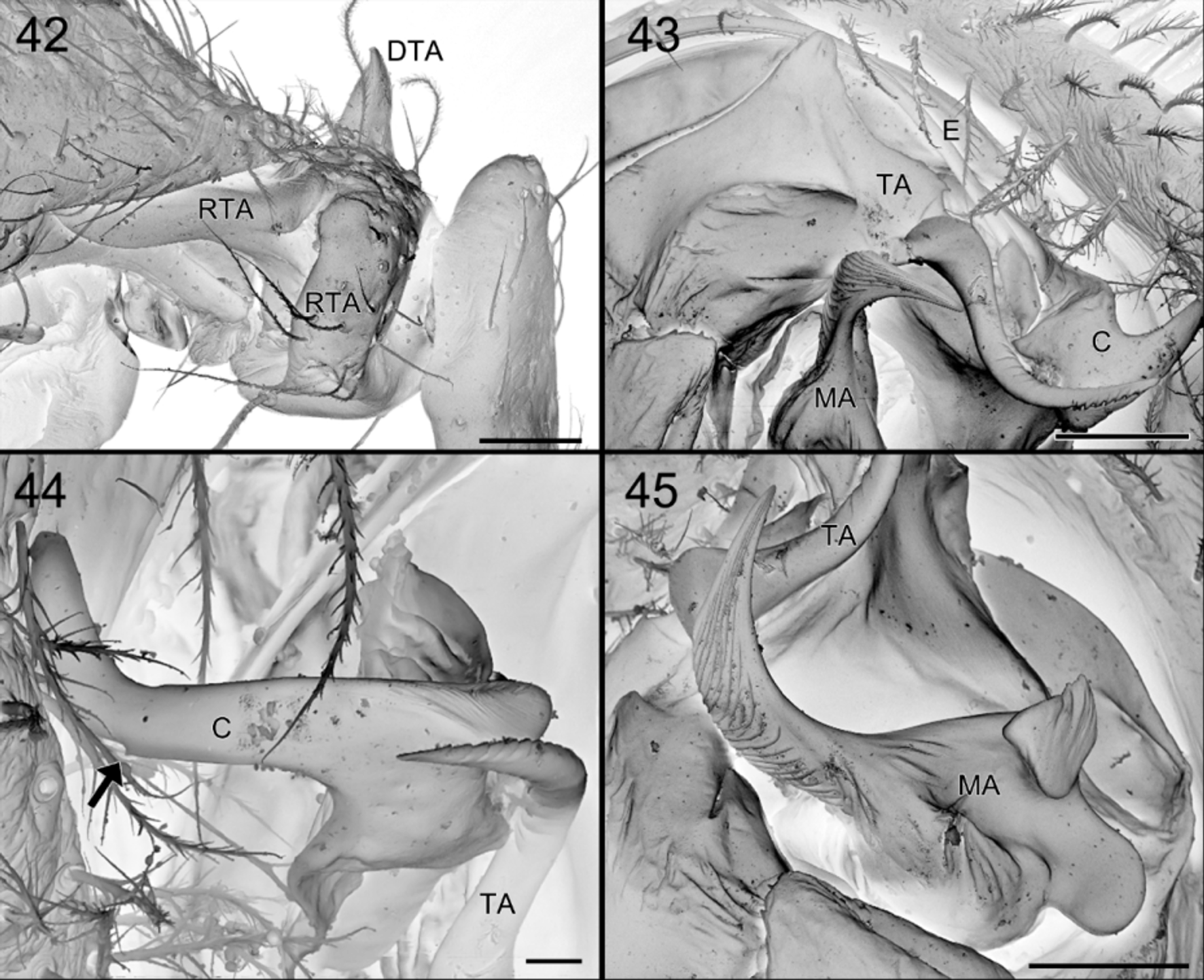
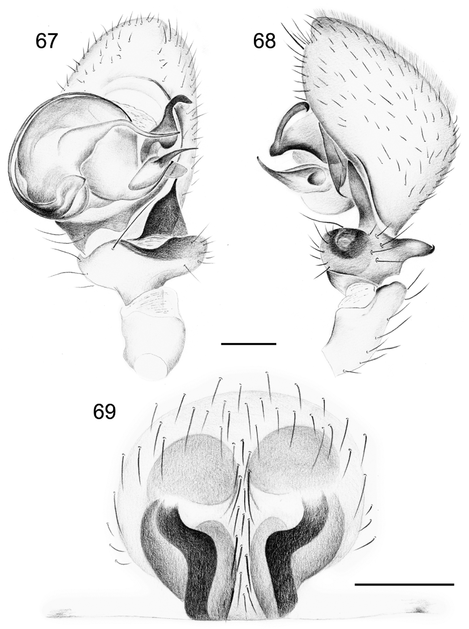
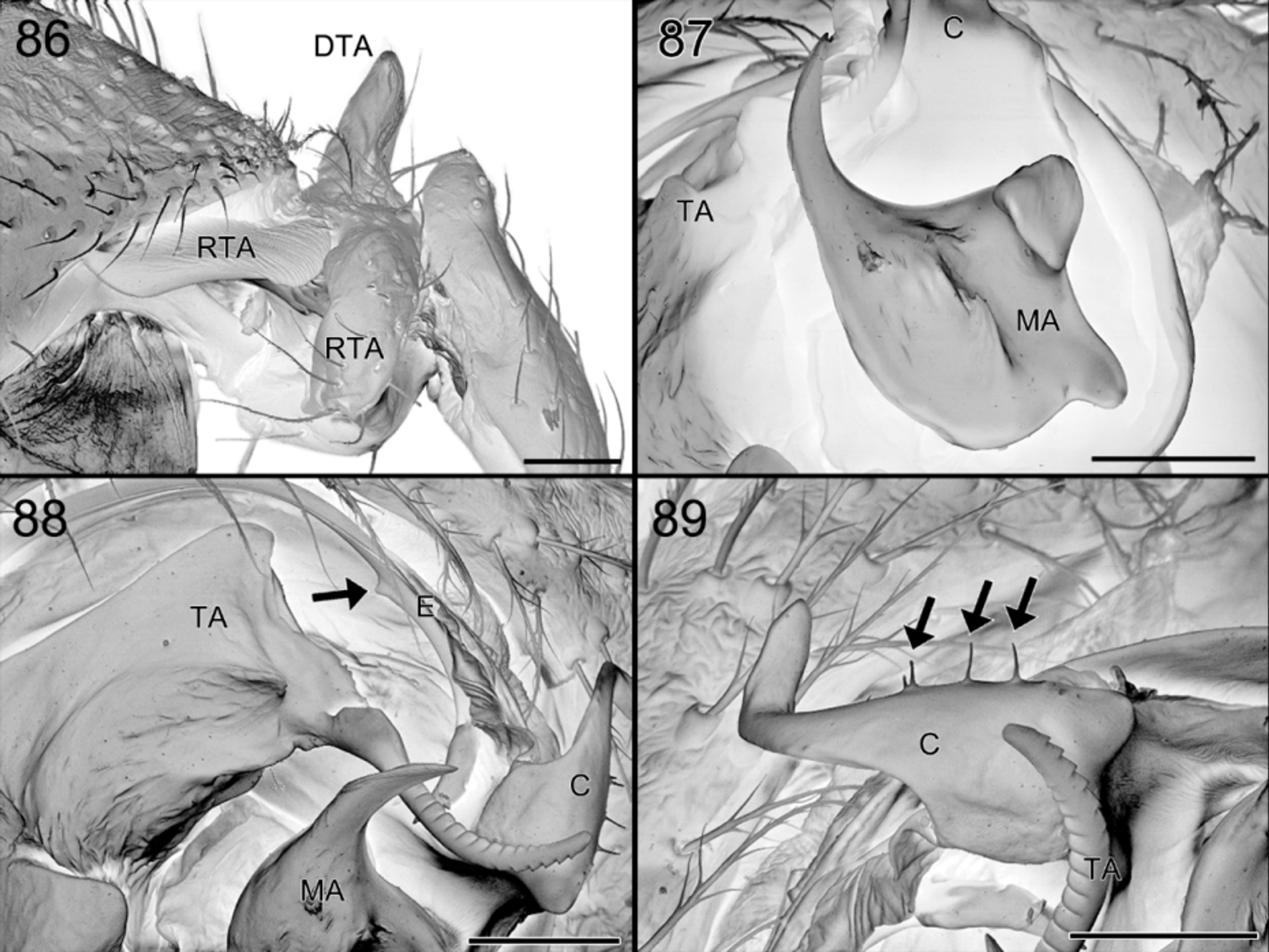
Male palp with patella provided with rounded dorsal extension, tibia with both DTA and RTA: DTA short, conical and sturdy, RTA with two parts: one fairly long anterior lamellar extension pointing forwards and one rounded with transparent window, the two parts separated by plate-shaped concavity ( Figs 16 View FIGURES 15 – 20 , 42 View FIGURES 42 – 45 , 86 View FIGURES 86 – 89 ). Cymbium domed, with basal concavity accommodating lamellar extension of RTA; with patch of chemosensitive setae; embolus originating on posterior part of tegulum ( Fig. 10 View FIGURES 10 – 14 ), cylindrical and grooved, long, curved, flexible, with small tooth at some distance from tip ( Figs 12 View FIGURES 10 – 14 , 88 View FIGURES 86 – 89 ); extremity truncated and folded ( Fig. 12 View FIGURES 10 – 14 ); MA ( Figs 11 View FIGURES 10 – 14 , 45 View FIGURES 42 – 45 , 87 View FIGURES 86 – 89 ) complex with different appendages: one prolateral, long, sharp and curved forwards and two retrolateral, rounded and stout; distal tegular apophysis harpoon-shaped ( Fig. 13 View FIGURES 10 – 14 ); conductor with basal groove accommodating embolus and distal part provided with curved, flat apophysis ( Figs 11, 14 View FIGURES 10 – 14 , 15 View FIGURES 15 – 20 , 44 View FIGURES 42 – 45 , 89 View FIGURES 86 – 89 ).
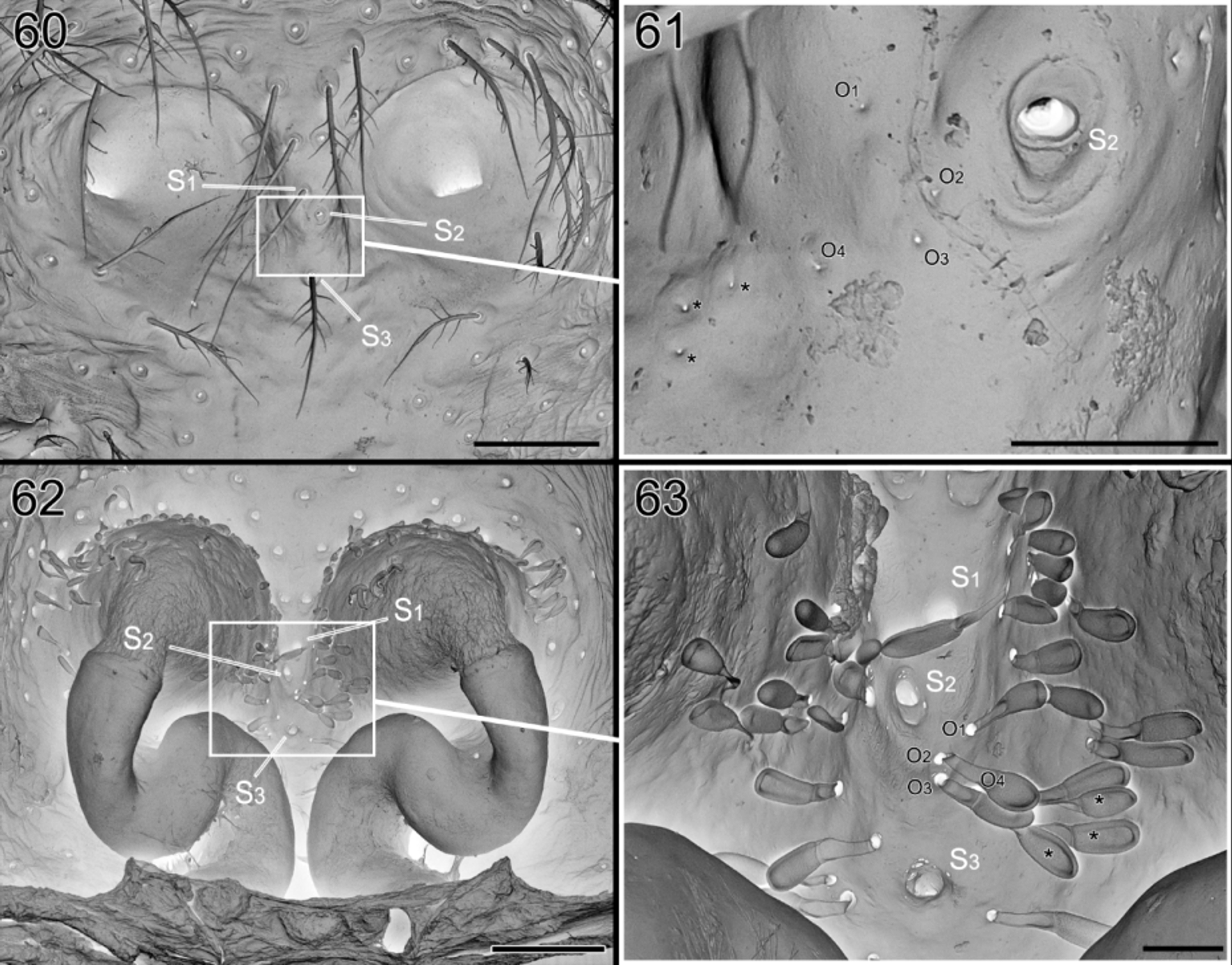
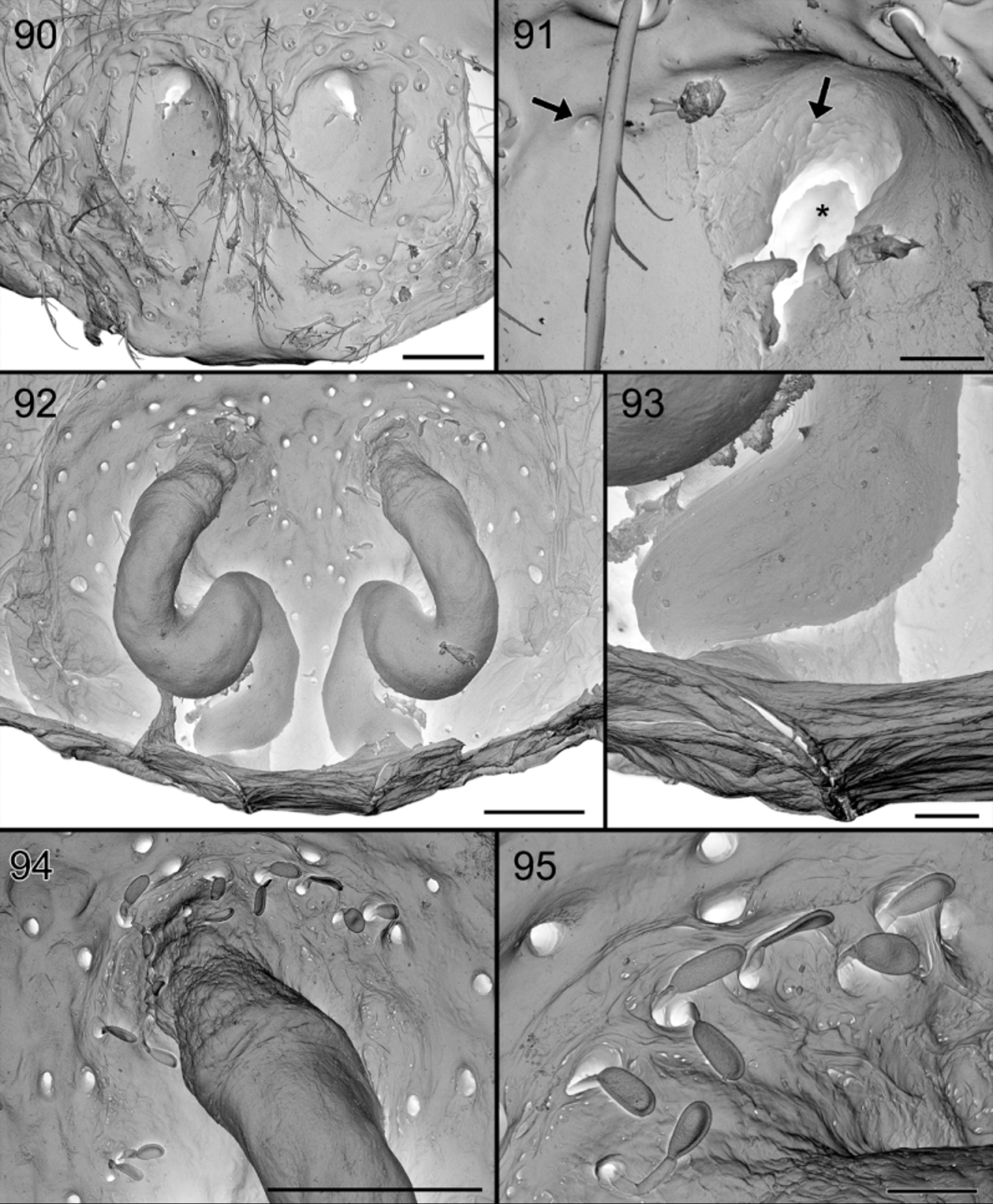
Epigyne ( Figs 30 View FIGURES 28 – 31 , 34 View FIGURES 32 – 34 , 54, 55 View FIGURES 54 – 59 , 60 View FIGURES 60 – 63 , 69 View FIGURES 67 – 69 , 72 View FIGURES 70 – 73 , 90, 91 View FIGURES 90 – 95 ) anteriorly with two membranous funnels ( Figs 58 View FIGURES 54 – 59 , 94 View FIGURES 90 – 95 ) connected to sinuous sclerotized copulation ducts, leading to poorly defined spermathecae ( Figs 31 View FIGURES 28 – 31 , 56, 57 View FIGURES 54 – 59 , 62 View FIGURES 60 – 63 , 73 View FIGURES 70 – 73 , 92, 93 View FIGURES 90 – 95 ). Funnels internally with numerous biarticulate glands ( Figs 59 View FIGURES 54 – 59 , 63 View FIGURES 60 – 63 , 95 View FIGURES 90 – 95 ), here called “lollipops”, opening through small holes on the outside ( Figs 55 View FIGURES 54 – 59 , 61 View FIGURES 60 – 63 , 91 View FIGURES 90 – 95 ).
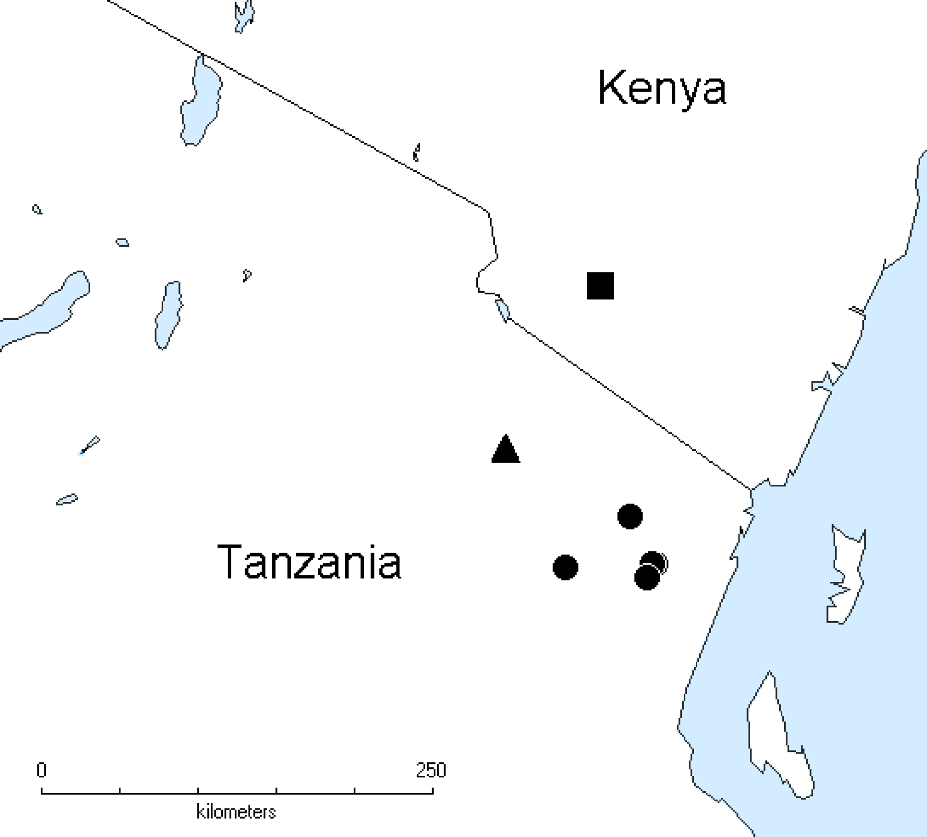
Distribution. Suffrica is found in the Eastern Arc Mountains and the adjacent Mkomazi Game Reserve ( Fig. 96 View FIGURE 96 ).


No known copyright restrictions apply. See Agosti, D., Egloff, W., 2009. Taxonomic information exchange and copyright: the Plazi approach. BMC Research Notes 2009, 2:53 for further explanation.