
Tytthosoceros lizardensis Newman & Cannon, 1996
|
publication ID |
https://doi.org/ 10.11646/zootaxa.3860.4.2 |
|
publication LSID |
lsid:zoobank.org:pub:3F59B0E9-F943-407C-A49B-6D05DBACCCEE |
|
DOI |
https://doi.org/10.5281/zenodo.6122994 |
|
persistent identifier |
https://treatment.plazi.org/id/DF12878D-FF99-FFB7-94A0-18B0F746FE2E |
|
treatment provided by |
Plazi |
|
scientific name |
Tytthosoceros lizardensis Newman & Cannon, 1996 |
| status |
|
Tytthosoceros lizardensis Newman & Cannon, 1996 View in CoL
( Figures 8–9 View FIGURE 8 View FIGURE 9 )
Type locality. Heron Island, southern Great Barrier Reef- Australia.
Other localities. Indonesia, Philippines, Papua New Guinea, South Africa ( Newman & Cannon 2005) and Iran ( Khalili et al. 2009).
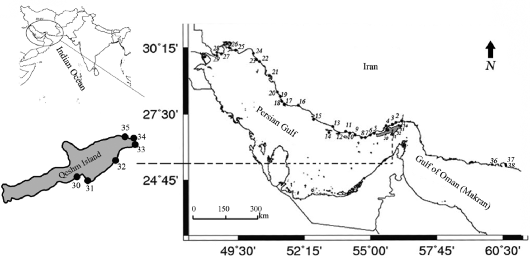
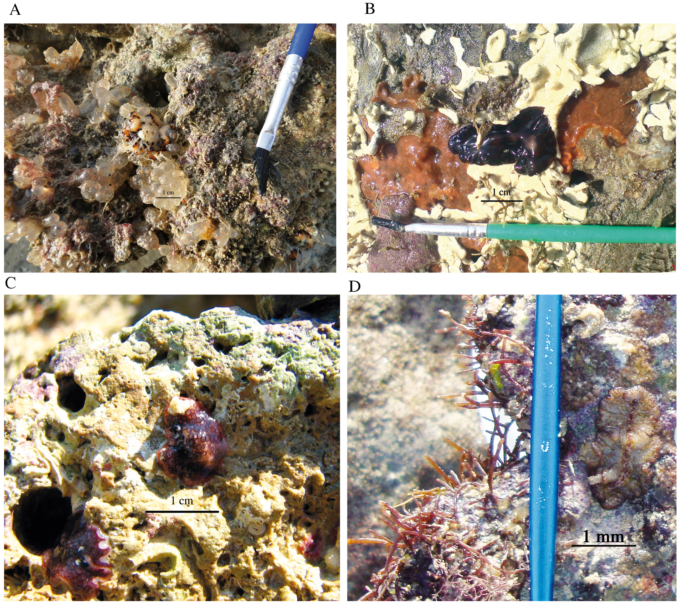
Location in Iran. Specimens were collected from two intertidal localities of Qeshm Island ( Fig.1 View FIGURE 1 stations 33 and 31). Both stations have sandy-rocky substrate with numerous small and large intertidal pools covered in red and brown algae, sponges and tunicates (Table 1).
Material examined. Eight individuals were examined: three mature specimens as ZUTC platy 1269-71 HS; five specimens as ZUTC platy 1272 S (Table 1). The following description is based on observation from ZUTC platy 1269, unless otherwise stated.
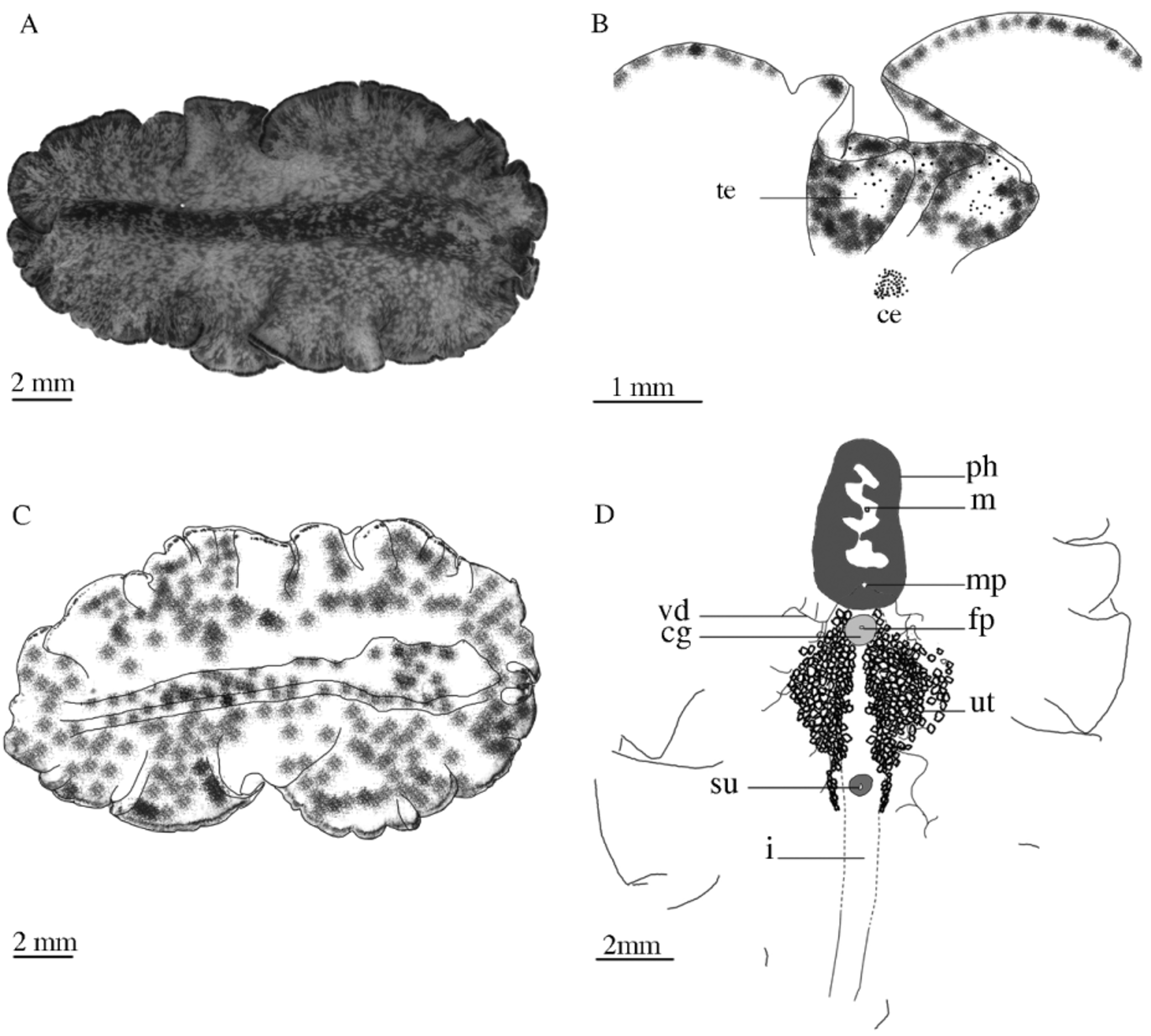
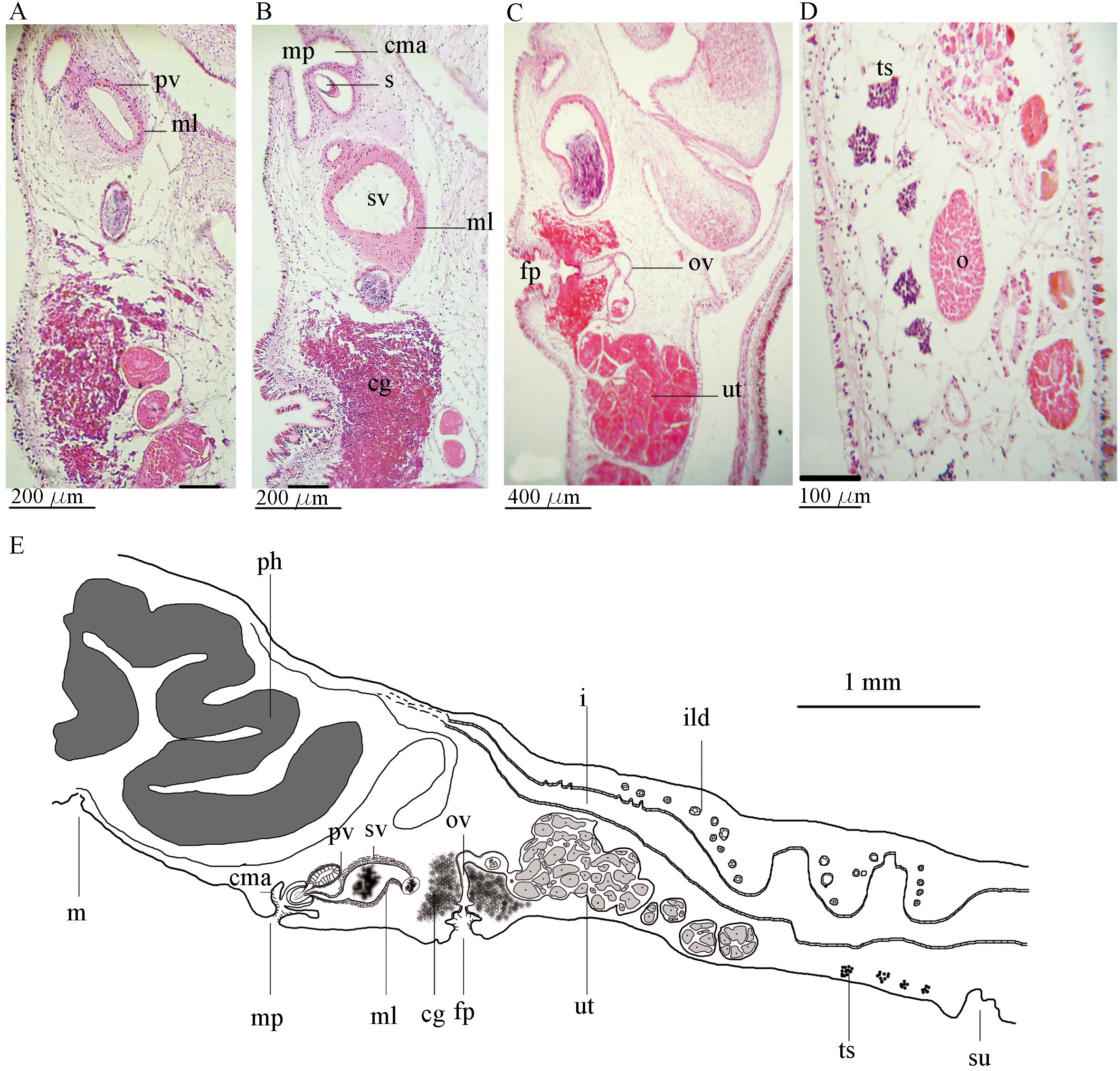
Description. External morphology. Body soft and delicate, long and oval, slightly tapered posteriorly and raised medially ( Figs. 8 View FIGURE 8 A, C). The average size of the fixed specimens is 22 × 13 mm, the largest and smallest specimens 30 × 1.7 and 16 × 10, respectively. Dorsal background darker on the raised middle region, with transverse streaks medially and laterally, mottled with a mixture of chocolate brown, reddish brown or light brown and cream. Body margin with outer narrow whitish grey band and inner black band, a light beige sub-marginal band may be visible ( Fig. 8 View FIGURE 8 A), body margin broken at the edge with short white transverse streaks of microdots. Pseudotentacles small, ear- like, with color pattern similar to the dorsal body, with white tips ( Figs. 8 View FIGURE 8 A, B); dorsal tentacular eyes scattered, with 33–37 eye spots; cerebral eyes horseshoe shaped, containing 55 to 90 ocelli on a whitish grey background. Ventral surface transparent cream, with a grey and black margin, internal structures such as uterus, vasa deferentia, genital pores and pharynx visible from the ventral surface. Pharynx 3.9 mm long, with five to six pairs of simple folds; mouth central ( Fig. 8 View FIGURE 8 D); main intestine extended posteriorly with lateral branches in histological sections ( Figs. 8 View FIGURE 8 D, 9E). One male gonopore located posterior to the pharynx, female pore 800 µm posterior to the male pore. Sucker located ventrally, about 3.5 mm posterior to the female pore, 272 µm long ( Figs. 8 View FIGURE 8 D, 9E). Dorsal epidermis with rhabdites, ventral epidermis without ( Fig. 9 View FIGURE 9 D).
Reproductive structures. Male reproductive system ( Figs. 9 View FIGURE 9 A–C, E) with true seminal vesicle. Muscular seminal vesicle, 44 µm thick, oblong, 390 µm long × 241 µm wide ( Figs. 8 View FIGURE 8 B–C, 9E). Prostatic vesicle ( Figs. 9 View FIGURE 9 A, E) small, oval, 198 µm × 102 µm, somewhat muscular, 21 µm thick, slightly oriented postero-dorsally, prostatic duct joints ejaculatory duct in the middle of the penis ( Fig. 9 View FIGURE 9 E); penis stylet 134 µm × 61 µm, stylet length to width ratio 1: 2.1; male atrium ciliated, wide (120 µm) and shallow (90 µm) ( Figs. 9 View FIGURE 9 B, E). Vas deferens forming a network of thin tubes ( Fig. 8 View FIGURE 8 D), testes scattered ventrally throughout the body ( Figs. 9 View FIGURE 9 D, E). Female system ( Figs. 9 View FIGURE 9 B, C, E) with deep and ciliated atrium; cement glands large; vagina thick and muscular; oviduct full of eggs ( Figs. 9 View FIGURE 9 B, C, E); female gonopore round, 230 µm in diameter ( Figs. 9 View FIGURE 9 B, C, E); ovaries distributed dorsally ( Fig. 9 View FIGURE 9 D).
Remarks. The genus Tytthosoceros belongs to the family Pseuodocerotidae, erected by Newman and Cannon (1996) based on specimens from the Central and Eastern Indo-Pacific (the Great Barrier Reef, Australia, Papua New Guinea, and the Philippines). Most species of the family occur in tropical and subtropical waters of the Indo- Pacific region ( Hyman 1954). So far, three species have been assigned to the genus Tytthosoceros : T. inca Baeza, Véliz, Pardo, Lohrmann and Guisado, 1997 ; T. lizardensis Newman & Cannon, 1996 ; T. nocturnus Newman & Cannon, 1996 .
Based on color and color patterns for pseudocerotids ( Newman & Cannon 1996), T. lizardensis belongs to the fourth group, whichis characterized by spots, dots, and mottling by spots ( Newman & Cannon 1994). With regard to the overall body shape, color patterns and anatomy of male reproductive system, our observations agree well with descriptions of Newman and Cannon (1996a) and Khalili et al. (2009).
The distribution of T. lizardensis is assumed to be rare on the reef crest and subtidal areas, on the other hand it is supposed to be abundant on intertidal areas ( Newman & Cannon 1996). Khalili et al. (2009) have already reported the presence of T. lizardensis in the Iranian waters of the Persian Gulf ( Fig. 1 View FIGURE 1 , stations 31 and 33). Six of the 38 stations ( Fig. 1 View FIGURE 1 , stations 30–35) studied here are same those of Khalili et al. (2009) where we again found T. lizardensis in two of the six stations ( Fig. 1 View FIGURE 1 , stations 31 and 33). Consequently, it’s reasonable to suppose that the distribution of this species on the southern coasts of Iran (the Persian Gulf and Gulf of Oman), for some reasons, is limited to the eastern side of Qeshm Island (Persian Gulf, Fig. 1 View FIGURE 1 , stations 31 and 33). In terms of the SACFOR abundance scale for animals greater than 15 cm ( Connor et al. 1997), this species was distributed frequently on the aforementioned Island.
Khalili et al. (2009) found their specimens associated with orange sponge ( Cliona vastifica ); we collected our specimens under rocks and in tidal pools, mostly covered with red algae (Solieria) and the sponge, Gelliodes carnosa ( Fig. 10 View FIGURE 10 D).
No known copyright restrictions apply. See Agosti, D., Egloff, W., 2009. Taxonomic information exchange and copyright: the Plazi approach. BMC Research Notes 2009, 2:53 for further explanation.