
Thysanozoon brocchii (Risso, 1818) Grube, 1840
|
publication ID |
https://doi.org/ 10.11646/zootaxa.3860.4.2 |
|
publication LSID |
lsid:zoobank.org:pub:3F59B0E9-F943-407C-A49B-6D05DBACCCEE |
|
DOI |
https://doi.org/10.5281/zenodo.6122992 |
|
persistent identifier |
https://treatment.plazi.org/id/DF12878D-FF9B-FFB3-94A0-1A76F4DEFCB3 |
|
treatment provided by |
Plazi |
|
scientific name |
Thysanozoon brocchii (Risso, 1818) Grube, 1840 |
| status |
|
Thysanozoon brocchii (Risso, 1818) Grube, 1840
( Figures 6 View FIGURE 6 , 7 View FIGURE 7 )
Type locality. Italy ( Prudhoe 1985)
Other localities. Cosmopolitan, temperate to cold seas, reported from both the Pacific and Atlantic Oceans ( Bahia et al. 2012; Brusa et al. 2009; Lang 1884; Marcus & Marcus 1968; Prudhoe 1989; Yeri & Kaburaki 1918).
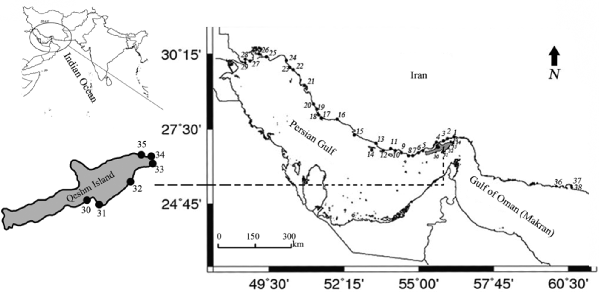
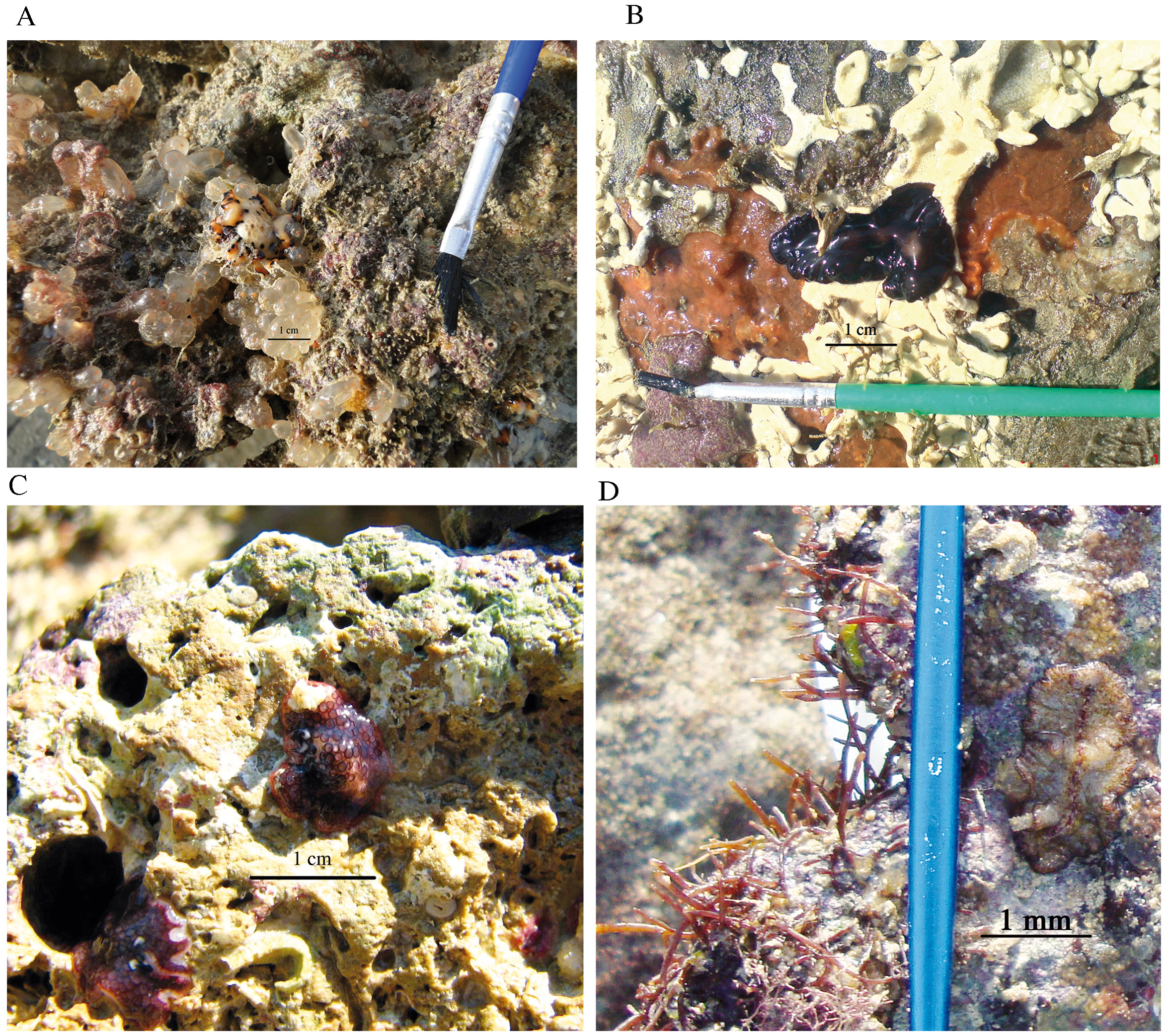
Location in Iran. Specimens of Thysanozoon brocchii in this study were collected from the Persian Gulf ( Fig. 1 View FIGURE 1 stations 12, 31, 33 and Table 1), and from the Gulf of Oman ( Fig. 1 View FIGURE 1 station 36 and Table 1). We collected our specimens mainly under stones and pebbles in close association with didemnid ascidians ( Fig. 10 View FIGURE 10 C).
Material examined and localities (Table 1). Fourteen specimens collected: Two mature specimens as ZUTC platy 1275-76 HS collected from Chiruyeh station, the Persian Gulf ( Fig. 1 View FIGURE 1 station 12); four specimens as ZUTC platy 1280 S (containing two specimens) and ZUTC platy 1277 S collected from Chiruyeh station; eight specimens ZUTC platy 1278 S, collected from Cinema- Darya station ( Fig. 1 View FIGURE 1 station 33) and ZUTC platy 1279 S, collected from Shib- Deraz station ( Fig. 1 View FIGURE 1 station 31). The following description is based on observations from ZUTC platy 1276, unless otherwise stated.
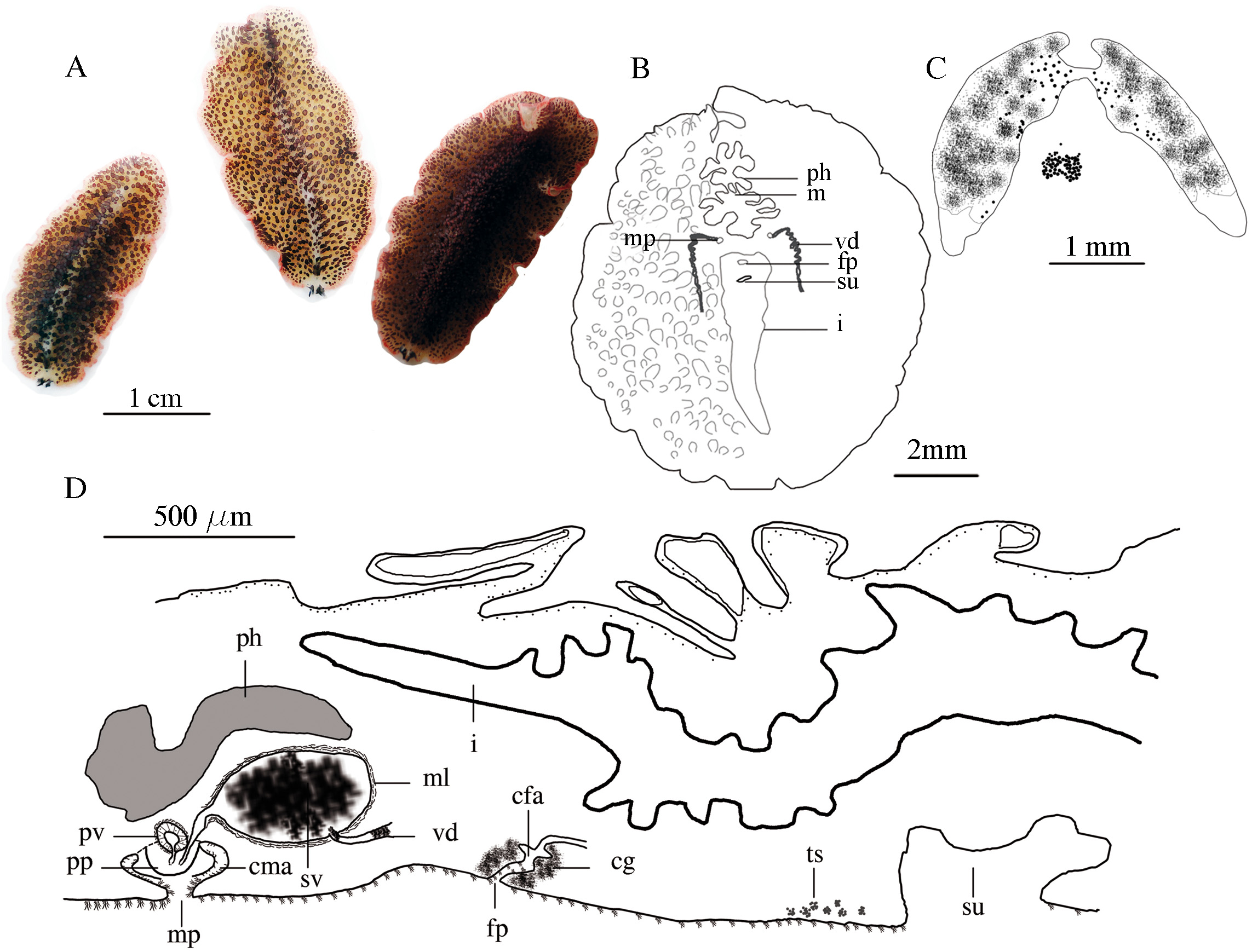
Generic diagnosis. Dorsal region adorned with conical papillae, papillae become smaller toward the margin; male apparatus doubled ( Fig. 6 View FIGURE 6 A).
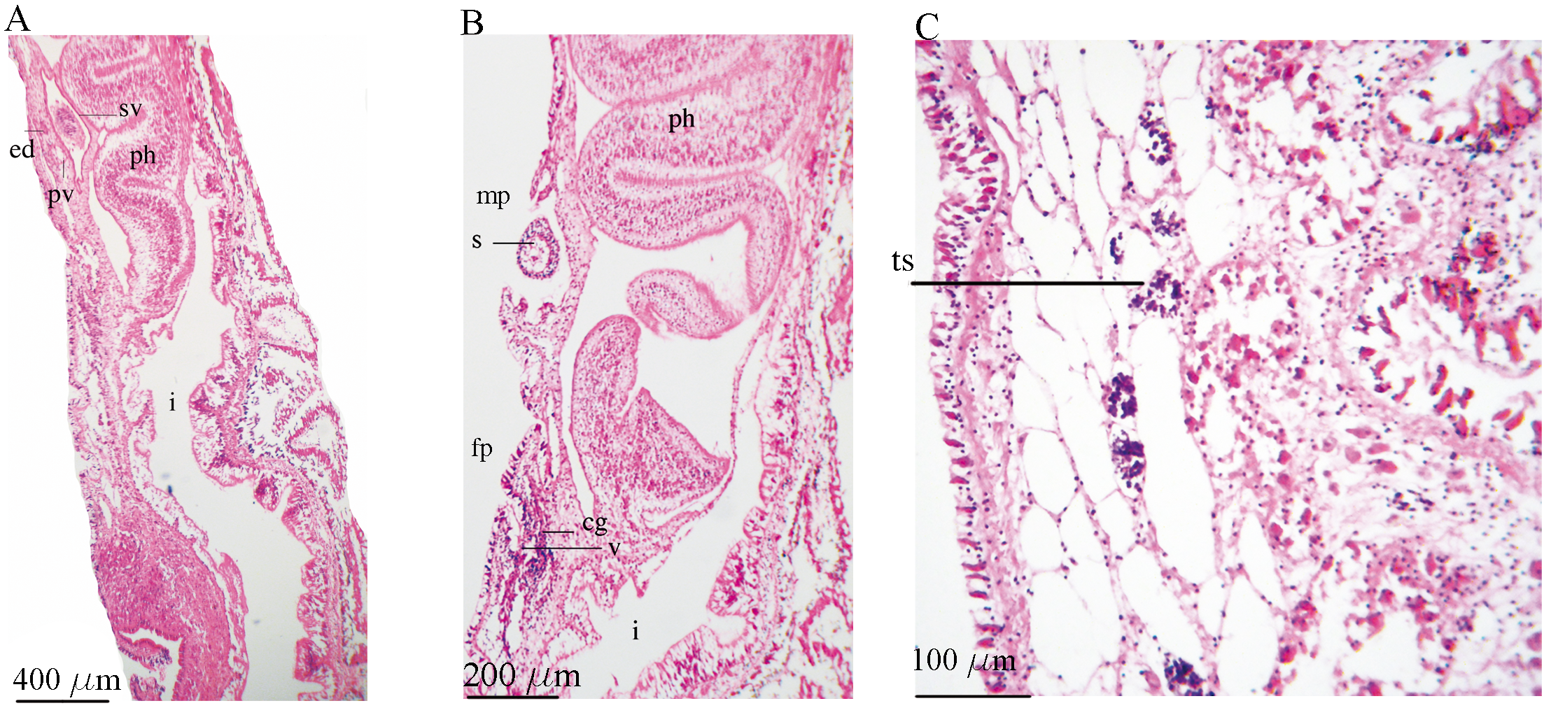
Description. External morphology. Body long, oval, soft and delicate, with slightly undulating margin ( Fig. 6 View FIGURE 6 A); the average size of the fixed specimens is 15.2 × 12.6 (SE± 1.4 × 1.4 mm), largest specimen 17.3 × 13.4 (ZUTC platy 1278). Dorsal surface of living specimens purplish cream or grey, with whitish or cream longitudinal median line, a purple marginal band ( Fig. 6 View FIGURE 6 A); papillae conical, slender, dark grayish or brownish with a tint of purple, except at the base of papilla which is colorless, with pointed and mostly dark apical part, a few papillae with white pointed tips, papilla size ranges from 850 µm (ZUTC platy 1275) to 1.04 mm, becoming larger medially, smaller towards margin and finally disappearing near the margin; in some of the specimens examined, papillae were more crowded on the median line of body ( Fig. 6 View FIGURE 6 A). Marginal tentacles 1.4–1.8 mm long, with white tips ( Figs. 6 View FIGURE 6 A, C). Cerebral eyespots horseshoe shaped, with 42 to 65 eyes, about 1.1 mm from the anterior margin ( Fig. 6 View FIGURE 6 C); the ventral pseudo-tentacular eyes are more numerous, about 50–70 ocelli per tentacle, arranged in four separate clusters, the dorsal pseudo-tentacular eyes are fewer, 25–32 ocelli, in two clusters ( Fig. 6 View FIGURE 6 C). Ventral surface translucent white, two male gonopores positioned immediately behind the pharynx, on both sides of the mid-ventral line; female gonopore immediately posterior to male gonopores, 500 µm apart (ZUTC platy 1275, Fig. 6 View FIGURE 6 B). The ventral body wall is about 50 µm thick ( Fig. 7 View FIGURE 7 C), thicker than the dorsal wall, with more developed muscular fibers including an outer longitudinal layer, followed by circular fibers and a well-developed innermost layer of diagonal muscles.
Reproductive structures. In ventral view, vasa deferentia appear as two coiled strips, parallel to mid-ventral line ( Fig. 6 View FIGURE 6 B), in sagittal view, each entering into the seminal vesicle from the ventral surface ( Fig. 6 View FIGURE 6 D). Male structures double, the seminal vesicle oval, large ( Figs. 6 View FIGURE 6 D, 7A), ranging from 450 Μm long × 250 Μm wide (ZUTC platy 1275) to 730 Μm × 370 Μm (ZUTC platy 1276); prostatic vesicle free and small, somewhat round and muscular, situated ventrally to the large seminal vesicle ( Figs. 6 View FIGURE 6 D, 7A), ranging from 93 Μm × 55 Μm (ZUTC platy 1275) to 140 Μm × 100 Μm (ZUTC platy 1276); penis stylet ( Fig. 7 View FIGURE 7 B), ranging from 110 µm × 80 µm (ZUTC platy 1275) to 126 µm × 91 µm (ZUTC platy 1276), stylet length to width ratio 1: 1.38. Numerous testes scattered ventrally in two rows ( Fig. 7 View FIGURE 7 C), 50 Μm in diameter. Female atrium, typical of pseudocerotids, long, oriented backwards and surrounded by cement glands ( Figs. 6 View FIGURE 6 D, 7B); uteri branched and dorsal to male structures.
Remarks. Grube (1840) established the genus Thysanozoon , within the family Pseudocerotidae . Occasionally, species determinations within the genus Thysanozoon can be made using coloration and color pattern ( Bolaños et al. 2007). The presence of the two external characters such as a papillated dorsal surface (unlike Pseudobiceros ) and two male gonopores (unlike Acanthozoon with one male pore) placed these worms in this genus. Thysanozoon brocchii is a cosmopolitan species distributed from the temperate to cold seas in both the Pacific and Atlantic Oceans ( Bahia et al. 2012; Brusa et al. 2009; Bulnes et al. 2011; Lang 1884; Marcus & Marcus 1968; Prudhoe 1989; Yeri & Kaburaki 1918).
With regard to the dorsal color, the species described here ( Fig. 6 View FIGURE 6 A) agrees well with the description provided by Yeri & Kaburaki (1918), but differs from the description of Bahia et al. (2012) and by Bursa et al. (2009). Their specimens were depicted as having a brown to yellowish dorsal region with a median longitudinal cream line, while those described here are purplish cream with a purple marginal band.
Thysanozoon sp. has been previously reported by Khalili et al. (2009) from Qeshm Island, Persian Gulf. Based on the figures 8–11 provided by Khalili et al. (2009), we believe that what they have illustrated might have been T. brocchii , particularly because we found specimens of that species from the same localities. Unlike the study of Khalili et al. (2009) that was limited only to one locality in the Persian Gulf, we expanded our sampling stations to the Iran coastline of the Persian Gulf and the Gulf of Oman ( Fig. 1 View FIGURE 1 ). T. brocchii was found in two stations of the Persian Gulf ( Fig. 1 View FIGURE 1 station 31& 33) and one station in the Gulf of Oman ( Fig. 1 View FIGURE 1 station 36). We found our specimens mostly associated with didemnid ascidians ( Fig. 10 View FIGURE 10 C), an association previously noted by Bahia et al (2012). We hereby report T. brocchii from the tropical Western Indo-Pacific, and from Iran coastal waters of the Persian Gulf and the Gulf of Oman, for the first time.
No known copyright restrictions apply. See Agosti, D., Egloff, W., 2009. Taxonomic information exchange and copyright: the Plazi approach. BMC Research Notes 2009, 2:53 for further explanation.
|
Kingdom |
|
|
Phylum |
|
|
Class |
|
|
Order |
|
|
Family |
|
|
Genus |