
Priceiella (Thescelovora) coleyae Gustafsson, Clayton
|
publication ID |
https://doi.org/ 10.11646/zootaxa.4382.3.1 |
|
publication LSID |
lsid:zoobank.org:pub:4BE1AB50-46E7-402D-9E72-A45D78352E2B |
|
DOI |
https://doi.org/10.5281/zenodo.5995565 |
|
persistent identifier |
https://treatment.plazi.org/id/E11BB55A-B758-FFC4-FF76-92E8FBDE6E12 |
|
treatment provided by |
Plazi |
|
scientific name |
Priceiella (Thescelovora) coleyae Gustafsson, Clayton |
| status |
|
Priceiella (Thescelovora) coleyae Gustafsson, Clayton , & Bush, new species
( Figs 36–42 View FIGURES 36–37 View FIGURES 38–42 )
Type host. Stachyris strialata tonkinensis Kinnear, 1938 —spot-necked babbler ( Timaliidae ).
Type locality. Jingxi County, Guangxi Province, China.
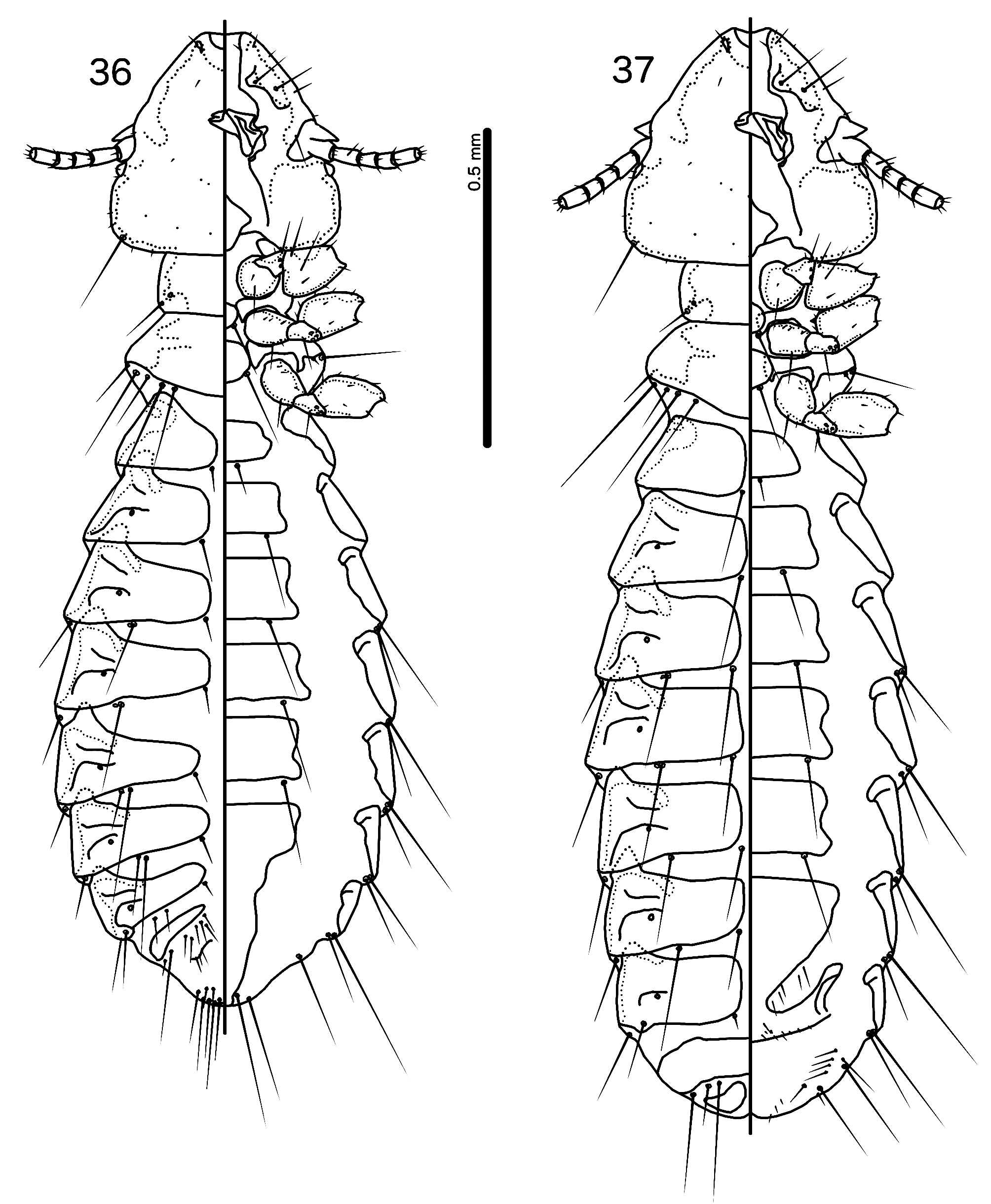
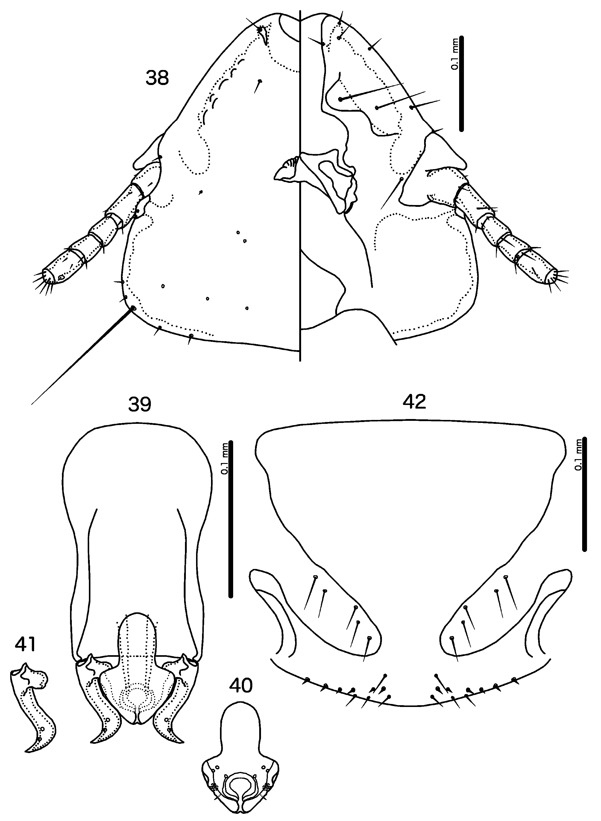
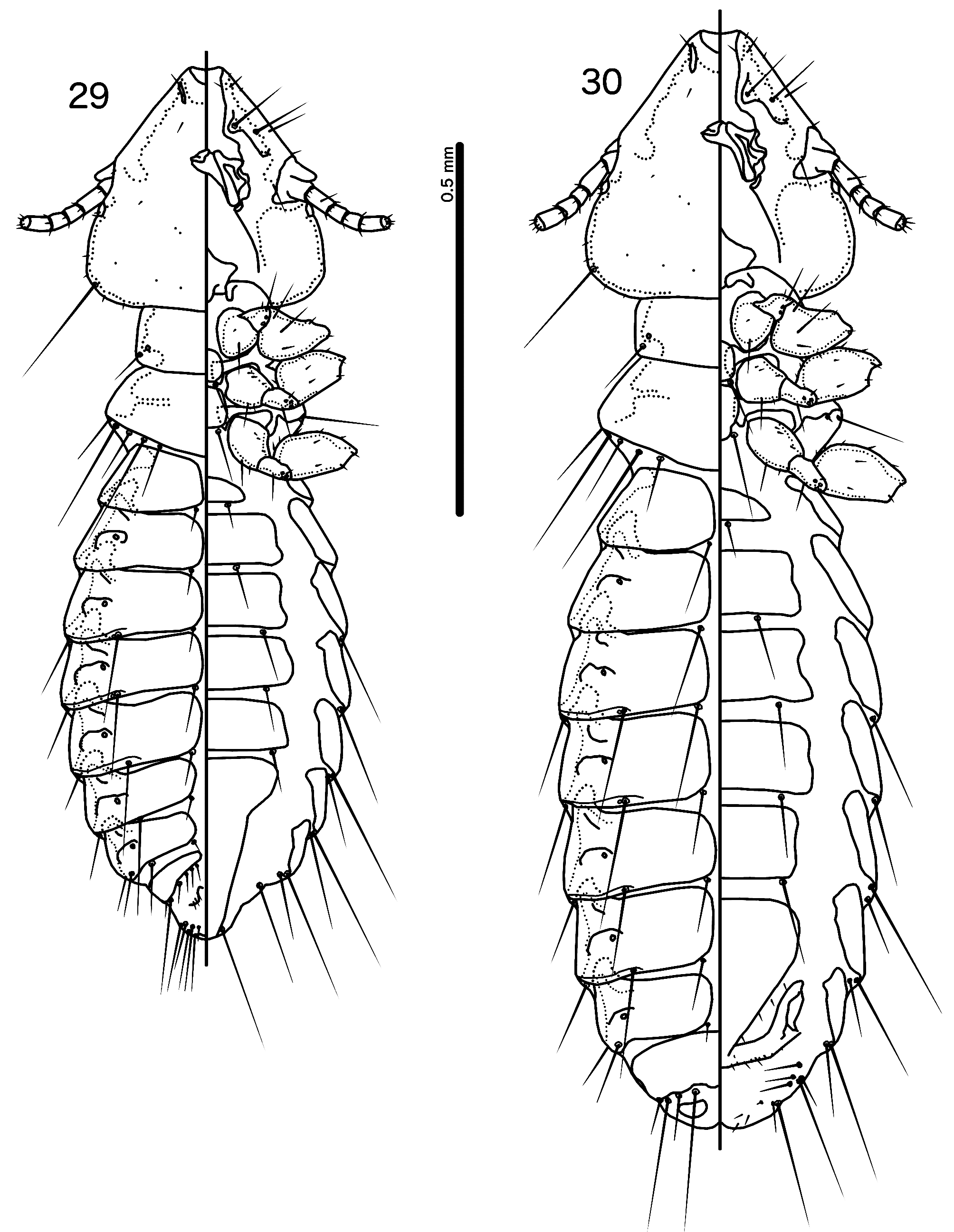
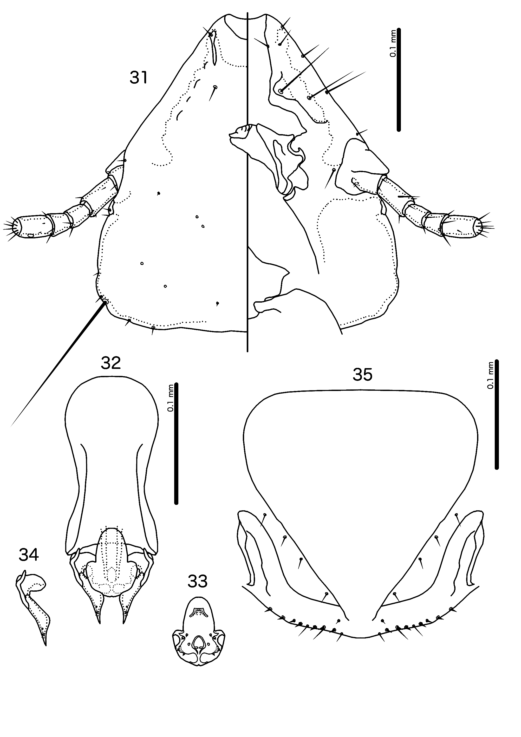
Diagnosis. Priceiella (Thescelovora) coleyae n. sp. ( Figs 36–42 View FIGURES 36–37 View FIGURES 38–42 ) is most similar to P. (T.) fuscicaena n. sp. ( Figs 29–35 View FIGURES 29–30 View FIGURES31–35 ), with which it shares the following characters: frons shallowly concave ( Figs 31 View FIGURES31–35 , 38 View FIGURES 38–42 ); proximal mesosome rounded ( Figs 33 View FIGURES31–35 , 40 View FIGURES 38–42 ); marginal thickening of mesosomal lobes broad ( Figs 33 View FIGURES31–35 , 40 View FIGURES 38–42 ). The two species can be separated on the following characters: lateral margins of preantennal area convex in P. (T.) coleyae ( Fig. 38 View FIGURES 38–42 ), but straight in P. (T.) fuscicaena ( Fig. 31 View FIGURES31–35 ); dorsal preantennal suture reaches at least half-way between dsms and ads in P. (T.) fuscicaena ( Fig. 31 View FIGURES31–35 ) but is much shorter in P. (T.) coleyae ( Fig. 38 View FIGURES 38–42 ); aps present on male tergopleurites VI– VII in P. (T.) coleyae ( Fig. 36 View FIGURES 36–37 ) but absent in P. (T.) fuscicaena ( Fig. 29 View FIGURES 29–30 ); basal apodeme slender, notably constricted at mid-length in P. (T.) fuscicaena ( Fig. 32 View FIGURES31–35 ) but broader and less or not constricted in P. (T.) coleyae ( Fig. 39 View FIGURES 38–42 ); distal mesosome convergent to medial point in P. (T.) coleyae ( Fig. 40 View FIGURES 38–42 ), but rounded in P. (T.) fuscicaena ( Fig. 33 View FIGURES31–35 ); proximal mesosome with ventral rugose area in P. (T.) fuscicaena ( Fig. 33 View FIGURES31–35 ) but without such area in P. (T.) coleyae ( Fig. 40 View FIGURES 38–42 ); parameres divergent distally in P. (T.) coleyae ( Fig. 41 View FIGURES 38–42 ), but parallel distally in P. (T.) fuscicaena ( Fig. 34 View FIGURES31–35 ); vos longer in P. (T.) coleyae ( Fig. 42 View FIGURES 38–42 ) than in P. (T.) fuscicaena ( Fig. 35 View FIGURES31–35 ) but vulval chaetotaxy otherwise similar.
Description. Both sexes. Head broad, dome shaped, with convex posterior margin ( Fig. 38 View FIGURES 38–42 ). Frons slightly concave. Lateral margins of preantennal head clearly convex. Dorsal preantennal suture present around dsms, does not reach even half-way to ads. Head chaetotaxy as in Fig. 38 View FIGURES 38–42 . Coni do not reach distal margin of scapes. Pteronotum with 5 mms on each side ( Figs 36–37 View FIGURES 36–37 ). Base pigmentation pale yellowish brown; marginal and temporal marginal carinae, head nodi, flagellomere II, proepimera, metepisterna and pleural incrassations dark brown; margins of antennal socket, mandibular framework, flagellomeres I and III, gular plate and subgenital and sternal plates IV–VI medium brown; sternal and subgenital plates darkening laterally.
Male. Abdominal plates and chaetotaxy as in Fig. 36 View FIGURES 36–37 ; aps present on tergopleurites VI–VII. Male genitalia as in Figs 39–41 View FIGURES 38–42 . Basal apodeme roughly rectangular, slightly or not constricted at mid-length ( Fig. 39 View FIGURES 38–42 ). Proximal mesosome slender, rounded ( Fig. 40 View FIGURES 38–42 ). Mesosomal lobes rounded triangular, distally convergent to blunt medial point. Lateral thickening of mesosome slightly sinuous, broad. Rugose nodi present. Gonopore open only distally; marginal thickening wide; 2 ames sensilla on each side near antero-lateral corners of mesosomal lobes; 1 pmes sensilla on each side of anterior end of gonopore; 1 pmes microseta laterally on each side distal to rugose nodi. Parameral heads rounded but slightly irregular in shape with slightly sinuous median margin ( Fig. 41 View FIGURES 38–42 ). Parameral blades clearly divergent distally; pst1–2 close together. Measurements ex Stachyris striolata tonkinensis (n = 11): TL = 1.35–1.54 (1.41); HL = 0.33–0.35 (0.34); HW = 0.34–0.36 (0.35); PRW = 0.20–0.22 (0.21); PTW = 0.29– 0.34 (0.31); AW = 0.43–0.53 (0.47).
Female. Abdominal plates and chaetotaxy as in Fig. 37 View FIGURES 36–37 . Vulval margin ( Fig. 42 View FIGURES 38–42 ) shallowly rounded, with 3 short, slender vms and 6 short thorn-like vss on each side; 5–6 long, slender vos on each side; distal vos near vss. Measurements ex Stachyris striolata tonkinensis (n = 18, except TL where n = 16 and AW where n = 15): TL = 1.57–1.79 (1.70); HL = 0.36–0.38 (0.37); HW = 0.37–0.41 (0.39); PRW = 0.22–0.24 (0.23); PTW = 0.33–0.36 (0.35); AW = 0.51–0.62 (0.55).
Etymology. This species is named for Phyllis D. Coley, University of Utah, in recognition of her distinguished contributions to ecology and her friendship and support of authors Bush and Clayton.
Type material. Ex Stachyris striolata tonkinensis : Holotype Ƌ, Jingxi County, Guangxi Province, China, 29 Sep. 2004, S.E. Bush, ATP-2004-129, P-364 ( NHML) . Paratypes: 2♂, 5♀, same data as holotype (PIPeR) ; 1♂, same data, ATP-2004-120, P-345 (PIPeR) ; 7♂, 13♀, same data, AN-434, P-351 (PIPeR).
| NHML |
Natural History Museum, Tripoli |
No known copyright restrictions apply. See Agosti, D., Egloff, W., 2009. Taxonomic information exchange and copyright: the Plazi approach. BMC Research Notes 2009, 2:53 for further explanation.