
Bolitophagus reticulatus
|
publication ID |
https://doi.org/ 10.11646/zootaxa.4111.3.1 |
|
publication LSID |
lsid:zoobank.org:pub:DAFF2F08-FF7D-499F-A6C0-2FA7FB080C73 |
|
DOI |
https://doi.org/10.5281/zenodo.5617803 |
|
persistent identifier |
https://treatment.plazi.org/id/E124B46C-DC52-8058-5BCA-6D24B0E8FB1E |
|
treatment provided by |
Plazi |
|
scientific name |
Bolitophagus reticulatus |
| status |
|
Description of mature larva and pupa of Bolitophagus reticulatus
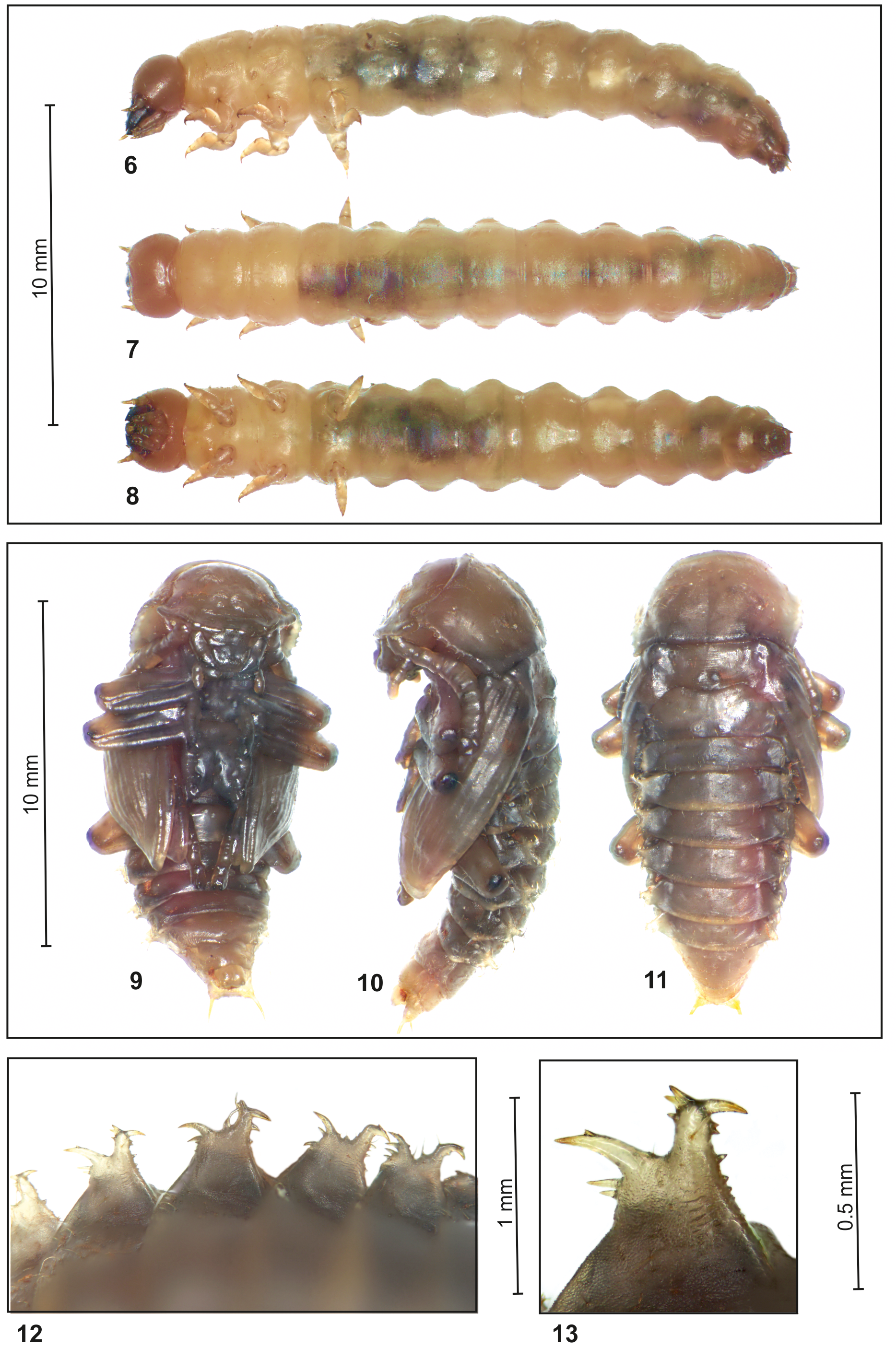
Mature larva. Coloration ( Figs. 6–8 View FIGURES 6 – 13 ). Head brown, thoracic and abdominal segments I–VI dark yellow to brown, remaining segments brown to dark brown; anal lobes dark brown; legs yellowish to brown. Thoracic and abdominal tergites more pigmented than remaining parts of the body. Cuticle densely, covered by fine asperities.
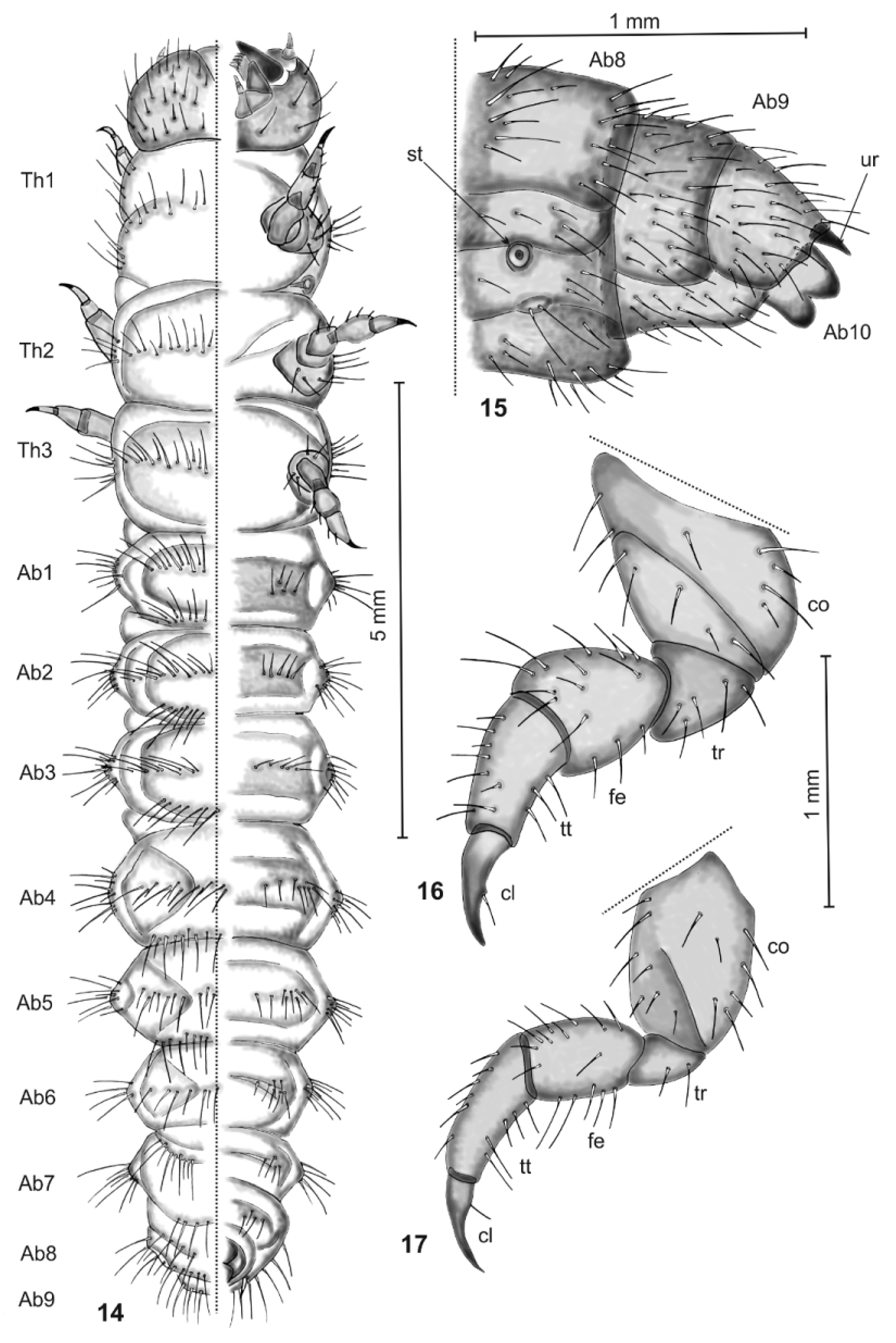
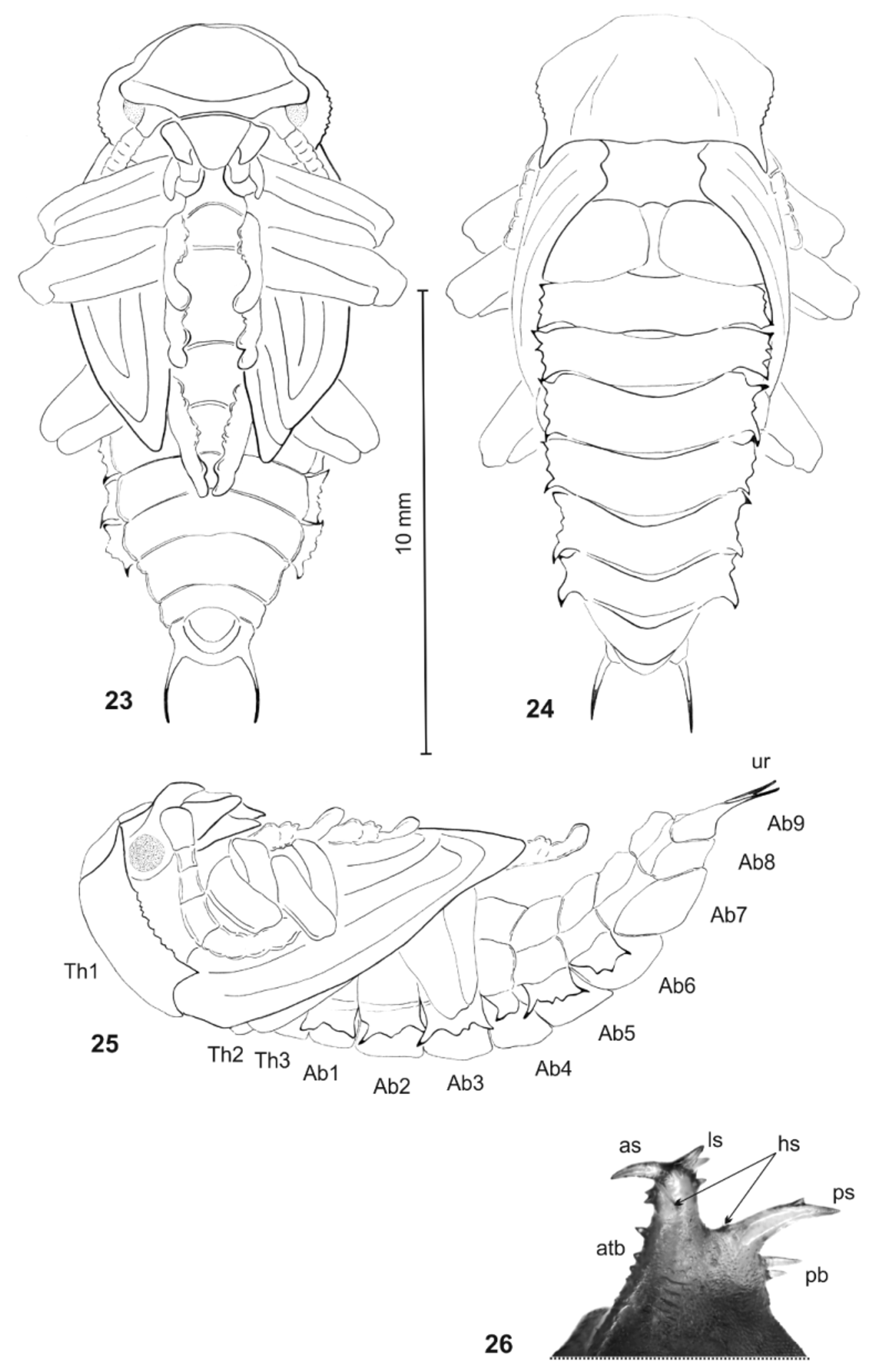
Body shape and chaetotaxy ( Fig. 14 View FIGURES 14 – 17 ). Body: length 9.5–12.5 mm, greatest width (at metathorax or abdominal segment I) 1.3–1.5 mm, shape moderately elongated, rounded in cross section, slightly curved. Chaetotaxy well developed, setae various in length, transparent or yellowish, always hair-like. Thorax: Prothorax almost as long as wide; both meso- and metathorax approximately 1.5 times broader than long. Prothorax with a row of long and medium long setae situated close to anterior margin; meso- and metathorax each with similar groups of setae situated dorso-medially; each of thoracic segments with groups of several setae of various length placed laterally. Legs ( Figs. 16, 17 View FIGURES 14 – 17 ): forelegs longer and more massive than next pairs (especially coxae of forelegs distinctly stout); trochanter short, conical, femur of foreleg conical, of mid- and hindlegs subcylindrical, tibiotarsus elongated, all covered irregularly with number of setae; claws elongated dark brown, with a seta placed ventrally; claws almost two times shorter than tibiotarsus. Abdomen: Abdominal segments I – IV almost of equal length, next abdominal segments decreasing gradually to the terminal parts of the body. Abdominal segment X reduced to four anal lobes of almost equal size. Anus located terminally. Tergites of segments I–VII with two rows of setae (first located along anterior margin, second situated medially). Long and short setae placed in both rows alternatively. Abdominal segments I–VII with a group of several, various in length setae placed laterally. Medial parts of abdominal segments I–VII with 4 – 6 equal in length setae. Setae of abdominal segment VIII well developed. Dorsal and lateral parts of abdominal segment IX densely covered by numerous setae of various length. Abdominal segment IX with a pair of small, conical urogomphi, strongly sclerotized, localized dorso-laterally, slightly curved to inside. Urogomphi and anal lobes without setae ( Fig. 15 View FIGURES 14 – 17 ).
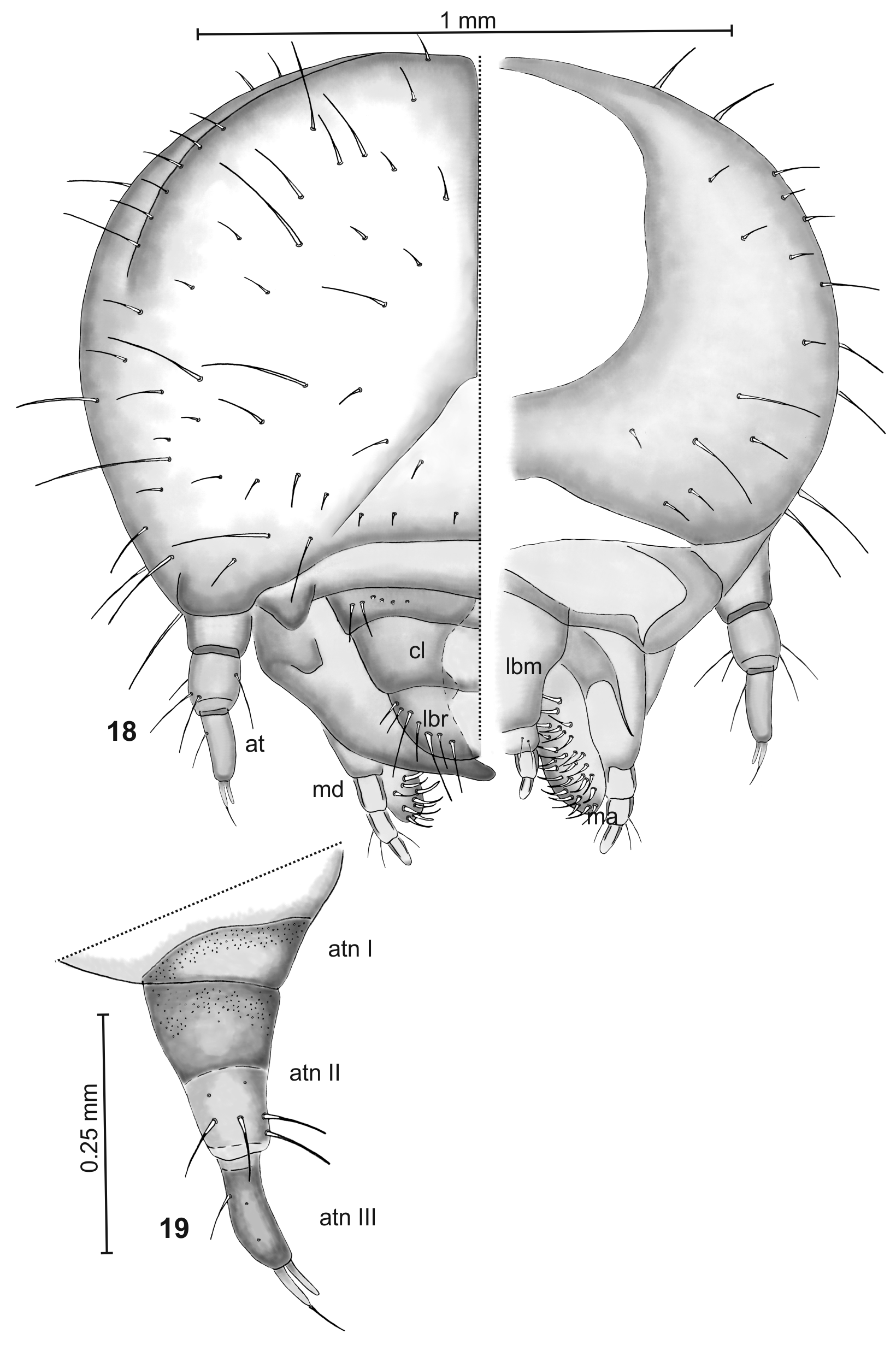
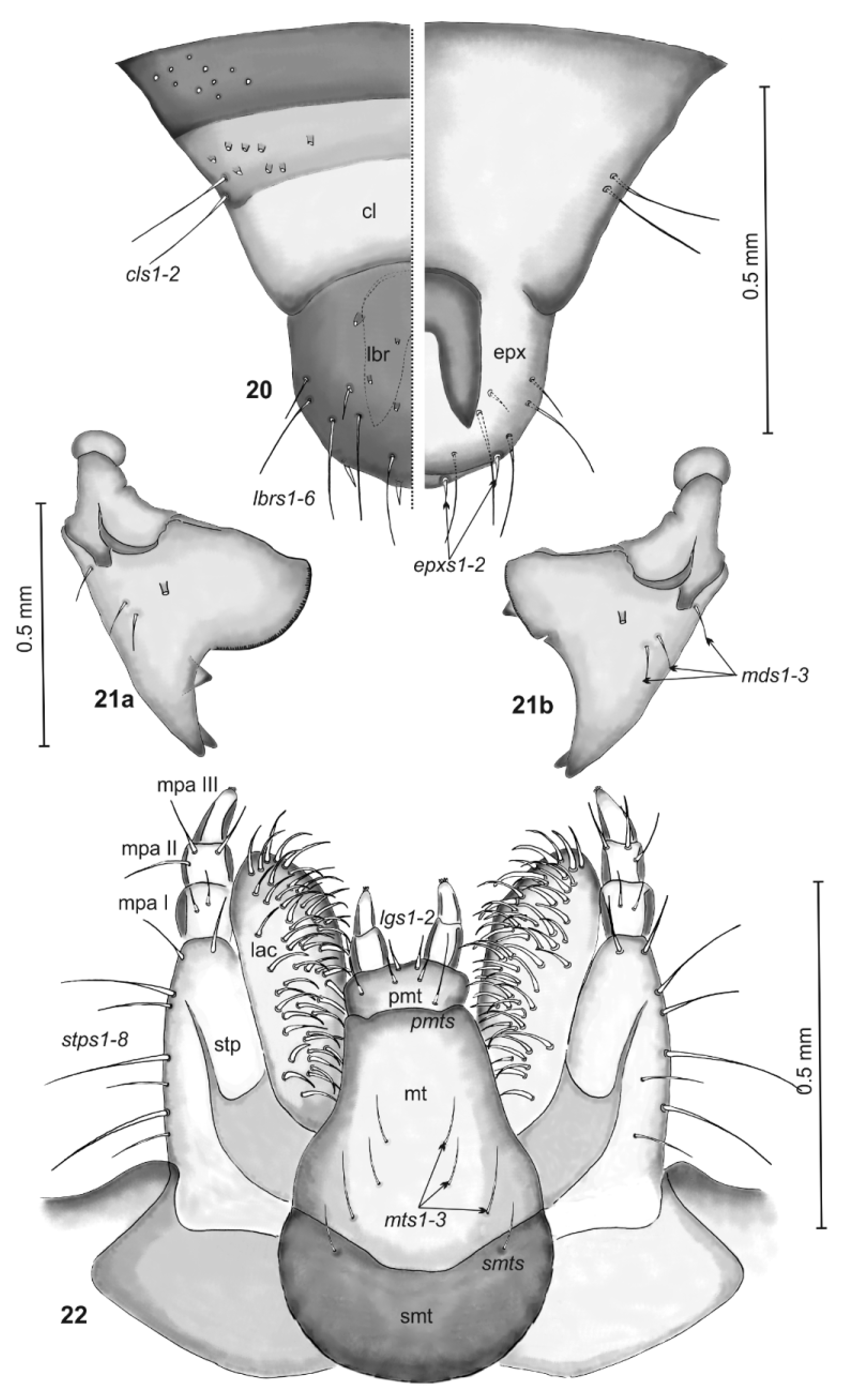
Head ( Fig. 18 View FIGURES 18 – 19 ), width 1.3–1.55 mm, hypognathous, almost oval, sutura coronalis weakly visible, sutura frontalis not reaching antenna, frons relatively small, triangular. Epicranium and frons covered by numerous hairlike setae of various length. Setae do not form any regular pattern. Antennae ( Fig. 19 View FIGURES 18 – 19 ) trimerous; antennomere I distinctly wider than long; antennomere II 1.7 times as long as wide, slightly narrowed in apical part, with 4 setae; antennomere III slender, finger-like, with one seta and two sensillae apically (one of them with a seta situated on the top). Basalpart of antennomere II more pigmented than the upper part, so whole antennomere seems to consist of two rings. Clypeus (Fig. 20—left side) convex in lateral view, transverse, trapezoidal, anterior margin strongly hollowed. Basal part of clypeus more pigmented then middle and distal parts. A pair of long setae located mediolateral on each side. Labrum (Fig. 20) almost as long as wide, with rounded anterior margin its dorsal surface flat in lateral view, with eight long and four short setae. All setae placed more or less marginally. Epipharynx (Fig. 20—right side) with four short marginal setae; two of them placed apically, next laterally. Mandibles ( Figs 21 View FIGURES 21 – 22 a – b) asymmetrical, strong sclerotized, bifid, in dorsal view with three long setae, placed laterally; right mandible wide, with well developed, semicircular molar part and small triangular tooth close to apical part; left mandible narrow, with short cut molar part and triangular tooth located basally. Each of mandibles bears dorso-laterally distinct conical process with oblate apex. Maxilla ( Fig. 22 View FIGURES 21 – 22 ): stipes bears eight setae various in length, palpus threesegmented, both segments I and II almost as long as wide, with two and three setae respectively, segment III longer than wide, sub-conical, with some sensillae apically. Lacinia densely covered by numerous thick, various in length, curved setae. Labium ( Fig. 22 View FIGURES 21 – 22 ) divided into prementum, mentum and submentum. Prementum almost oval-shaped, with a pair of setae placed antero-laterally; mentum subcylindrical, tapering in apical part, with three pairs of setae equal in length, located medio-laterally; submentum centrally, almost two times wider than longer, with a pair of long setae medially; ligula concave with two pairs of short, thick setae placed apically. Labial palpi two-segmented.
Pupa. Coloration ( Figs 9 – 11 View FIGURES 6 – 13 ). Body yellowish to greyish (changing its color during preservation into brown), claws dark brown, lateral processes more pigmented yellow with brown spines.
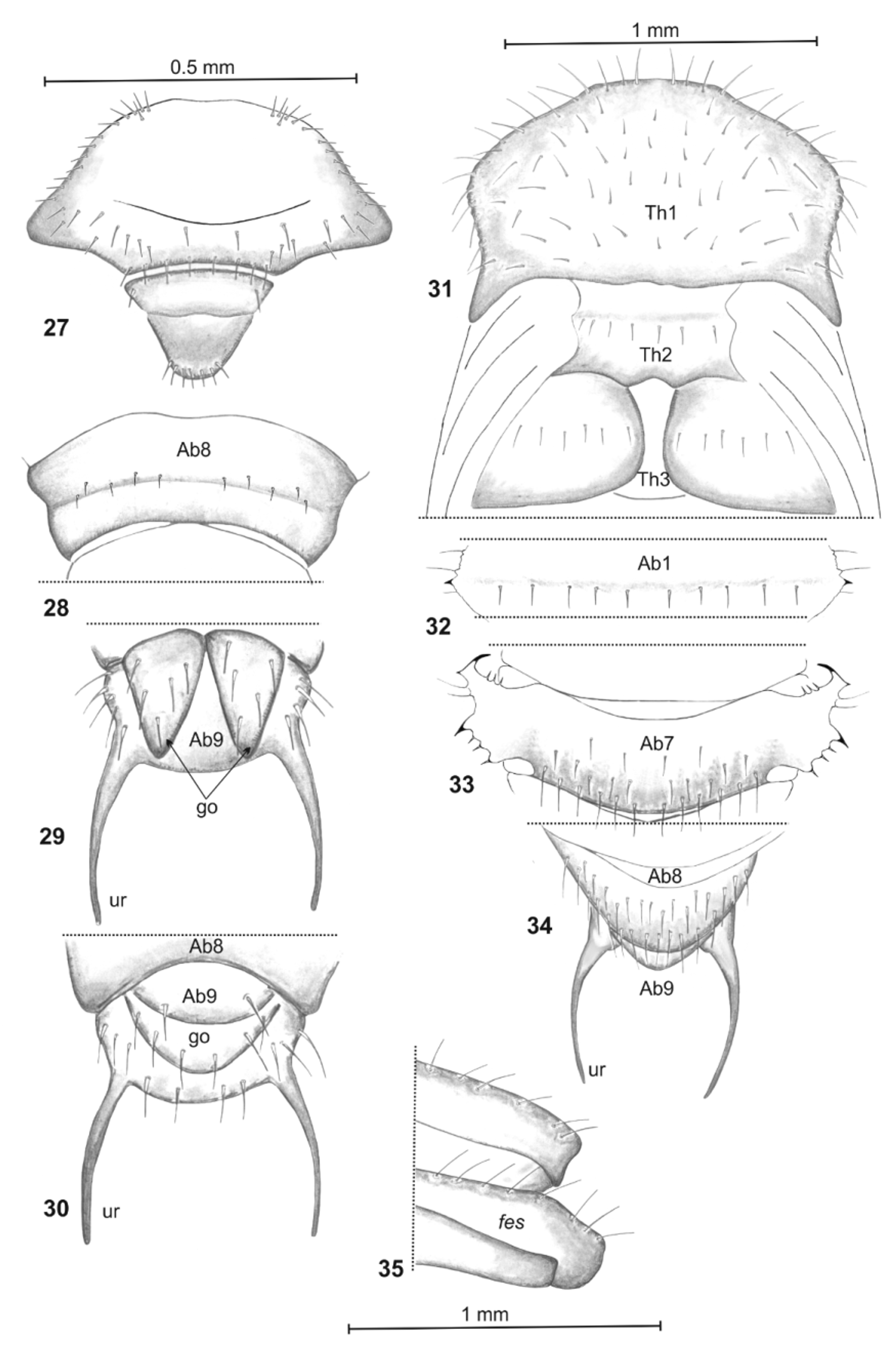
Shape, chaetotaxy and lateral processes. Body ( Figs 23 – 25 View FIGURES 23 – 26 ) rather elongated, slightly curved, oblate dorsoventrally, length: 5.5–7.7 mm, width (measured on the level of second pair of legs): 3.0–4.0 mm, chaetotaxy developed, setae hair-like, straight, yellow, various in length. Gin traps of abdominal tergites I – VI well developed ( Figs 12, 13 View FIGURES 6 – 13 , 26 View FIGURES 23 – 26 ); on the first abdominal segment, triangular, bear apical spine and some very fine setae; on abdominal segments II – VI conical, divided apically, consist of anterior spine, lateral spine and massive posterior spine; anterior borders possess 4 – 6 various in size triangular teeth; posterior borders with 2 – 3 long spines and two hair sensillae. Abdominal segments I – VII decreasing gradually, segment VII conical, IX strongly reduced. Tergite IX with a pair of elongated slightly curved to inside urogomphi, with sclerotized apical parts. Head ( Fig. 27 View FIGURES 27 – 35 ) wide, eyes well visible, black; marginal parts of head irregular covered by numerous short setae; medial part ridged, without setae, clypeus transversely wrinkled, with single setae on each side; labrum trapezium-shaped with seven setae along its anterior margin. Pronotum ( Fig. 31 View FIGURES 27 – 35 ) transversely wrinkled with serrated, dark sclerotized lateral parts; lateral angles elongated, central part of pronotum regular, covered by numerous short setae, borders bear distinctly longer setae; hypomeron glabrous; elytral and metathoracic wing sheaths smooth, mesonotum slightly shorter than metanotum, both almost as long as first abdominal tergum; each of them with a horizontal row of setae medially. Spiracles seven pairs, first elongated, placed between pro- and mesonotum, next six pairs rounded, located medio-laterally on abdominal segments I – VI. Abdominal tergites I – VI ( Fig. 32 View FIGURES 27 – 35 ) with rows of weakly visible short setae, situated medially, tergite VII ( Fig. 33 View FIGURES 27 – 35 ) with numerous of various in length setae located medially and postero-medially; tergite IX ( Fig. 34 View FIGURES 27 – 35 ) covered by numerous elongated setae; abdominal ventrites I – VIII ( Fig 28 View FIGURES 27 – 35 ) with weakly visible, short setae, distributed medially, ventrite IX with some elongated setae laterally. Gonotheca ( Figs 29 – 30 View FIGURES 27 – 35 ) of female divided, elongated, covered by elongated setae; gonotheca of male undivided with groups of very long setae. Urogomphi without setae. Femora ( Fig. 35 View FIGURES 27 – 35 ) bear several elongated setae; remaining parts of legs without setae.
No known copyright restrictions apply. See Agosti, D., Egloff, W., 2009. Taxonomic information exchange and copyright: the Plazi approach. BMC Research Notes 2009, 2:53 for further explanation.