
Neomida haemorrhoidalis
|
publication ID |
https://doi.org/ 10.11646/zootaxa.4111.3.1 |
|
publication LSID |
lsid:zoobank.org:pub:DAFF2F08-FF7D-499F-A6C0-2FA7FB080C73 |
|
DOI |
https://doi.org/10.5281/zenodo.5617805 |
|
persistent identifier |
https://treatment.plazi.org/id/E124B46C-DC5A-8041-5BCA-6BBAB46FFEA3 |
|
treatment provided by |
Plazi |
|
scientific name |
Neomida haemorrhoidalis |
| status |
|
Description of mature larva and pupa of Neomida haemorrhoidalis
Mature larva. Coloration ( Figs 36 – 38 View FIGURES 36 – 43 ). Head and thoracic segments dark brown, abdominal segments I – II light brown, next segments light brown or dark yellow; anal lobes yellowish; legs yellowish to brown. Tergites of thoracic segments distinctly sclerotized. Tergites and sternites of abdominal segments I – VIII more pigmented than lateral parts, so body seems to be covered by stipes. Cuticle smooth.
Body shape and chaetotaxy ( Fig. 44 View FIGURES 44 – 47 ). Body: length 12.0– 14.5 mm, width in the widest place (metathorax or abdominal segment I) 1.3–1.5 mm, shape distinctly elongated, slightly oblate dorso-ventrally in cross section, curved. At the first sight body seems to be completely void of setae. In fact chaetotaxy is well developed but weakly observed. Setae hair-like, slim, various in length, extremely transparent. Thorax: Prothorax almost as long as wide; meso- and metathorax each approximately 1.5 times broader than long. Prothorax with two horizontal rows of 6 – 8 medium long setae each first situated close to anterior margin, the second along posterior margin; mesothorax with 6 – 8 short setae placed anteriorly, next 8 very long and 4 – 6 short setae located posteriorly. Chaetotaxy of metathorax similar to mesothorcical. A pair of long setae placed latero-ventrally on each side of all thoracic segments. Legs ( Figs. 46, 47 View FIGURES 44 – 47 ) thin, elongated: forelegs some stouter than next pairs; coxa of first pair subcylindrical, of next pairs conical, trochanters, conical, more or less elongated, femur of foreleg rather massive, of mid- and hindlegs thin, tibiotarsus elongated, all sparsely covered with setae various in length; claws very thin, dark brown, with a seta placed ventrally; less than two times shorter than tibiotarsus. Abdomen: Abdominal segments I – V almost of equal length, next abdominal segments decreasing gradually to the terminal parts of the body. Abdominal segment X reduced to four anal lobes of unequal size. The ventral lobe, some elongated, bigger than remaining ones. Anus located ventrally. Tergites of segments I–VII with two horizontal rows of setae (first located along anterior margin, second situated posteriorly). First row consists only of 6 – 8 short setae, second with 6 – 8 long and similar number of short setae. Long and short setae of second rows located by turns. Sternites of abdominal segments I–VIII with 6 long setae, distributed along posterior margin of each segments. Abdominal segment IX with 4 very long and several short setae dorsally and 10 long setae placed ventro-laterally. Abdominal segment IX with a single short, conical urogomphus, weakly sclerotized, localized dorso-medially. Each of anal lobes with a very short seta ( Fig. 45 View FIGURES 44 – 47 ).
Head ( Fig. 48 View FIGURES 48 – 49 ), width 1.30–1.40 mm, prognathous, almost oval slightly widened posteriorly, distinctly oblate dorso-ventrally. Sutura coronalis well visible, sutura frontalis reached almost to antenna, frons triangular, relatively wide. Epicranium and frons more pigmented than postepicranial and lateral area. Setae of head sparse, very long distributed, dorso-laterally in two vertical rows on each side of head. There are also several long setae placed ventrally. Antennae ( Fig. 49 View FIGURES 48 – 49 ) trimerous; antennomere I conical, wide; antennomere II cylindrical as long as wide; antennomere III very slender, elongated, with one elongated and two small sensillae apically. Down parts of antennomeres I and II more pigmented than the upper parts, so both antennomeres seem to be fixed from four rings. Clypeus ( Fig. 50 View FIGURES 50 – 52 –left side) convex in lateral view, transverse, trapezoidal, anterior margin strongly hollowed. Basal part of clypeus more pigmented then distal parts. A pair of long setae located postero-lateral on each side. Labrum ( Fig. 50 View FIGURES 50 – 52 –left side) almost as twice long as wide, with sinuated anterior margin. Labrum with 14 very long setae dorsally. All setae placed more or less marginally. Epipharynx ( Fig. 50 View FIGURES 50 – 52 —right side) with six short marginal setae; two of them placed apically, next four laterally. Mandibles ( Figs. 51 View FIGURES 50 – 52 a – b) asymmetrical, strong sclerotized, bifid, in dorsal view with five setae, placed laterally; right mandible wide, with well developed, semicircular molar part and small triangular tooth close to apical part; left mandible narrow, with short cut molar part and triangular tooth located basally. Maxilla ( Fig. 52 View FIGURES 50 – 52 ): stipes bears five setae equal in length, hair-like and one short, stout seta. Palpus three-segmented, segment I almost as long as wide, with short seta, segments II and III distinctly longer than wide, cylindrical, segment II with two long setae, segment III with some sensillae apically. Lacinia densely covered by numerous setae of various length. Setae on lacinia straight or with curved apical part. Labium ( Fig. 52 View FIGURES 50 – 52 ) divided into prementum, mentum and submentum. Prementum almost as long as wide with slightly rounded lateral parts, with two pairs of setae placed antero-laterally; mentum subcylindrical, tapering in apical part, with two pairs of setae equal in length, located laterally; submentum centrally slightly wider than long, with a pair of long setae distally; ligula concave with two pairs of setae of various length, thick, placed apically. Labial palpi two-segmented.
Pupa. Coloration ( Figs. 39 – 41 View FIGURES 36 – 43 ). Body grayish to yellowish (changing its color during preservation into brown), claws dark brown, lateral processes light yellow with brown spines.
Shape, chaetotaxy and lateral processes ( Figs 53 – 55 View FIGURES 53 – 56 ). Body elongated, slightly curved, oblate dorsoventrally, length: 6.4–7.3, width (measured on the level of second pair of legs): 2.4–2.6 mm, chaetotaxy weakly developed, setae hair-like, straight, yellow, various in length. Gin traps of abdominal tergites I – VII well developed ( Figs 42, 43 View FIGURES 36 – 43 , 56 View FIGURES 53 – 56 ); on abdominal segments II – VI consist of well-developed apical spine (absent on abdominal segment I), lateral and posterior spines; serrated anterior and posterior borders with triangular, strongly teeth sclerotized and some hair sensillae (one close to lateral spine strongly elongated, much bigger than others). Lateral processes of tergite VII triangular, anterior and posterior spines absent, anterior border smooth, posterior border with some setae. Abdominal segments I – VII decreasing gradually, segment VII semicircular, IX strongly reduced. Tergite IX with a pair of elongated urogomphi, with sclerotized apical parts. Head ( Fig. 57 View FIGURES 57 – 65 ) semicircular, eyes weakly visible; head irregular covered by numerous short setae; head of female medially with a pair of small tubercles, of male with a pair of very elongated tubercles, always covered by relatively long setae, clypeus transversely wrinkled, with single setae on each side; labrum wide, trapezium-shaped without setae. Pronotum ( Fig. 61 View FIGURES 57 – 65 ) trapezium-shaped transversely wrinkled, its central part irregular, covered by numerous short setae, borders bear with elongated setae; hypomeron glabrous; elytrals with some short setae distributed irregular; metathoracic wing sheaths smooth, mesonotum almost twice shorter than metanotum, mesonotum with a row of very short setae medially, metanotum with two rows of weakly visible setae medially and posteriorly. First abdominal tergum as wide as mesonotum; Abdominal tergites I – VII ( Figs 62, 63 View FIGURES 57 – 65 ) with horizontal rows of weakly visible short setae, situated posteriorly, tergite VII with several long setae located along posterior margin; tergite IX ( Fig. 64 View FIGURES 57 – 65 ) bears some short setae; abdominal ventrites I – VII ( Fig. 58 View FIGURES 57 – 65 ) with weakly visible, very short setae, distributed posteriorly, ventrite VIII with elongated setae located postero-laterally. Gonotheca ( Figs 59, 60 View FIGURES 57 – 65 ) of female divided, strongly elongated, curved to outside; gonotheca of male undivided. Urogomorpi possess several short setae. Femora ( Fig. 65 View FIGURES 57 – 65 ) bear three elongated setae; remaining parts of legs without setae.
Some information on the larval stages of B. reticulatus was presented in a comprehensive work by Gilarov (1964). This included the habitus of the larva (lateral view), the shape of the antenna, urogomphi, labrum, epipharynx, labium, and maxilla, and the chaetotaxy of the dorsal part of the abdominal segment. Unfortunately, all figures provided by Gilarov (1964) differed in varying degrees from our observations. Some of these differences are presented in Table 1 View TABLE 1 .
Some of the characteristic features listed by Matthews et al. (2010) for the Bolitophagini tribe, such as reduced urogomphi or their absence, were contrary to the information reported by Gilarov (1964) but found confirmation in our observations. Moreover, Matthews et al. (2010) also stated that their larvae have a very lightly sclerotized body with an asymmetrical head and mandibles, which corresponds with our description of B. reticulatus larvae.
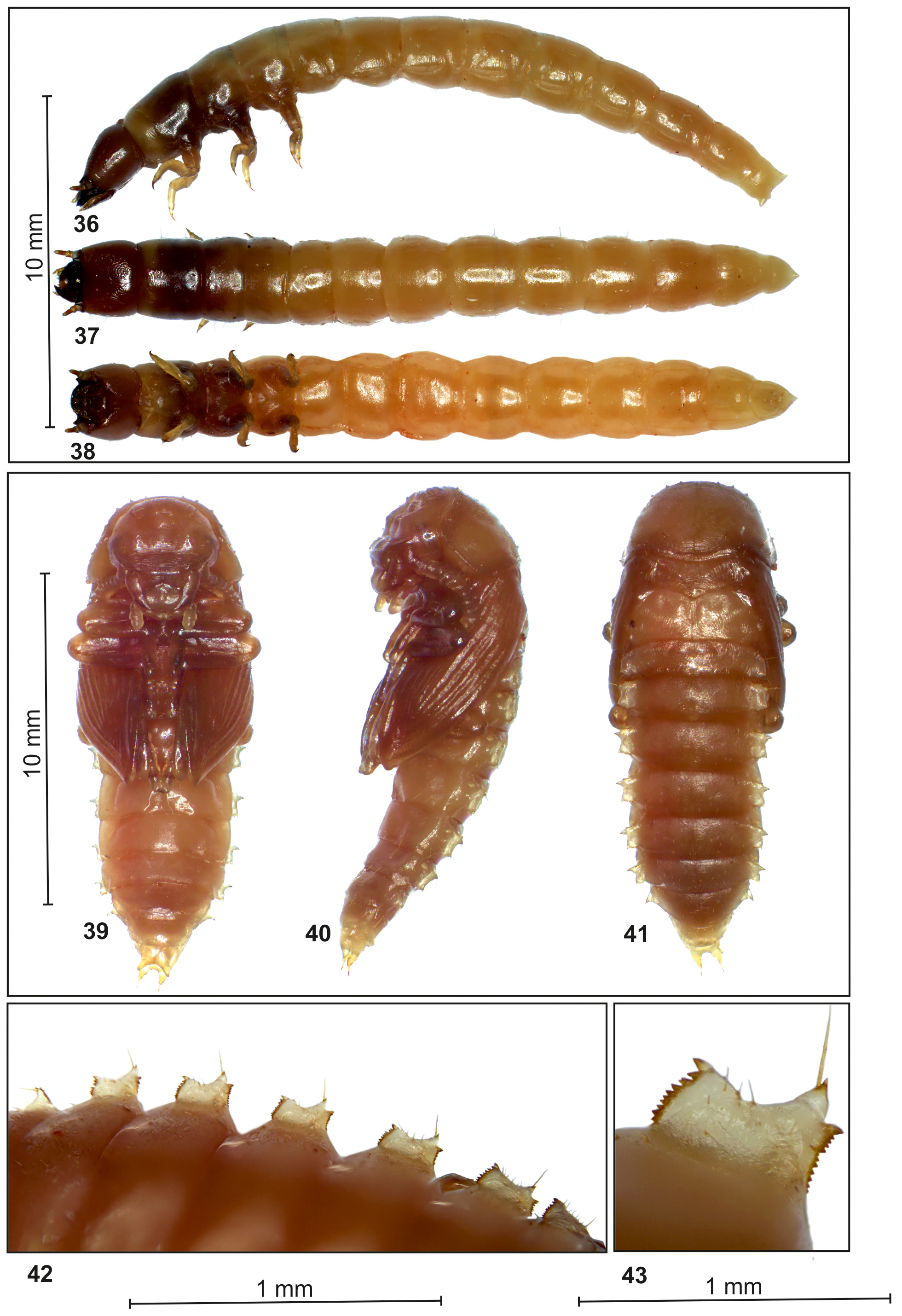
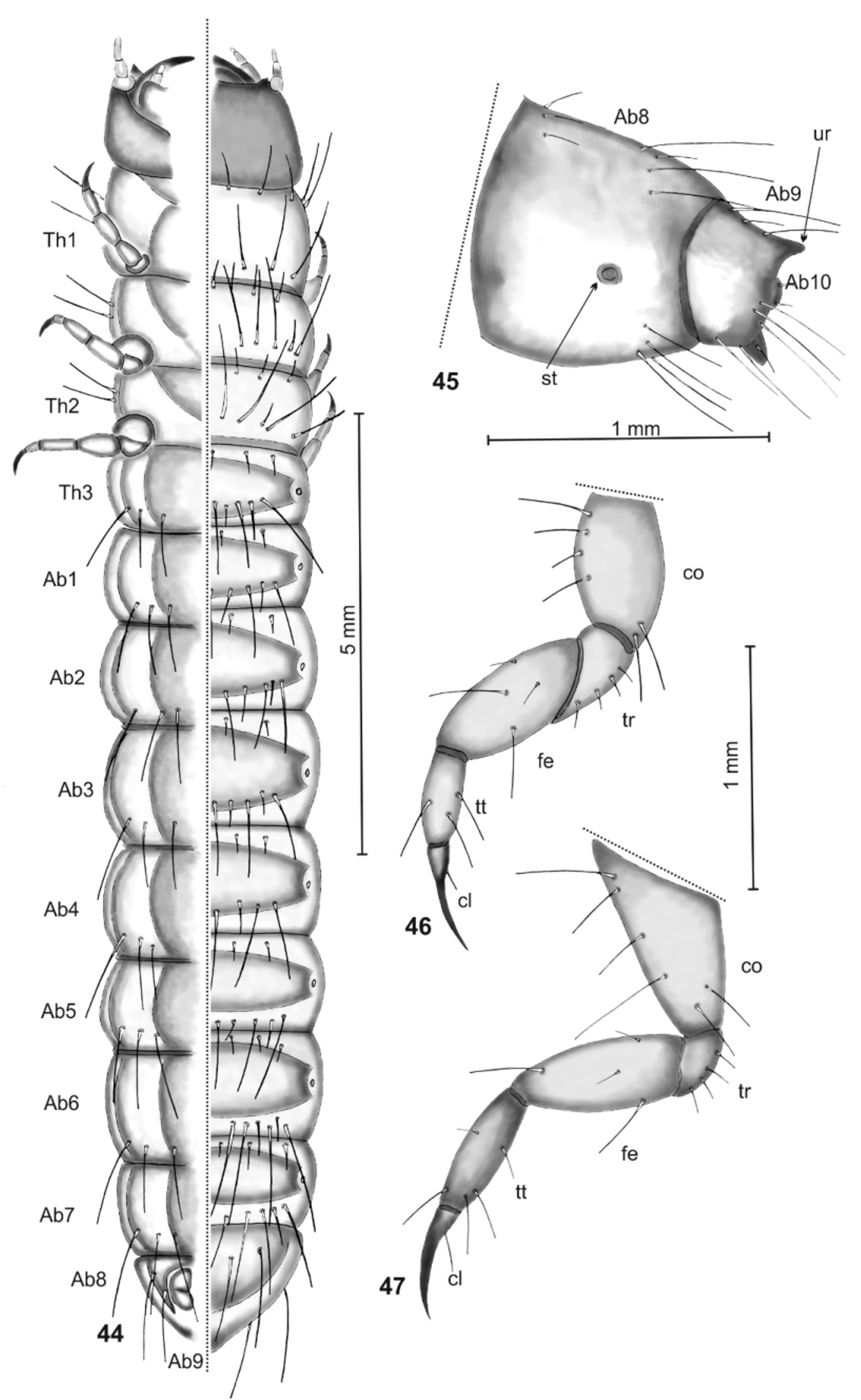
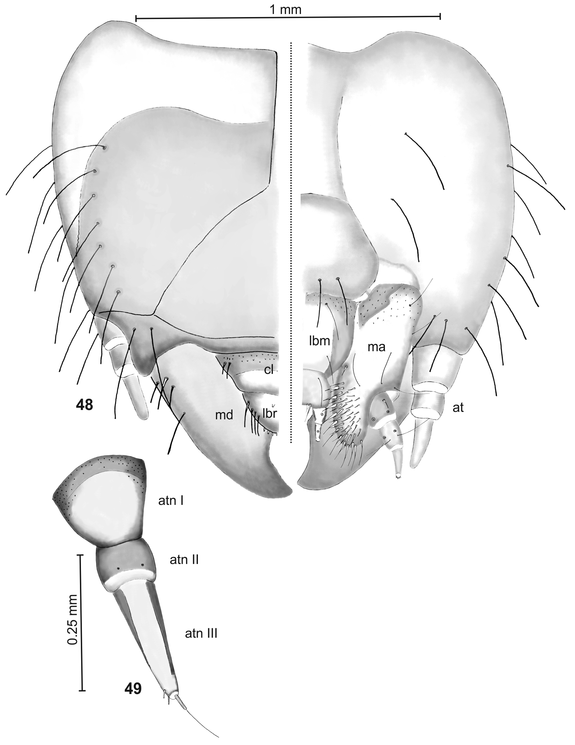
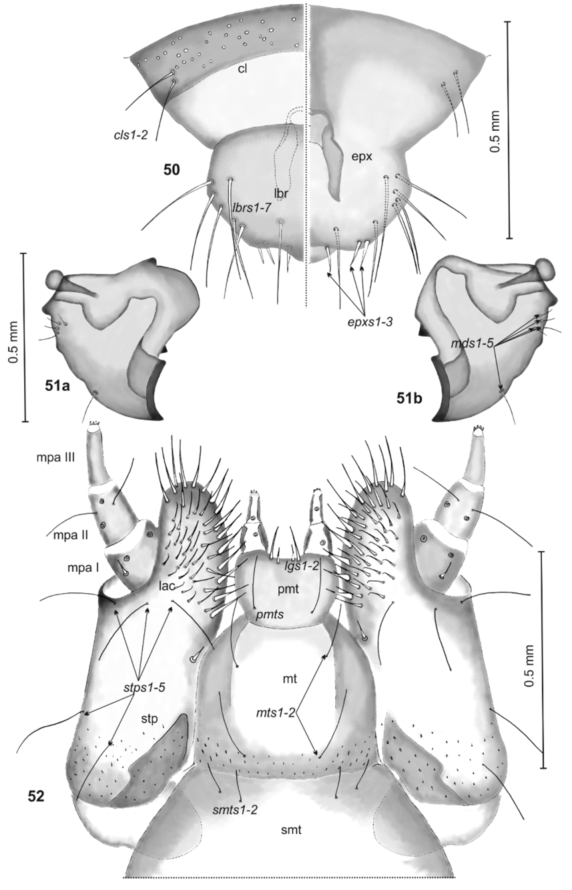
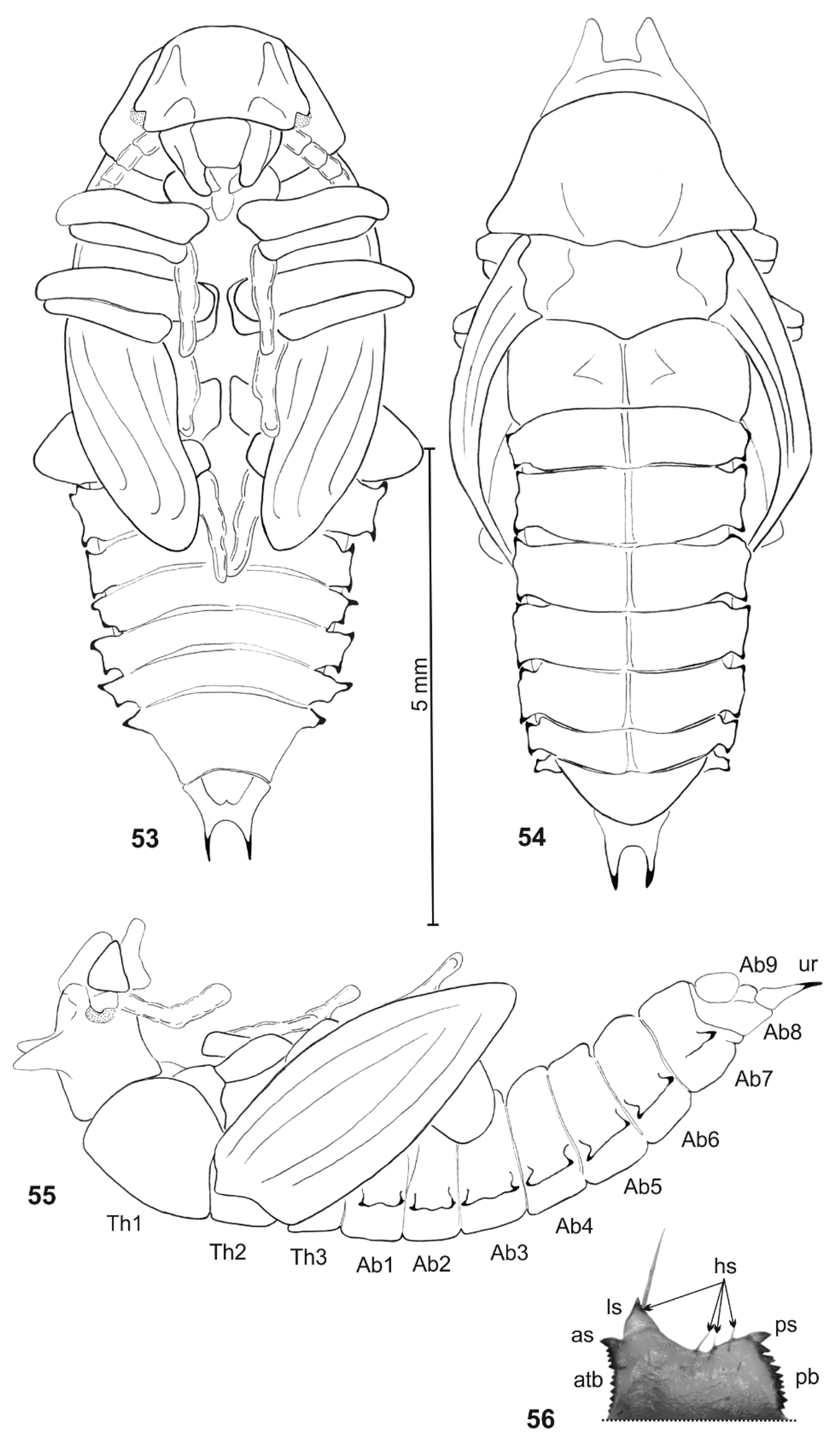
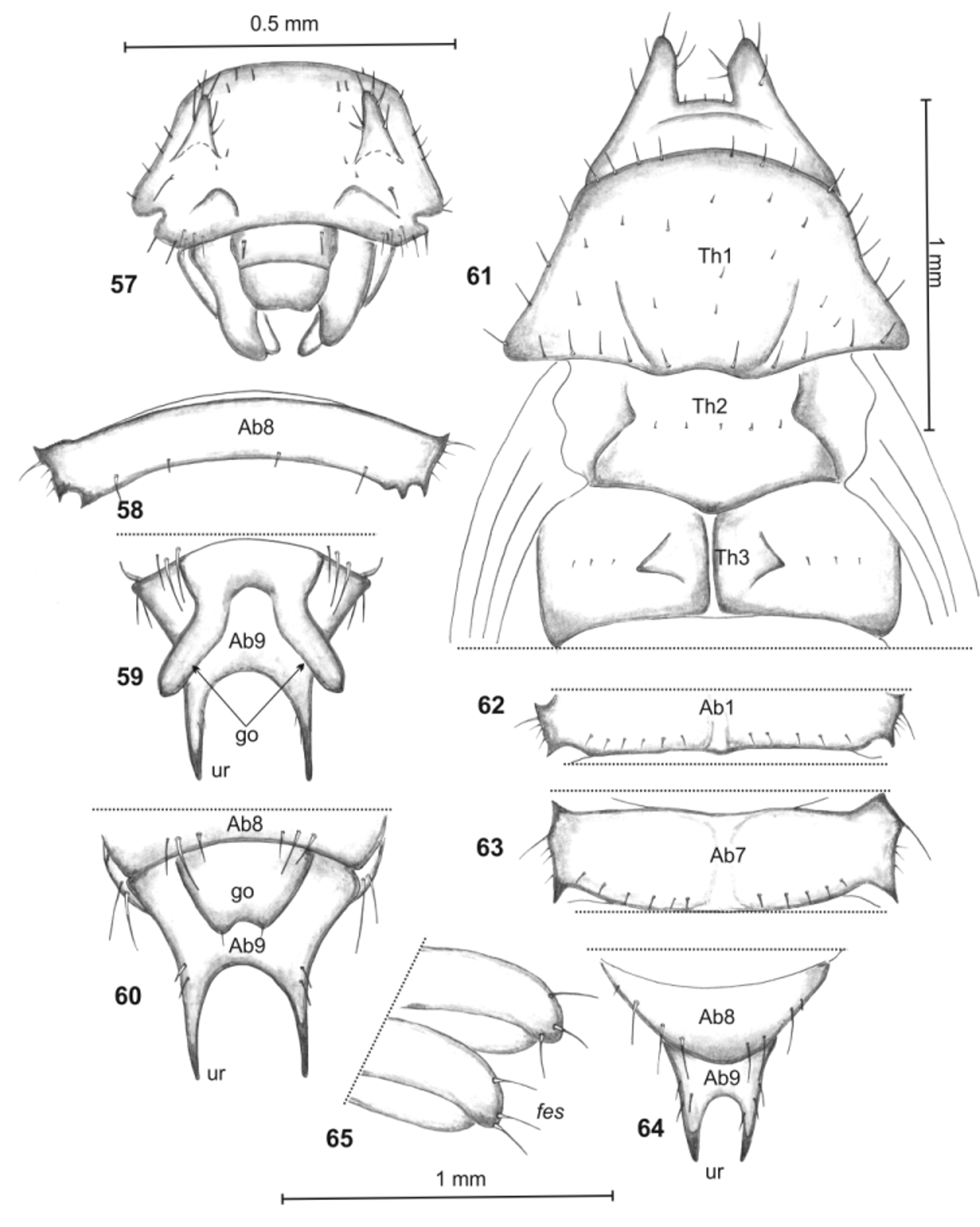
TABLE 1. Differences between the Bolitophagus reticulatus larva described by Gilarov (1964) and in this paper.
| Feature | B. reticulatus larva | |
|---|---|---|
| Gilarov (1964) | presented paper | |
| Body shape | slightly oblate dorso-ventrally | rounded in cross section |
| Urogomphi | pronounced, thorn-like, distinctly curved upwards | small, slightly curved backward |
| Chaetotaxy of the body | visible only on dorsal part of 9th abdominal segment | well-developed |
| Head | distinctly oblate dorso-ventrally | oval |
| Second antennomeres | shortened and widened, with only 1 seta | elongated, with 4 setae. |
| Sensillae on apical antennomere | distinctly different in size and shape | almost the same |
| Labrum | with 4 setae | with 6 setae |
| Epipharynx setae | 4, conical, distinct | 2, thorn-like, rather small |
| Maxilla | only margin of lacinia covered by setae | whole lacinia covered by setae |
| Maxillary palpus | without setae | with 5 setae |
| Ligula | conical, rounded, almost as high as labial palpi | straight, not extruded from labial palpi. |
| Mentum | square | trapezium-shaped |
| Submentum | strongly elongated | rounded |
No known copyright restrictions apply. See Agosti, D., Egloff, W., 2009. Taxonomic information exchange and copyright: the Plazi approach. BMC Research Notes 2009, 2:53 for further explanation.