
Temnaspis puae
|
publication ID |
https://doi.org/ 10.11646/zootaxa.3737.4.3 |
|
publication LSID |
lsid:zoobank.org:pub:7CC5D919-FA22-4768-8BB6-6DACFEAEE8D1 |
|
DOI |
https://doi.org/10.5281/zenodo.6154978 |
|
persistent identifier |
https://treatment.plazi.org/id/E1666158-2F14-FF9B-F8E9-F08BA2B4AD0F |
|
treatment provided by |
Plazi |
|
scientific name |
Temnaspis puae |
| status |
|
Temnaspis puae Li & H.B. Liang, sp. nov.
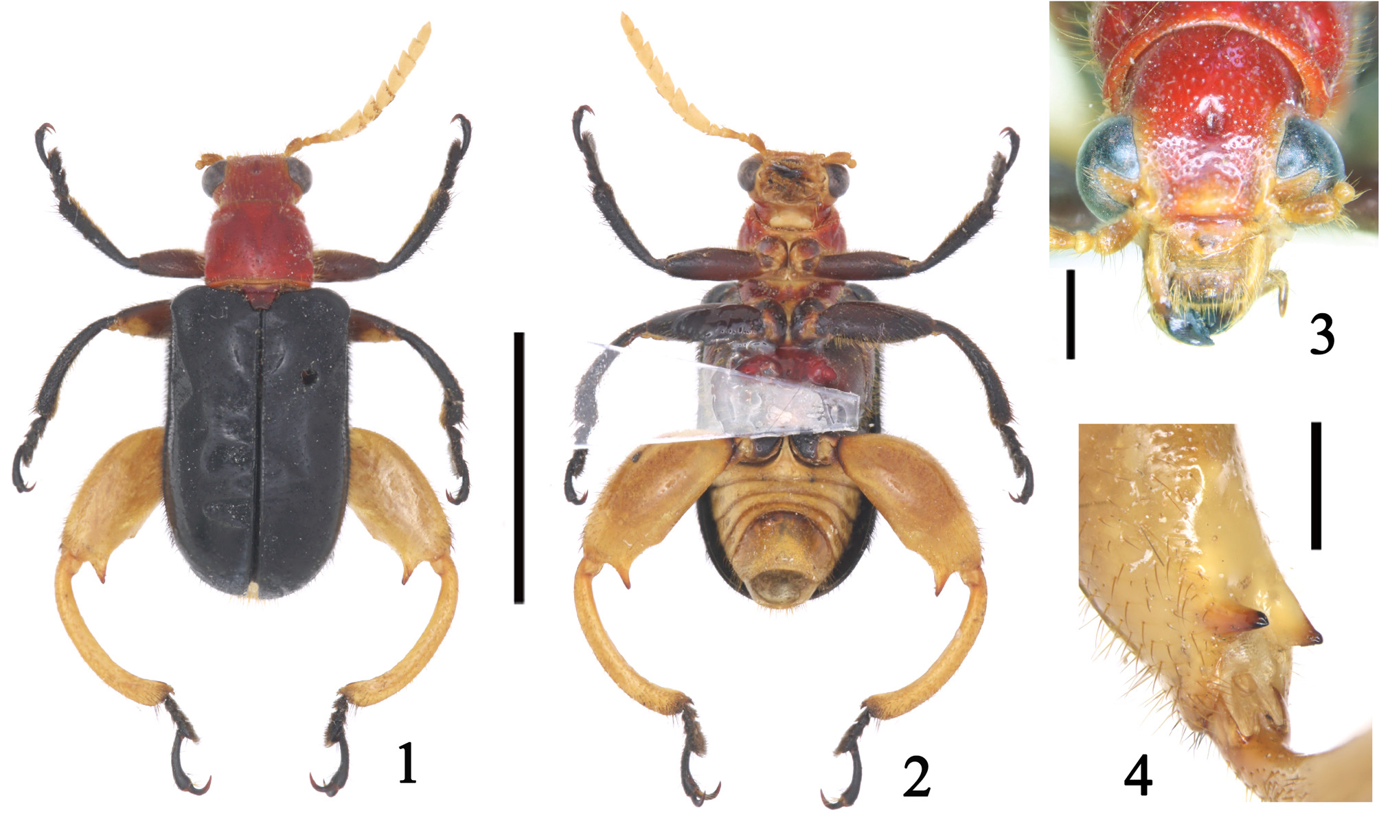
( Figs 1–14 View FIGURES 1 – 4 View FIGURES 5 – 9 View FIGURES 10 – 14 , 39 View FIGURES 39 – 44 , 51)
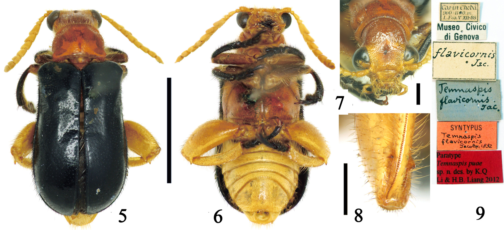
Material examined. Types. Holotype: m (IZCAS) / CHINA, Yunnan Province, Ruili City, Nongdao Town, Alongba, 900m, 2003. XI.17, Jinxin Song coll. / Holotype Temnaspis puae sp. n., des. by K.Q. Li & H.B. Liang, 2012 [red label] /; paratype (paralectotype of T. flavicornis Jacoby, 1892 ): 1 f (MSNG), Carin Chebá, 900–1100m, L. Fea, V XII–88 / flavicornis Jac. / Temnaspis flavicornis Jac. [blue label] / Syntype Temnaspis flavicornis Jacoby, 1892 [red label] / Museo Civico di Genova / Paratype Temnaspis puae sp. n., des. by K.Q. Li & H.B. Liang, 2012 [red label] /.
Description. BL = 13.5–14.0 mm, BW = 5.0– 5.3 mm. Head, pronotum, scutellum, prosternum, meso-, and metaventrite red; labrum, clypeus, antennae, abdomen, metafemora and metatibiae yellow; pro- and mesofemora brown; elytra, tarsi, pro- and mesotibiae black; pubescence on head and pronotum yellow, on elytra black.
Head: eyes prominent, inner margin with distinct canthus; vertex coarsely and densely punctate, with small deep median fovea ( Figs 3 View FIGURES 1 – 4 , 7 View FIGURES 5 – 9 ); occiput constricted; clypeus trapezoid, punctate and pubescent laterally; labrum rectangular, densely punctate and pubescent; antennae reaching slightly beyond elytral bases, antennomeres 1–4 cylindrical with sparse long setae, 5–11 strongly flattened and widened, densely pubescent, length of first antennomere 1.3x third antennomere.
Thorax: PW/PL = 1.3–1.4, PBW/PAW = 1.1; basal and apical pronotal margins nearly straight; anterior transverse pronotal groove deep laterally, obsolete medially, posterior groove absent; pronotal disc convex, slightly impressed along midline, sparsely punctate and pubescent, punctures sparser than on vertex; lateral margins nearly parallel; each basal angle produced as an indistinct tubercle. Scutellum trapezoid, slightly emarginate at apex, sparsely punctate and pubescent.
EL/EW = 1.6–1.7; elytral humeri broad, projecting antero-laterally; elytra laterally slightly widened from humeri to posterior third, then convergent to suture; disc convex, shiny, finely punctate and pubescent; epipleura densely punctate and pubescent. Metaventrite sparsely punctate and pubescent, each side raised to a prominent conical tubercle. Hind femora swollen, with two large ventral teeth at apex, on inner and outer surfaces, outer slightly longer than inner ( Figs 4 View FIGURES 1 – 4 , 8 View FIGURES 5 – 9 ); hind tibiae curved in male ( Figs 1–2 View FIGURES 1 – 4 ), less so in female.
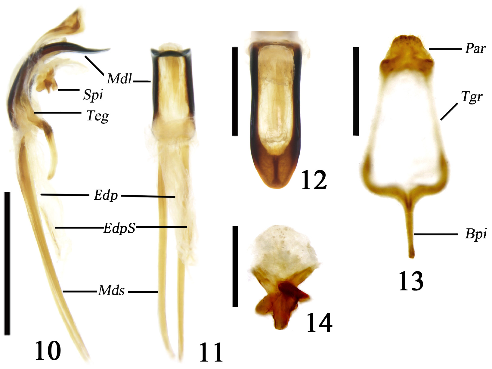
Abdomen: apex of last abdominal sternite concave in female, rounded in male. Median lobe flattened, strongly curved in lateral view ( Fig. 10 View FIGURES 10 – 14 ), sides and apex strongly sclerotised, dorsocentral portion membranous ( Figs 10– 12 View FIGURES 10 – 14 ), sides parallel, apex rounded ( Fig. 12 View FIGURES 10 – 14 ); median struts rod-shaped, widely separated from each other, 1.5–2.0 times as long as median lobe ( Figs 10–11 View FIGURES 10 – 14 ); tegmen trapezoid, basal piece rod-like, tegminal ring gradually narrowed towards parameres, apical margin of parameres with dense setae ( Fig. 13 View FIGURES 10 – 14 ); endophallus membranous, with paired elongate stick shaped sclerites at base in repose (EdpS, Figs 10–11 View FIGURES 10 – 14 ). Spiculum relictum short, basal portion strongly sclerotised, apical portion rounded, weakly sclerotised ( Fig. 14 View FIGURES 10 – 14 ). Ovipositor long (Fig. 51), base broad, apex narrow, divided into two vaginal palpi, two baculi at each side connected at their bases, the outer short, the inner long, coxite strongly sclerotised and sub-quadrate, apical margin with long setae, stylus small and distinct (Fig. 52).
Distribution. China (Yunnan); Myanmar (Kayin).
Etymology. The specific name puae is proposed in memory of Professor Fuji Pu, who greatly contributed to taxonomy of Chinese Megalopodidae .
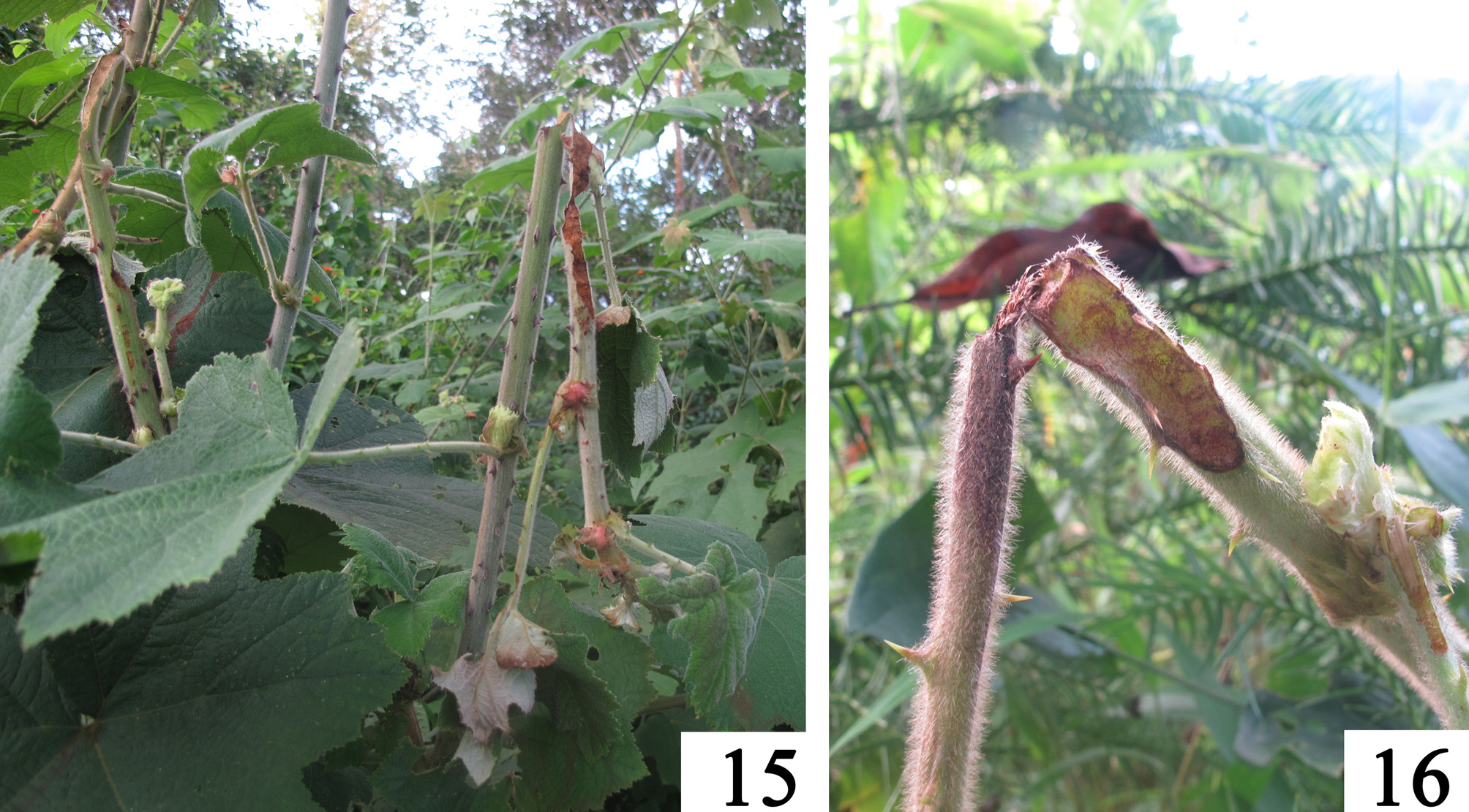
Remarks. On October 2, 2012, H.B. Liang went to Alongba, the type locality of the new species, where he found a few damaged shoots of Rubus alceaefolius ( Figs 15–16 View FIGURES 15 – 16 ). From what is known of other Temnaspis species (Yu 1977; Yu & Yang 1994), this was probably caused by Temnaspis adults. No adults were found on this or the succeeding two days. From October 21 to November 7, 2012, our colleague Mr. X. Huang looked for Temnaspis in the same area. He found only one Megalopodinae larva inside a shoot of R. alceaefolius , almost certainly of T. puae , since no other Megalopodidae are known from the area
The two syntypes of Temnaspis flavicornis Jacoby, 1892 , in MSNG belong to different species. One syntype, with two outer teeth on the hind femora ( Figs 18, 20 View FIGURES 17 – 24 ), fits the original description of T. flavicornis , and is designated lectotype of T. flavicornis (see below). However, the other, with a single outer tooth on the hind femora, is identical to the holotype of T. puae sp. nov. ( Figs 5–9 View FIGURES 5 – 9 , 51), and is designated a paratype of T. puae sp. nov., as well as being a paralectotype of T. flavicornis .
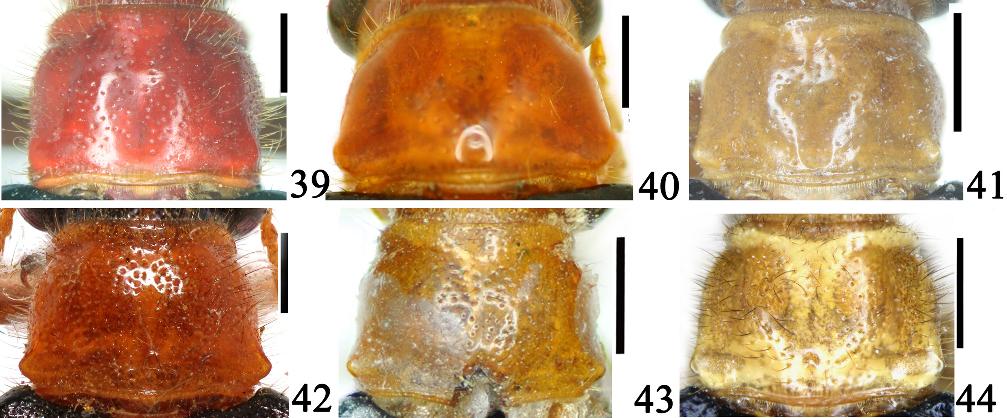
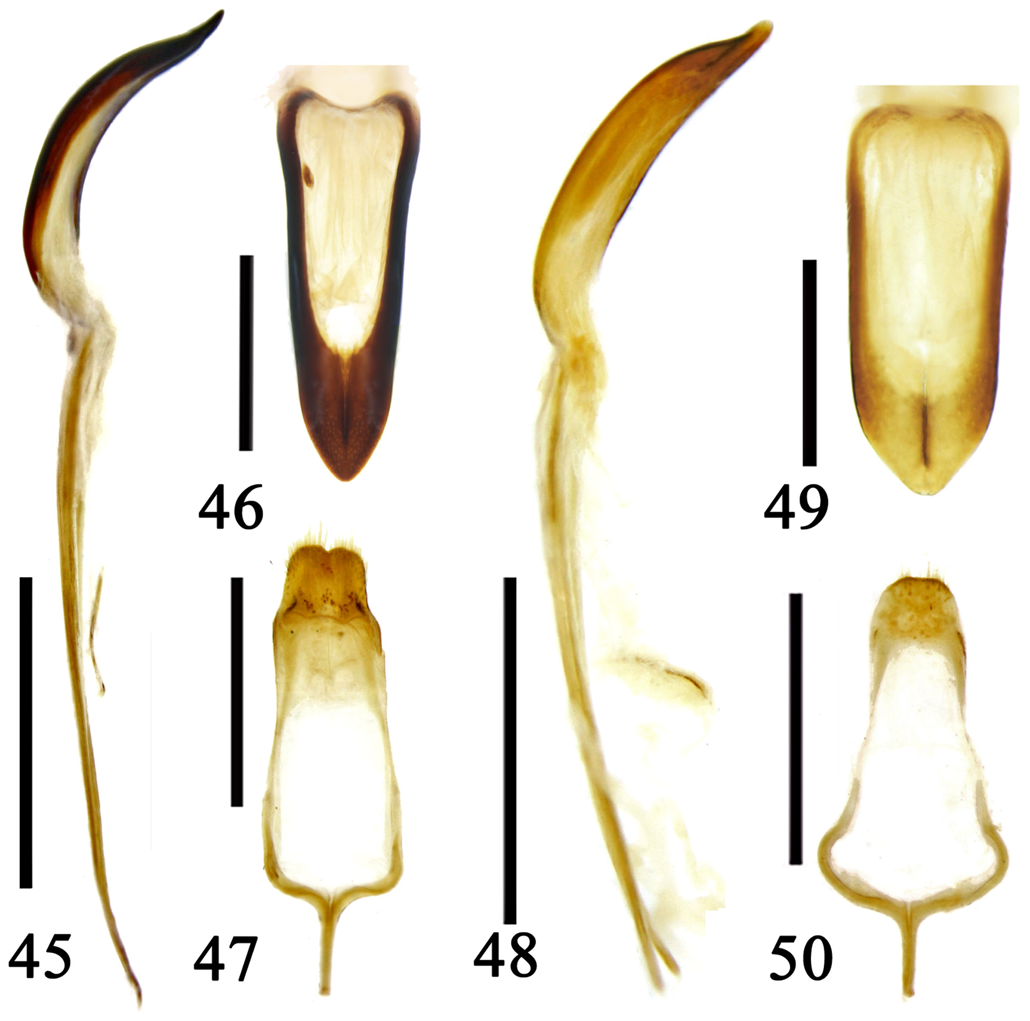
The new species differs from T. flavicornis by: the apex of the femora with single outer and inner teeth (two outer teeth and no inner tooth in T. flavicornis , Figs 18, 20, 23 View FIGURES 17 – 24 ); pronotum sub-quadrate ( Fig. 39 View FIGURES 39 – 44 ) with apex slightly constricted laterally (pronotum trapezoid with apex distinctly constricted in T. flavicornis , Fig. 40 View FIGURES 39 – 44 ); median lobe of aedeagus more curved ( Figs 45–46 View FIGURES 45 – 50 ), sides parallel and apex rounded (median lobe less curved, sides convergent from base to apex, and apex acute in T. flavicornis , Figs 45–46 View FIGURES 45 – 50 ); ovipositor bases and outer baculi (Fig. 51) strongly sclerotised (weakly sclerotised in T. flavicornis , Fig. 52). Photographs of the holotype of Temnaspis bidentata Pic, 1922 , show that is it correctly placed as a junior synonym of T. flavicornis ( Figs 22–23 View FIGURES 17 – 24 ).
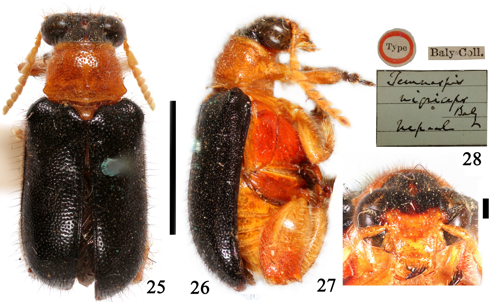
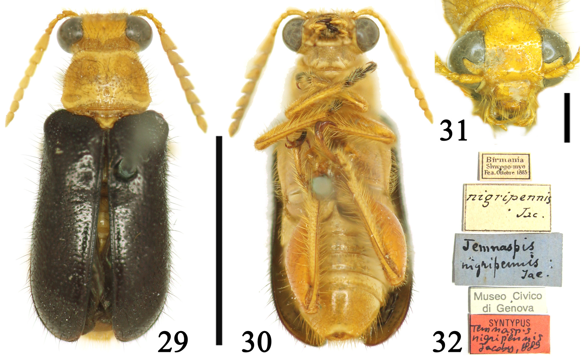
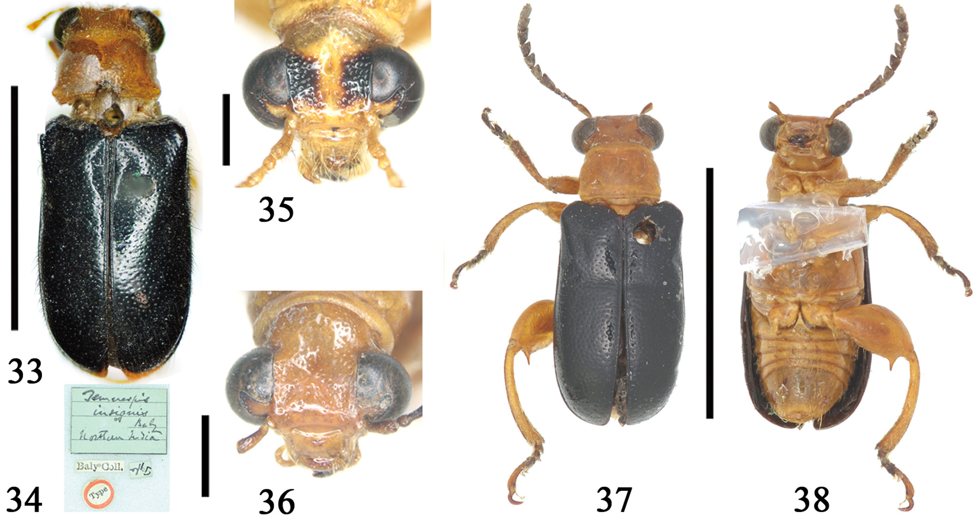
Temnaspis nigriceps also has black elytra and one ventral outer tooth on the femora, and occurs in China and Myanmar (Medvedev 2002; Yu & Liang 2002). Photographs of type material of T. nigriceps and its two junior synonyms (Medvedev 2002): T. insignis Baly, 1859 , and T. nigripennis Jacoby, 1889 , ( Figs 25–35 View FIGURES 25 – 28 View FIGURES 29 – 32 View FIGURES 33 – 38 , 48–50 View FIGURES 45 – 50 ), show that this is a different species. Temnaspis puae differs from T. nigriceps by having: pronotum sub-quadrate, with tubercle of basal angle less protruding (pronotum trapezoid with tubercle of basal angle more protruded in T. nigriceps ); eyes less prominent (eyes more prominent in T. nigriceps Baly ); median lobe strongly curved with longer apical lamella (median lobe less curved with shorter apical lamella in T. nigriceps Baly ).
Finally, T. puae differs from Temnaspis omeiensis (Gressitt 1942) by antennae yellowish-brown, pronotum with tubercle of basal angle less protruding, head nearly as wide as pronotum (antennae blackish-brown, tubercle of basal angle more protruding, head wider than pronotum in T. omeiensis, Figs View FIGURES 33 – 38 36–38, 41 View FIGURES 39 – 44 ).

No known copyright restrictions apply. See Agosti, D., Egloff, W., 2009. Taxonomic information exchange and copyright: the Plazi approach. BMC Research Notes 2009, 2:53 for further explanation.