
Sarsiella japonica, Karanovic, Ivana & Soh, Ho-Young, 2015
|
publication ID |
https://doi.org/ 10.11646/zootaxa.3947.4.1 |
|
publication LSID |
lsid:zoobank.org:pub:E0151A24-761D-4465-BB71-DAC4FA5B9550 |
|
DOI |
https://doi.org/10.5281/zenodo.5658723 |
|
persistent identifier |
https://treatment.plazi.org/id/E2798D5D-4A7A-FFDA-C8D7-507D2AE5F803 |
|
treatment provided by |
Plazi |
|
scientific name |
Sarsiella japonica |
| status |
|
Sarsiella japonica Hiruta, 1977 View in CoL
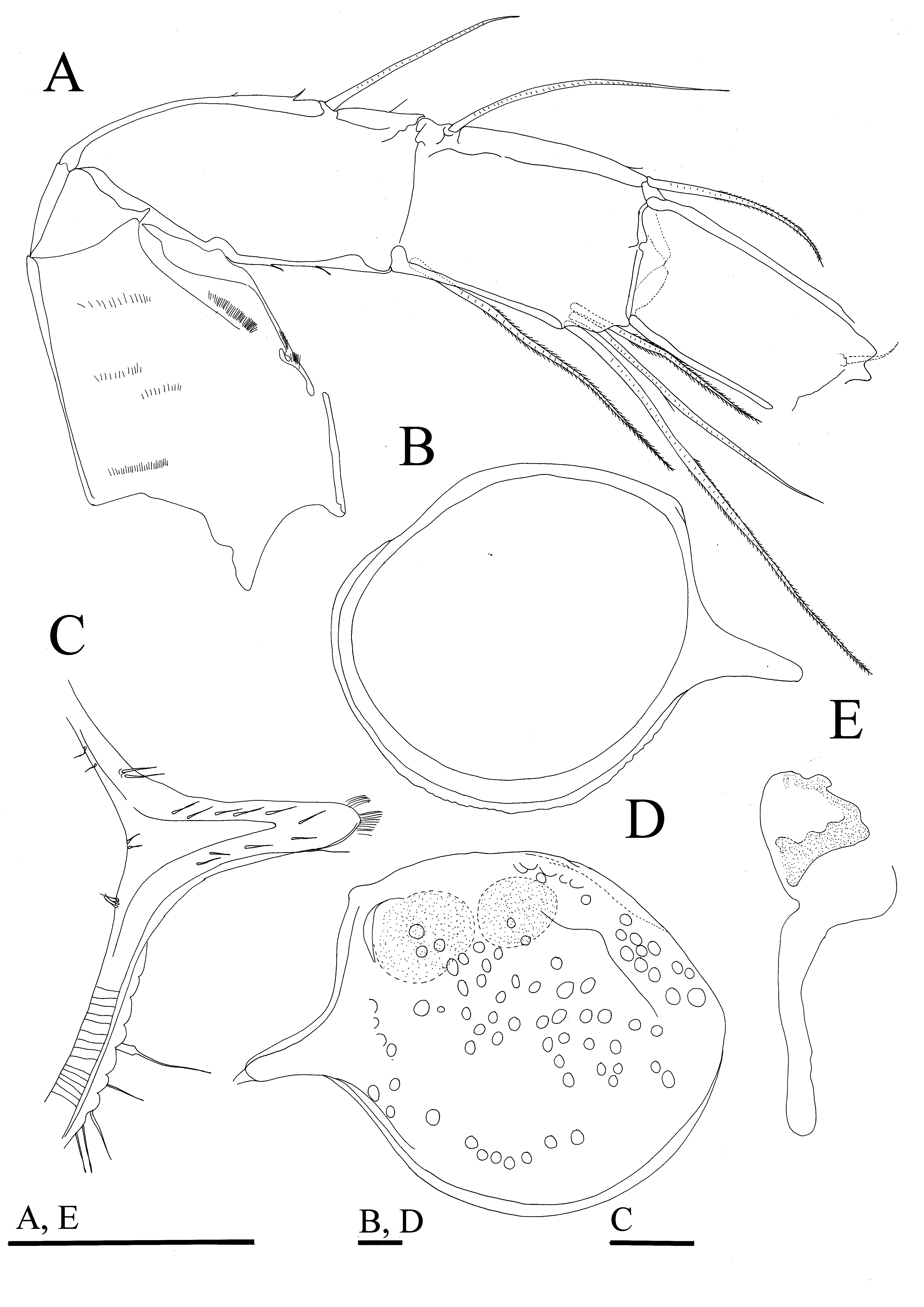
( Figures 8–12 View FIGURE 8 View FIGURE 9 View FIGURE 10 View FIGURE 11 View FIGURE 12 )
Synonymy. Sarsiella japonica n. sp. — Hiruta 1977: p. 45, Figs 1–12 View FIGURE 1 View FIGURE 2 View FIGURE 3 View FIGURE 4 View FIGURE 5 View FIGURE 6 View FIGURE 7 View FIGURE 8 View FIGURE 9 View FIGURE 10 View FIGURE 11 View FIGURE 12 , Plate 4.
Material examined. Two females dissected on one two slides, shells on micropaleontological slides ( NIBR IV 0 0 0 0 287236 and NIBR IV 0000287237); one female on SEM stub; three females and seven juveniles in ethyl alcohol ( NIBR IV 0 0 0 0 287238, and NIBR IV 0000287239), 10 paratypes (two females and eight juveniles) in ethyl alcohol ( NIBR IV 0000287240), and three females used for DNA sequencing from South Korea, East China Sea, Maemul Island, Station 3, 34°32’00”N 128°43’54.43”E, 25 July 2011, collector H. Y. Soh.
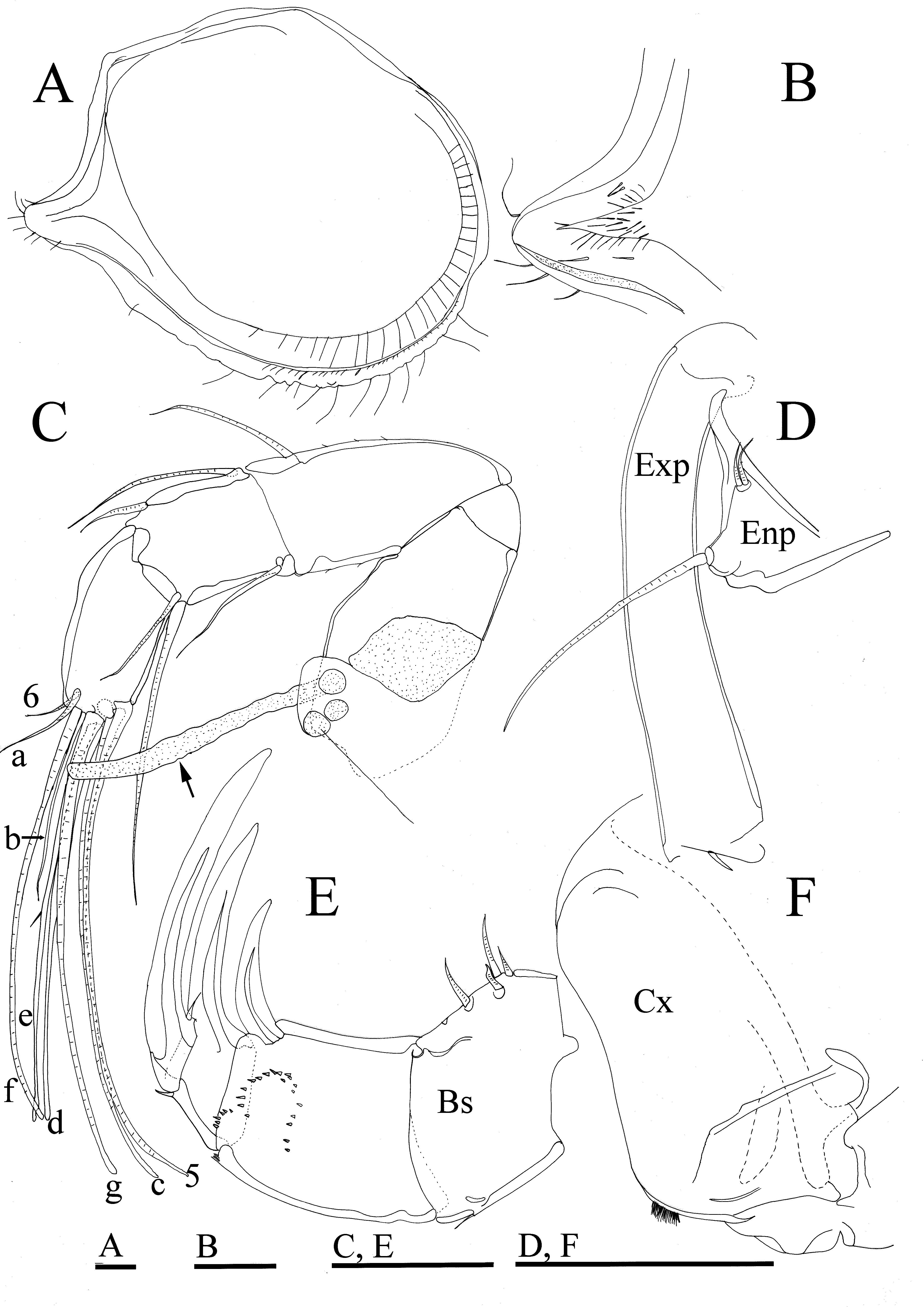
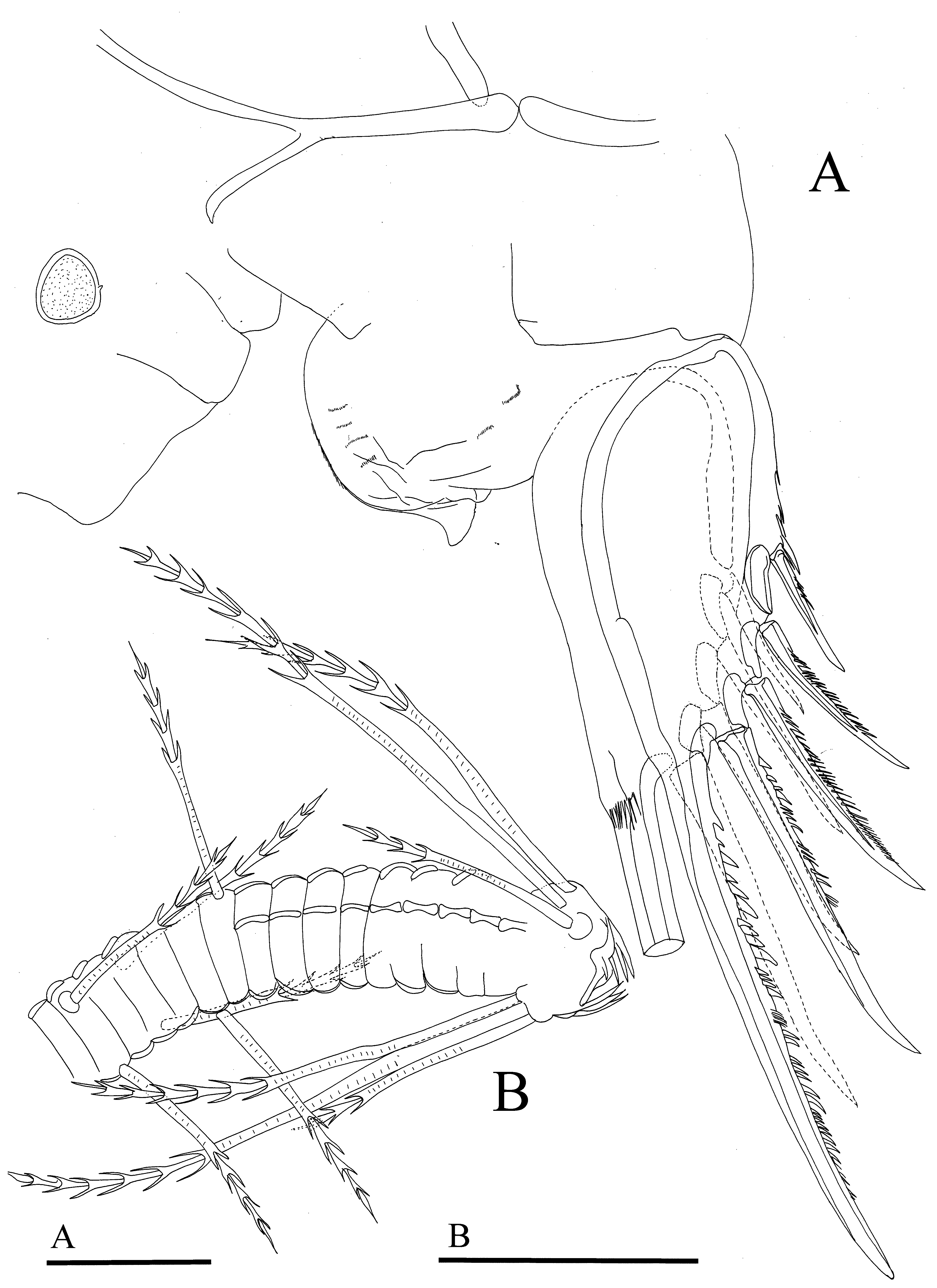
Description. Female. Shell ( Figures 8 View FIGURE 8 A, 9B, D) with one dorsal and one medio-ventral longitudinal ridge and several small perpendicular ridges starting from medio-ventral one. Surface covered with wide, shallow pits (fossae), and minute punctuations ( Figure 8 View FIGURE 8 B, D, E). Caudal process very long and distally pointed ( Figure 8 View FIGURE 8 B, C). Postero-dorsal bulge slightly over passing dorsal margin, and posteriorly framed by dorsal and medio-ventral ridge, otherwise not clearly outlined. Dorsal margin of carapace straight in posterior part, rounded in medial and anterior parts. Ventral margin rounded. Long and short individual bristles distributed on lateral surface of valves ( Figure 8 View FIGURE 8 B, D, E). Long pore canals lead from posterior process to inside of valves, shorter ones present along entire anterior and ventral margin ( Figure 9 View FIGURE 9 C). Anterior infold without bristles. Infold of caudal process with several short bristles running along caudal process, and several laying proximally to it. Shell L (including caudal process)= 1.1 mm.
A1 ( Figures 9 View FIGURE 9 A, 10B). First segment long and bare, armed only with short setulae. Second segment with few dorsal and ventral spines and annulated, bare, dorsal bristle reaching 1/3 of following segment. Third and fourth segment fused; third with annulated, bare dorsal bristle reaching 1/3 of following segment and one longer and pappose ventral bristle (exceeding distal margin of 3+4 segment). Fourth segment with pappose, annulated dorsal bristle not reaching distal end of following segment, and three, annulated bristles (one bare) ventrally (only one reaching distal margin of following segment, bare one slightly exceeding it, and last one almost two times longer than shortest). Fifth and sixth segments fused. Bristle of fifth segment long, annulated and bare; seta on sixth segment short, also bare. Seventh segment: a-bristle short (about four times longer than sixth segment’s bristle); bbristle slender, and about as long as a-bristle, this bristle with pointed, sensory-like tip; c-bristle longer than bristle of fifth segment. Eight segment: d- and e-bristles equally long, bare and shorter than c-bristle; f- and g-bristles slightly longer than fifth segment bristle, both annulated.
Bellonci Organ ( Figure 9 View FIGURE 9 E). Cylindrical, non segmented and short.
Eyes. Both medial and lateral eyes present. Medial eye with scattered pigment, lateral eye with four ommatidia.
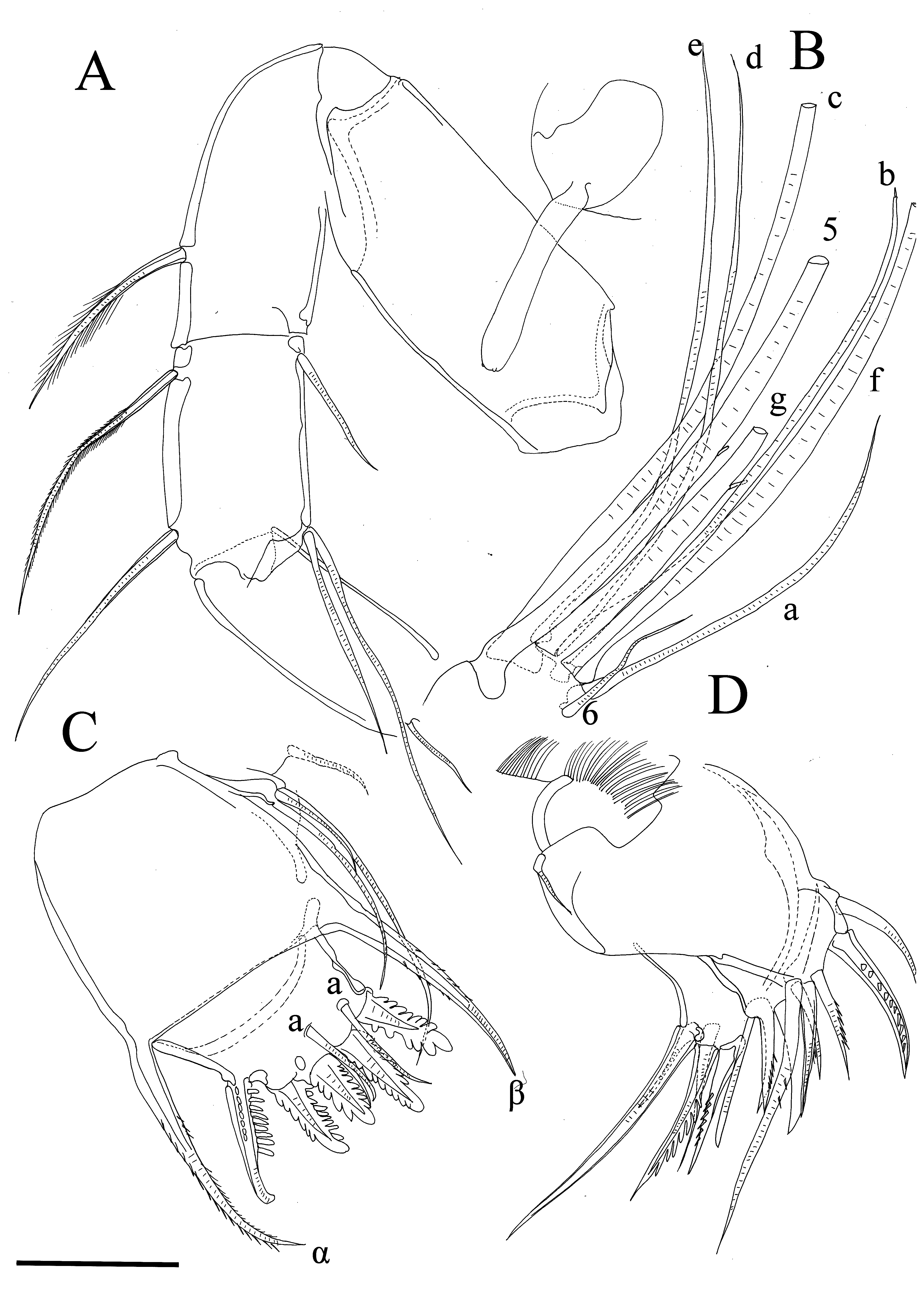
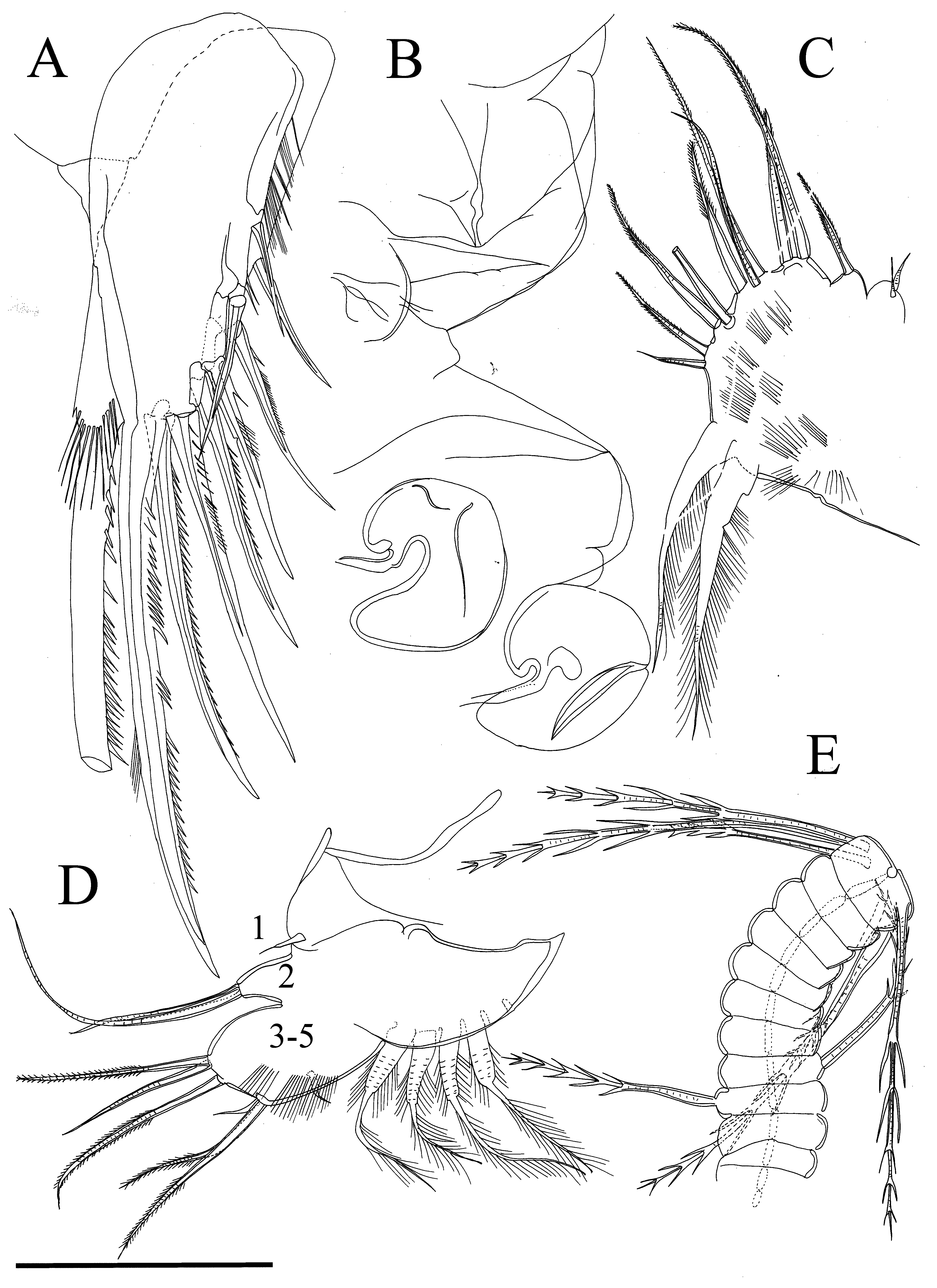
A2 ( Figure 10 View FIGURE 10 A). Prp bare. Enp with one short basal bristle (annulated and bare), and no bristles situated on terminal node. Enp covered with short spines. Exp: first segment with small terminal medial recurved tubular bristle; bristle on second segment with thick proximal ventral spines and distal swimming setae; bristles on segments 3–8 without thick spines proximally and with swimming setae distally; ninth segment with two bristles (one short, bare and other longer with swimming setae).
Md ( Figure 11 View FIGURE 11 A, B). Cx endite elongated and with short spine-like setae, Cx with long slender spines near ventral margin. Bs: ventral margin with six short bristles, dorsal margin with short marginal slender bristle. First Enp segment with short spines and with one stout claw distally; second Enp segment with strong claw ventrally; third Enp segment with stout terminal claw and small dorsal and ventral spines.
Mxl ( Figure 11 View FIGURE 11 C, D). Cx with long, bare dorsal bristle. Endite I with three pectinate claws (only one shown in Figure 11 View FIGURE 11 D) and two ringed bristles; endite II with four pectinate claws and four ringed bristle; endite III with three pectinate claws and four ringed bristles. Exp with two bristles: one long and one short. First Enp segment with spinous alpha- and beta-bristles. Second Enp segment with five pectinate claws, two a- and one c-bristle.
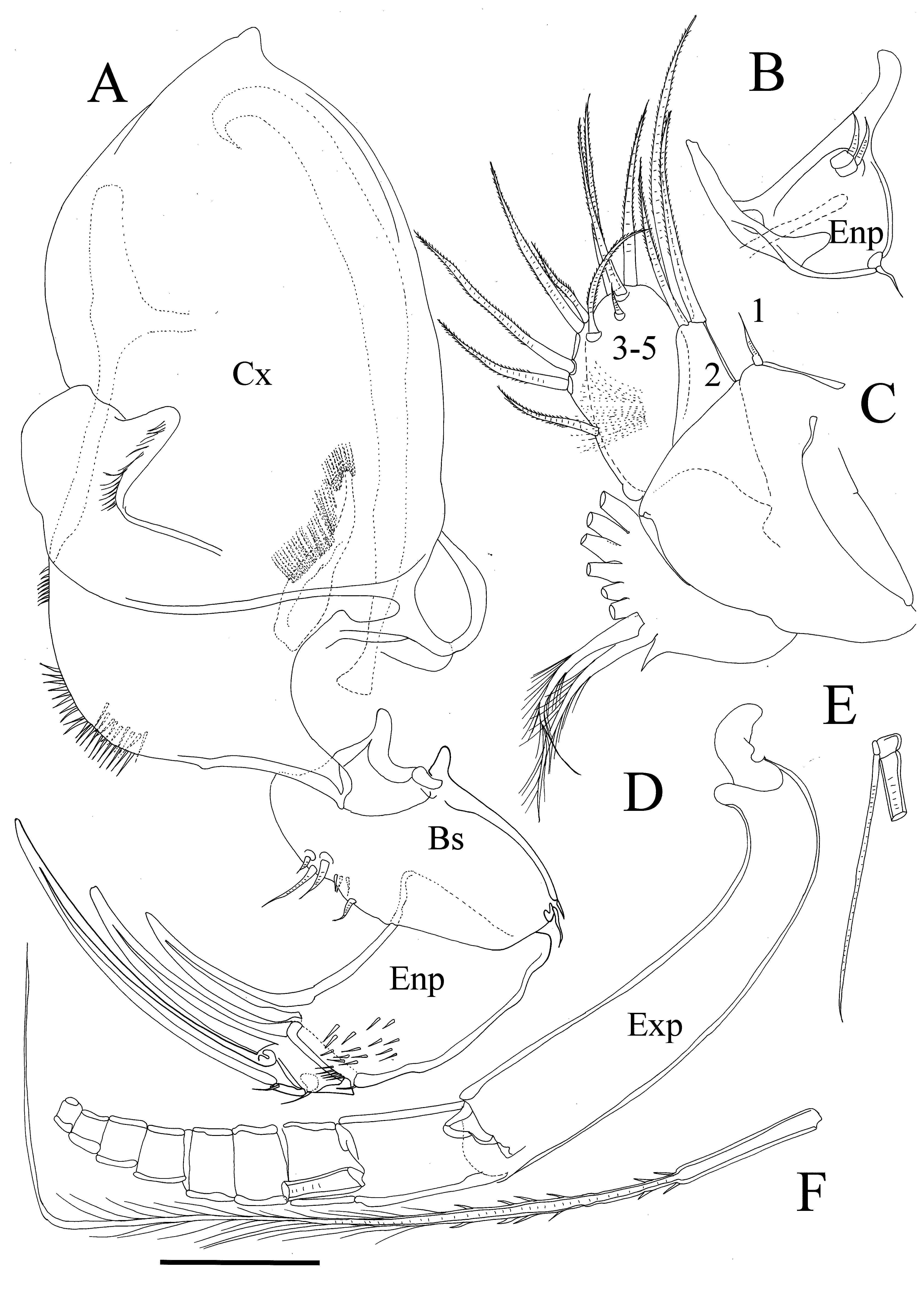
L5 ( Figure 12 View FIGURE 12 D). Lobe one with one short bristle; lobe two with two bristles (one longer than other); lobes 3–5 fused and with total of seven bristles, five long and annulated and with setulae, two bare, short bristle. Entire appendage hirsute.
L6 ( Figure 12 View FIGURE 12 C). One endite with two bristles, following one with one ringed pappose bristle. terminal segment with nine annulated bristles, each with short setulae, and almost all of different length. Nine bristles followed by space and two long plumose bristles. Entire appendage covered with fine, short setulae.
L7 ( Figure 12 View FIGURE 12 E). Proximal group with two bristles (one on each side) each with five bells. Terminal group with three bristles on each side, each with five bells. Terminus without opposing combs.
UL ( Figure 12 View FIGURE 12 A). Each lamella with five claws. Only claw one non-articulated; claws with long and short teeth along posterior edge. Right lamella with group of long, slender spines dorsal to first claw, left lamella with slender setulae following terminal claw. Genital segment shown in Figure 12 View FIGURE 12 B.
Remarks. Only one A1 of one dissected female had three bristles ventrally on the fourth segment ( Figure 9 View FIGURE 9 A) while the other one had only two bristles, as well as both A1 of the second dissected female. The only clear difference noted between the original description of S. japonica from Japan ( Hiruta 1977) and this population in Korea is that the Japanese specimens seem to have a more rounded dorsal margin of carapace, less bristles on the posterior infold, and a slightly shorter caudal process. All other morphological characters, including the number and length ratio of setae on each appendage, seem to be identical. Hiruta (1977) reported males, while males were not collected during our study.


No known copyright restrictions apply. See Agosti, D., Egloff, W., 2009. Taxonomic information exchange and copyright: the Plazi approach. BMC Research Notes 2009, 2:53 for further explanation.