
Pseudourostyla cristatoides, Jung, Jae-Ho, Park, Kyung-Min & Min, Gi-Sik, 2012
|
publication ID |
https://doi.org/10.5281/zenodo.208731 |
|
DOI |
https://doi.org/10.5281/zenodo.5631826 |
|
persistent identifier |
https://treatment.plazi.org/id/E55CDE7D-FF96-0D6C-B1F8-ED64FA6EDD07 |
|
treatment provided by |
Plazi |
|
scientific name |
Pseudourostyla cristatoides |
| status |
|
Description of Pseudourostyla cristatoides n. sp. ( Tables 1–2 View TABLE 1 View TABLE 2 and Figs. 1–18 View FIGURES 1 – 5 View FIGURES 6 – 14 View FIGURES 15 – 18 , 24–42 View FIGURES 24 – 31 View FIGURES 32 – 42 ).
Size 220–265 × 85–125 μm in vivo ( Figs. 1, 2 View FIGURES 1 – 5 , 24–28 View FIGURES 24 – 31 ), on average 243.8 × 89 μm in protargol preparations ( Table 1 View TABLE 1 and Figs. 3–5 View FIGURES 1 – 5 ; 32, 33). Body flexible and slightly contractile; cell colour grayish to colourless. Extrusomes trichocyst type, densely arranged all over cell surface ( Figs. 28, 29 View FIGURES 24 – 31 , arrows) and observed in some protargolimpregnated specimens ( Fig. 44 View FIGURES 43 – 47 , arrowhead); extruded extrusomes are observed from methyl green-pyronin and occasionally from protargol impregnated specimens and the extruded ones are ca. 5–10 μm in length, with oval shape of distal end ( Figs. 30, 31 View FIGURES 24 – 31 ). Two contractile vacuoles on left side of cell at 25% and 75% of body length, about 14 μm in diameter when fully extended; contractile vacuoles with anterior and posterior collecting canals about 180–200 μm in length. The two contractile vacuoles observed in either anterior or posterior or both positions ( Figs. 1, 2 View FIGURES 1 – 5 ; 24–26, arrows). On average 78 ellipsoidal macronuclear nodules; ca. four micronuclei of oval shape ( Figs. 4 View FIGURES 1 – 5 , 32–34 View FIGURES 32 – 42 ). Crawling moderately fast on bottom of Petri dish.
*Numbered from left to right, ** Numbered from inside to outside, DK - dorsal kineties, TC - transverse cirri, CV - coefficient of variation in
%, Max - maximum, Min - minimum, n - number of specimens investigated, SD - standard deviation, SE - standard error of arithmetic mean.
Measurements in μm.
* Number from Fig. 110 9 in Kahl (1932), ** Number from Fig. 150h in Berger (2006), *** Number of cirral pairs including frontal and midventral cirri from Kumar et al. (2010), **** Number of cirri including frontal and midventral cirri from Fig. 2 View FIGURES 1 – 5 a in Wiackowski (1988), ***** The positions of the two contractile vacuoles are estimated from Fig. 1 View FIGURES 1 – 5 b in Paiva and Silva-Neto (2006), ****** According to Berger (2006), all species in the genus Pseudourostyla likely have trichocyst type of extrusomes, FVT anlagen – fronto-ventral-transverse cirral anlagen, n/a – unavailable.
All cirri relatively fine, mostly 10–15 μm long; frontal cirri in a bicorona 13 μm long; midventral, marginal and frontoterminal cirri 10 μm in length; transverse cirri 15 μm in length ( Figs. 1 View FIGURES 1 – 5 , 27 View FIGURES 24 – 31 ). 20–30 frontal cirri in a bicorona and 17–25 midventral pairs; distinct gap between frontal and midventral cirri near posterior frontoterminal cirrus; one buccal cirrus. Invariably two frontoterminal cirri at distal end of adoral zone. Two pretransverse ventral and 6–12 transverse cirri. 5–7 left, 4–5 right marginal rows, number of cirri in each row decreasing gradually from inside to outside row. 10–13 complete dorsal kineties ( Figs. 5 View FIGURES 1 – 5 , 33 View FIGURES 32 – 42 , arrows). Number of dorsal bristles gradually decreases from outside to inside row and leftmost row of dorsal bristles have distinctly higher number of bristles, about 51 ( Table 1 View TABLE 1 ). Dorsal cilia about 3 μm long ( Fig. 29 View FIGURES 24 – 31 ).
Adoral zone of membranelles about 33% of body length in fixed specimens, base of largest membranelles about 12 μm long, cilia of membranelles about 15 μm long. Distal end of adoral zone extends far posteriorly ( Figs. 3 View FIGURES 1 – 5 , 32 View FIGURES 32 – 42 ). Paroral and endoral membranes are rather short, slightly curved and almost parallel ( Fig. 3 View FIGURES 1 – 5 ).
Morphogenesis.
Stomatogenesis and development of fronto-ventral-transverse cirral anlagen. The oral primordium of the opisthe close to the left of midventral pairs ( Figs. 6, 7 View FIGURES 6 – 14 , arrows) and the proter’s oral primordium is formed on the dorsal wall of the buccal cavity ( Fig. 7 View FIGURES 6 – 14 , asterisk). The buccal cirrus begins to dedifferentiate ( Figs. 6, 7 View FIGURES 6 – 14 , arrowheads) and the patches of the opisthe’s oral primordium form an ordinary longitudinal primordium ( Fig. 8 View FIGURES 6 – 14 ). The posterior cirri of the rear corona dedifferentiate and participate in fronto-ventral-transverse cirral anlagen (FVT anlagen) development ( Fig. 8 View FIGURES 6 – 14 ).
In the next stage, the FVT anlagen have grown into long oblique streaks and each FVT anlagen of proter and opisthe form about 33 streaks ( Fig. 8 View FIGURES 6 – 14 , asterisks; Fig. 9 View FIGURES 6 – 14 ). The leftmost frontal cirrus originates from the anterior end of undulating membranes anlagen (FVT I anlagen; Fig. 9 View FIGURES 6 – 14 , asterisks) and both anlagen for undulating membranes begin to differentiate ( Fig. 9 View FIGURES 6 – 14 ). The proximal portion of the old adoral membranelles will be gradually replaced by a new one and the opisthe’s adoral membranelles begin to differentiate posteriorly ( Figs. 8, 9 View FIGURES 6 – 14 ). The anterior end of opisthe’s adoral membranelles arches to the right. Later, the anterior part of the FVT anlagen begins to fragment and form new cirri. All FVT anlagen develop into two cirri, except posterior anlagen, which form three or four cirri comprising of midventral pairs, pretransverse, transverse, and frontoterminal cirri. The rearmost anlage (streak n) forms two frontoterminal cirri along with one pretransverse, and one transverse cirrus. The other (streak n-1) forms one midventral pair as well as one pretransverse and one transverse cirrus. The posterior cirrus of FVT anlagen II forms the buccal cirrus and migrates posteriorly ( Fig. 11 View FIGURES 6 – 14 , asterisks).
In late dividers, the new adoral membranelles will be completely developed in both daughter cells and the anterior parts of the membranelles arch to the right ( Figs. 13 View FIGURES 6 – 14 , 15, 17 View FIGURES 15 – 18 ). The new frontoterminal cirri migrate anteriad ( Figs. 13 View FIGURES 6 – 14 , 15, 17 View FIGURES 15 – 18 , arrows). The parental cirri will be resorbed and newly developed ones migrate towards their final positions. The undulating membranes anlagen split into endoral and paroral membranes in both dividers.
Marginal and dorsal anlagen. In the early stage, a few cirri of the anterior and middle part of the innermost left and outermost right marginal row likely begin to dedifferentiate and old cirri are incorporated into the primordium ( Fig. 8 View FIGURES 6 – 14 , arrows, arrowheads). Later, each primordium forms 4–7 anlagen and elongates posteriorly. The posterior part of the proter’s left marginal anlagen is distinctly curved to the left.
The dorsal kinety anlagen intrakinetally develop within the parental dorsal kineties. Within each parental row, two anlagen form at the anterior and posterior quarters of the body ( Figs. 10 View FIGURES 6 – 14 , arrows, 12, 14, 16, 18). The anlagen are arranged parallel and obliquely, that is, the anterior positions of each parallel dorsal kinety anlage are gradually arranged posteriorly from left to right. Each anlage stretches in both directions and the parental dorsal kineties are incorporated or resorbed.
Division of nuclear apparatus. Nuclear division proceeds in the usual way of urostylid. Briefly, during middle division all macronuclear nodules fuse to form a single and rod-shaped mass ( Fig. 10 View FIGURES 6 – 14 ). This mass is likely formed during first division after complete fusion. The single mass of macronuclei is continuously separated during the entire process of morphogenesis. The micronuclei are inflated and located at the left side of the cell ( Figs. 10, 12 View FIGURES 6 – 14 ). In late dividers, the micronuclei are finished division ( Fig. 16 View FIGURES 15 – 18 ).
Physiological reorganisation ( Figs. 19–23 View FIGURES 19 – 23 , 43–47 View FIGURES 43 – 47 ). Middle late reorganizers are observed and the main process of reorganisation is similar to that of the dividers. Briefly, the new adoral membranelles arise via dedifferentiation of the old ones. Posterior cirri of the rear corona are incorporated into FVT anlagen. Each marginal row anlage develops from the anterior parts of innermost left and outermost right marginal row ( Figs. 19 View FIGURES 19 – 23 , arrow; 20, arrowhead) and extend posteriorly. The differentiation of all FVT anlagen is completely identical to that of the dividers, that is, anlagen I–n develop into undulating membranes and frontal, buccal, midventral, pretransverse, transverse, and frontoterminal cirri ( Figs. 21, 22 View FIGURES 19 – 23 ). The frontoterminal cirri migrate anteriorly ( Fig. 22 View FIGURES 19 – 23 , arrow). Each dorsal kinety anlage originates on the center of the parental row and extends in both directions ( Figs. 20, 23 View FIGURES 19 – 23 ). The posterior cirrus of FVT anlage II becomes the buccal cirrus and migrates posteriorly ( Fig. 21 View FIGURES 19 – 23 , asterisk).
Molecular phylogeny ( Table 3 View TABLE 3 and Fig. 48 View FIGURE 48 ). The 18S rRNA gene sequence of the new species was 1,674 bp in length ( JN887467 View Materials ) and the range of pairwise distances using the Kimura (1980) two-parameter model was 0.93–1.00% between P. cristatoides n. sp. and the three Asian populations of P. cristata . Intra-specific distances among three P. cristata populations were ranged between 0.07–0.12% ( Table 3 View TABLE 3 ). The pairwise distances between Pseudourostyla and H. franzi , recently transferred from Pseudourostyla , were 4.41–4.67%. In the phylogenetic analysis, H. franzi represented a distinct clade from Pseudourostyla . Three Asian populations of P. cristata and Korean population of P. cristatoides were clustered monophyly with a high bootstrap value and posterior probability ( Fig. 48 View FIGURE 48 ).
* A gap, 108 bp in length, in the conserved middle portion of the SSU rRNA gene sequence was identified by multiple sequence alignment.
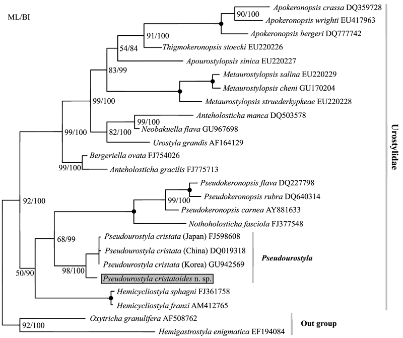
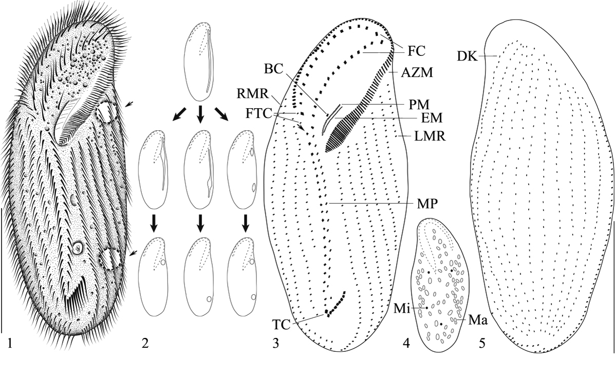
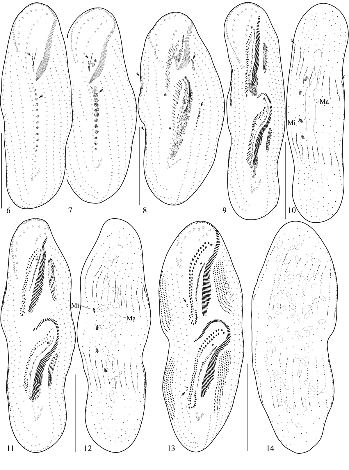
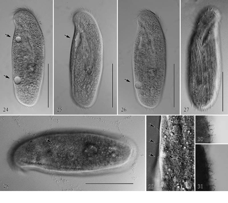


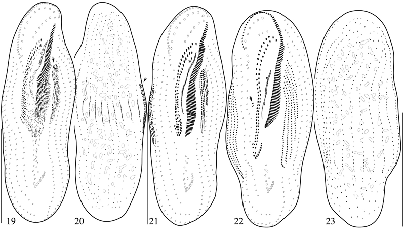
TABLE 1. Morphometric data on the protargol-impregnated specimens of Pseudourostyla cristatoides n. sp.
| Characteristics | Min | Max | Mean | SD | CV | n |
|---|---|---|---|---|---|---|
| Body, length Body, width Adoral zone of membranelles, length Adoral membranelles, number | 210 65 75 84 | 290 125 115 115 | 243.8 89.0 99.0 97.4 | 20.5 13.2 11.5 8.3 | 8.4 14.8 11.6 8.5 | 25 24 25 25 |
| Buccal cirri, number Frontal cirri, number Frontoterminal cirri, number | 1 20 2 | 1 30 2 | 1.0 24.0 2.0 | 0.0 2.4 0.0 | 0.0 9.9 0.0 | 18 25 25 |
| Midventral pairs, number Transverse cirri, number Macronucleus nodules, length Macronucleus nodules, width Macronuclear nodules, number | 17 6 6.4 2.4 30 | 25 12 20 6.4 106 | 20.9 9.4 9.8 4.1 77.7 | 2.3 1.3 3.2 1.1 17.3 | 10.8 13.4 32.3 25.6 22.2 | 25 25 25 25 18 |
| Micronuclei, length Micronuclei, with Micronuclei, number Posterior end of cell to rear most TC, distance | 3.2 3.2 3 17.6 | 4.8 4.8 5 35.2 | 3.9 3.5 4.1 27.9 | 0.9 0.6 0.7 4.4 | 22.0 17.8 16.7 15.7 | 7 7 7 25 |
| Dorsal kineties, number DK 1, number of bristles* DK 2, number of bristles* | 10 38 34 | 13 63 58 | 11.3 51.4 43.5 | 0.8 6.5 6.1 | 7.5 12.7 14.0 | 25 25 25 |
| DK 3, number of bristles* DK 4, number of bristles* DK 5, number of bristles* DK 6, number of bristles* | 22 24 23 23 | 39 38 36 37 | 30.8 29.5 28.9 29.3 | 4.2 4.1 3.9 4.0 | 13.7 13.8 13.4 13.7 | 25 25 25 25 |
| DK 7, number of bristles* DK 8, number of bristles* DK 9, number of bristles* | 22 22 23 | 36 35 39 | 28.8 29.0 30.3 | 3.8 3.6 4.3 | 13.3 12.4 14.1 | 25 25 25 |
| DK 10, number of bristles* DK 11, number of bristles* DK 12, number of bristles* DK 13, number of bristles* | 23 28 22 36 | 47 48 43 38 | 32.2 33.8 34.0 - | 5.4 5.3 5.8 - | 16.9 15.6 17.0 - | 25 21 9 2 |
| Left marginal rows, number Left marginal row 1, number of cirri** Left marginal row 2, number of cirri** Left marginal row 3, number of cirri** Left marginal row 4, number of cirri** | 5 32 31 30 22 | 7 47 46 45 37 | 6.0 36.9 37.9 36.9 29.9 | 0.4 3.5 3.8 4.2 3.8 | 6.8 9.5 10.0 11.4 12.8 | 25 25 25 25 25 |
| Left marginal row 5, number of cirri** Left marginal row 6, number of cirri** Left marginal row 7, number of cirri** | 12 4 7 | 30 20 9 | 21.3 11.9 - | 4.7 4.1 - | 21.9 34.3 - | 25 23 2 |
| Right marginal rows, number Right marginal row 1, number of cirri** Right marginal row 2, number of cirri** Right marginal row 3, number of cirri** | 4 8 27 35 | 5 31 41 44 | 4.2 21.3 33.1 38.3 | 0.4 6.3 3.6 2.5 | 10.3 29.7 10.9 6.6 | 25 25 25 25 |
| Right marginal row 4, number of cirri** Right marginal row 5, number of cirri** | 37 39 | 48 49 | 41.0 43.4 | 2.9 3.8 | 7.0 8.9 | 25 5 |
TABLE 3. Pairwise similarity (%) based on the Kimura (1980) two-parameter distance option of the 18 S rRNA gene sequence between Pseudourostyla cristatoides n. sp. and the congeneric species with Hemicycliostyla franzi.
| P. cristata (China, DQ019318 View Materials ) | Pseudourostyla cristatoides n. sp. 99.00 | P. cristata (China) | P. cristata (Japan) | P. cristata (Korea) * |
|---|---|---|---|---|
| P. cristata (Japan, FJ598608 View Materials ) | 99.00 | 99.88 | ||
| P. cristata (Korea, GU942569 View Materials ) H. franzi ( AM412765 View Materials ) | 99.07 95.59 | 99.93 95.46 | 99.93 95.46 | 95.33 |
No known copyright restrictions apply. See Agosti, D., Egloff, W., 2009. Taxonomic information exchange and copyright: the Plazi approach. BMC Research Notes 2009, 2:53 for further explanation.
|
Kingdom |
|
|
Phylum |
|
|
Class |
|
|
Order |
|
|
Family |
|
|
Genus |