
Guimaraesiella caligogularis, Gustafsson & Najer & Zou & Bush, 2022
|
publication ID |
https://doi.org/ 10.5852/ejt.2022.800.1683 |
|
publication LSID |
lsid:zoobank.org:pub:213B577F-867D-4ECD-AD2C-48ACA71801B5 |
|
DOI |
https://doi.org/10.5281/zenodo.6483467 |
|
persistent identifier |
https://treatment.plazi.org/id/7ED99A0B-945C-42B5-9A9D-7431C5BF1A1D |
|
taxon LSID |
lsid:zoobank.org:act:7ED99A0B-945C-42B5-9A9D-7431C5BF1A1D |
|
treatment provided by |
Felipe |
|
scientific name |
Guimaraesiella caligogularis |
| status |
sp. nov. |
Guimaraesiella caligogularis sp. nov.
urn:lsid:zoobank.org:act:70132E02-5ED3-4988-9EBC-7E1DD2F40FEE
Figs 36–42 View Figs 36–37 View Figs 38–42
Diagnosis
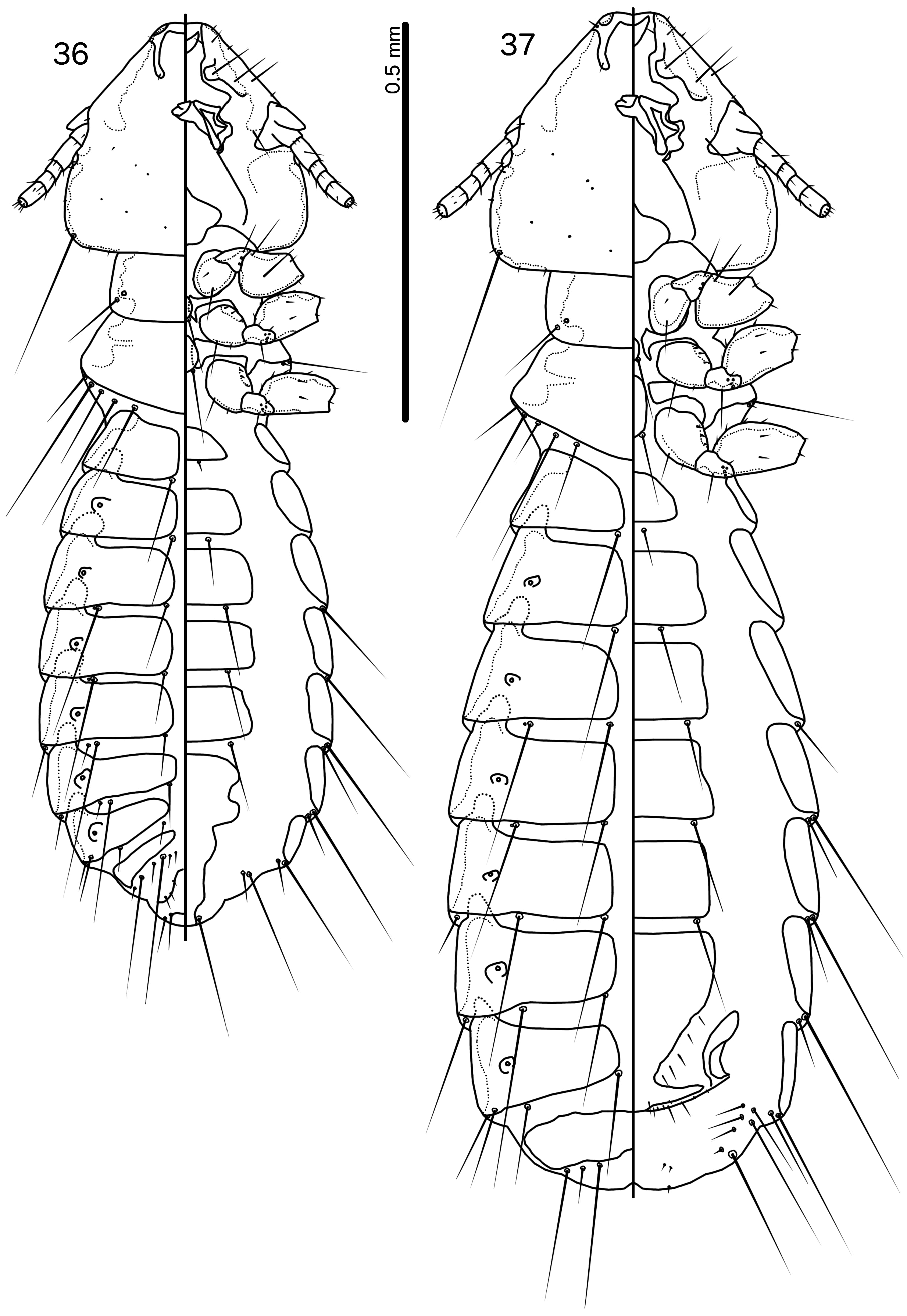
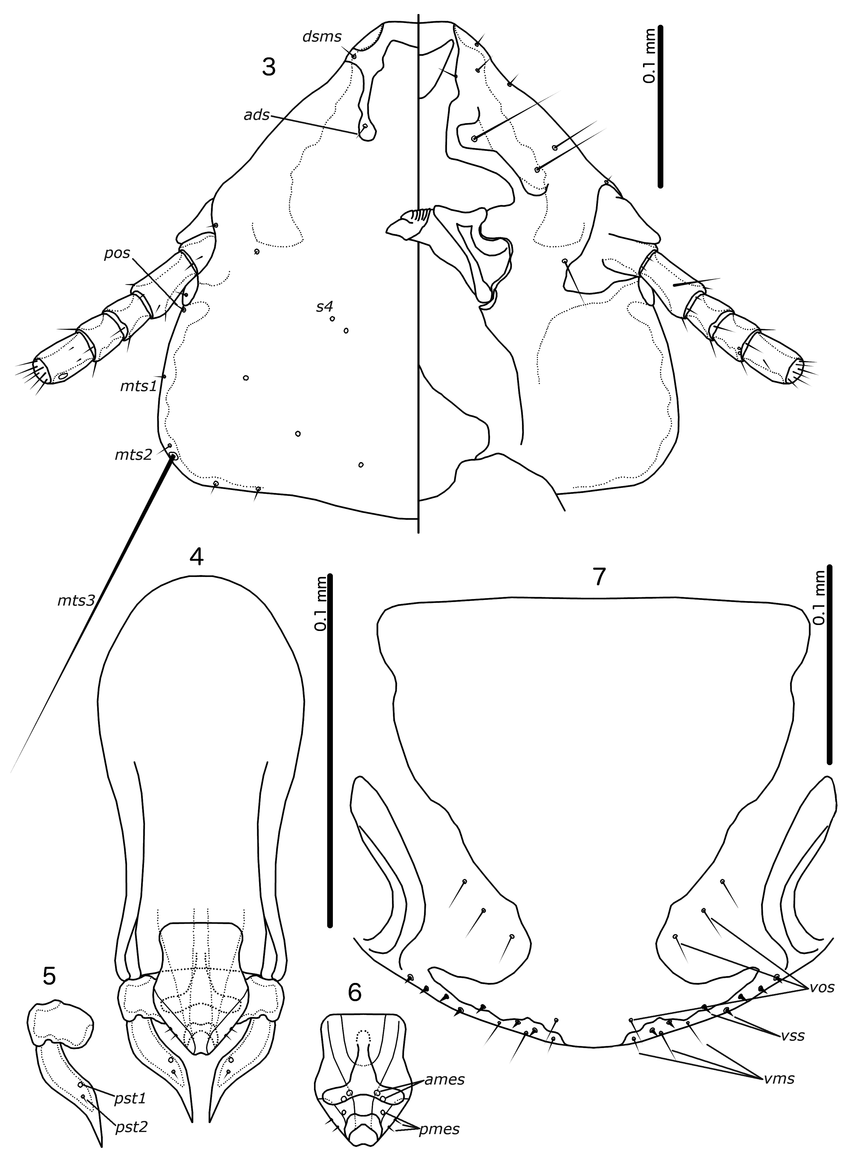
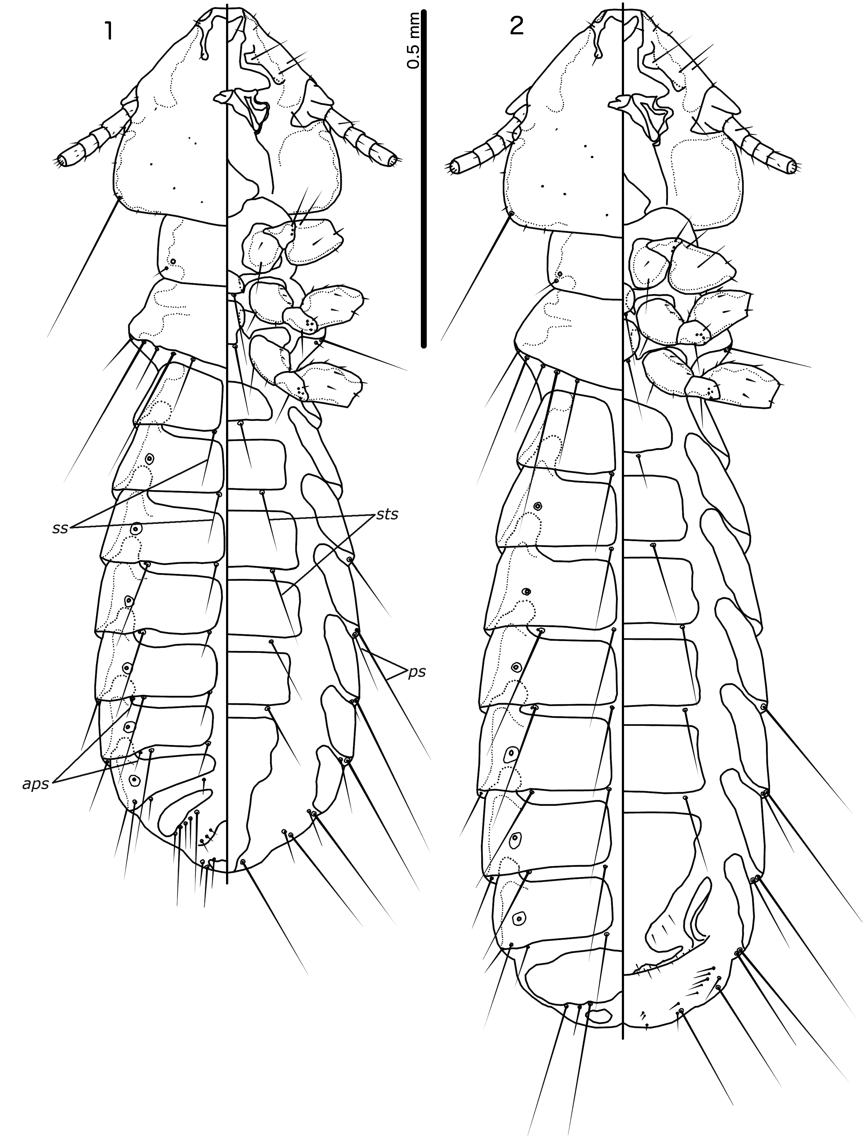
Guimaraesiella caligogularis sp. nov. is most similar to Guimaraesiella cinnamomea sp. nov., with which it shares the following characters: dorsal preantennal suture reaches ads ( Figs 3 View Figs 3–7 , 39 View Figs 38–42 ); male tergopleurites IV–V without aps, but tergopleurite VI with aps ( Figs 1 View Figs 1–2 , 36 View Figs 36–37 ); male tergopleurite VIII with 1 tps on each side ( Figs 1 View Figs 1–2 , 36 View Figs 36–37 ); proximal mesosome only slightly constricted distally ( Figs 6 View Figs 3–7 , 41 View Figs 38–42 ).
These two species can be separated by the following characters: female abdominal segment IV with 1 ps on each side in Gu. caligogularis sp. nov. ( Fig. 37 View Figs 36–37 ), but without ps in Gu. cinnamomea sp. nov. ( Fig. 2 View Figs 1–2 ); male abdominal segment V with 2 ps on each side in Gu. cinnamomea sp. nov. ( Fig. 1 View Figs 1–2 ), but with 1 ps on each side in Gu. caligogularis sp. nov. ( Fig. 36 View Figs 36–37 ); anterior extension of ventral sclerite broad in Gu. caligogularis sp. nov. ( Fig. 41 View Figs 38–42 ), but narrow in Gu. cinnamomea sp. nov. ( Fig. 6 View Figs 3–7 ); distal mesosome more broadly rounded in Gu. caligogularis sp. nov. ( Fig. 41 View Figs 38–42 ) than in Gu. cinnamomea sp. nov. ( Fig. 6 View Figs 3–7 ).
Etymology
The species name is derived from Latin ‘ caligo ’ for ‘dark’, and ‘ gula ’ for ‘throat’, referring to the dark gular plate.
Material examined
Holotype (ex Pycnonotus plumosus plumosus) MALAYSIA • ♂; [Petaling District], Subang [Jaya]; 1 Mar. 1962; M-00947 ; NHML.
Non-type material (ex Alophoixus bres tephrogenys [as Criniger bres ]) MALAYSIA • 1 ♂, 1 ♀; Terengganu [as Trengganu]; 102º 37′ E, 5º25′ N, 2600 ft a.s.l.; 3 Mar. 1974; Gn. Lawit exped.; Brit. Mus. 1974-2; NHML GoogleMaps .
Type host
Pycnonotus plumosus plumosus Blyth, 1845 – olive-winged bulbul.
Other host
Alophoixus bres tephrogenys (Jardine & Selby, 1833) – gray-cheeked bulbul.
Description
Both sexes
Head pentagonal ( Fig. 38 View Figs 38–42 ), lateral margins of preantennal head straight, frons slightly concave. Marginal carina moderate, narrowing anteriorly, median margin slightly irregular. Dorsal anterior plate roughly square-shaped, with shallowly concave anterior margin and slightly convex lateral margins. Ventral anterior plate crescent shaped. Dorsal preantennal suture reaches ads and lateral margins of head. Preantennal nodi extended medianly. Head chaetotaxy as in Fig. 38 View Figs 38–42 . Coni with convex margins do not reach distal margin of scapes. Temples rounded. Marginal temporal carina moderate, irregular. Thoracic and abdominal segments as in Figs 36–37 View Figs 36–37 . Base pigmentation pale brown with slight reddish tint, except preantennal nodi, gular plate, proepimera, metepisterna, and lateral sections of tergopleurites a medium reddish brown. Reddish tint absent in material from Alophoixus bres tephrogenys , which is also generally paler.
Male
Thoracic and abdominal chaetotaxy as in Fig. 36 View Figs 36–37 . Basal apodeme short, slender, widening distally, slightly constricted at mid-length ( Fig. 39 View Figs 38–42 ). Proximal mesosome broad, trapezoidal ( Fig. 41 View Figs 38–42 ), anterior margin slightly concave, lateral margins converging slightly distally. Ventral sclerite broad, anterior end rounded and nearly reaching proximal margin of mesosome. Mesosomal lobes broad, distal end broadly flattened; 3 ames sensilla on each side; 2 pmes microsetae on lateral margins of mesosome. Gonopore crescent shaped, lateral margins slightly rugose. Parameral heads as in Fig. 40 View Figs 38–42 . Parameral blades broad, narrowing in distal third; pst1–2 as in Fig. 40 View Figs 38–42 . Measurements as in Table 1 View Table 1 .
Female
Thoracic and abdominal chaetotaxy as in Fig. 37 View Figs 36–37 . Subgenital plate broad distally ( Fig. 42 View Figs 38–42 ), and lateral submarginal extensions almost reach lateral ends of vulval margin. Vulval margin gently rounded ( Fig. 42 View Figs 38–42 ), with 2–3 short, slender vms and 5 short, thorn-like vss on each side; 3–6 short, slender vos on each side of subgenital plate; distal 1 vos median to vss. Measurements as in Table 1 View Table 1 .
Remarks
The male specimen from Al. b. tephrogenys is indistinguishable from the male from the type host in all characters except pigmentation patterns; the specimens from Al. b. tephrogenys are paler than the holotype. We consider these specimens to be conspecific.
A female specimen from the type host (NHML) has a distorted head, and different pigmentation patterns compared to the female from the non-type host, and can therefore not with certainty be identified as Gu. caligogularis sp. nov. Illustrations of the female Gu. caligogularis sp. nov. are therefore based on the female from Al. b. tephrogenys.

No known copyright restrictions apply. See Agosti, D., Egloff, W., 2009. Taxonomic information exchange and copyright: the Plazi approach. BMC Research Notes 2009, 2:53 for further explanation.
|
Kingdom |
|
|
Phylum |
|
|
Class |
|
|
Order |
|
|
Family |
|
|
Genus |