
Halichoanolaimus ossilagulus, Leduc, 2020
|
publication ID |
https://doi.org/ 10.5852/ejt.2020.726.1175 |
|
publication LSID |
lsid:zoobank.org:pub:3099C8E5-38D0-4985-90AE-B8AD4CB66D98 |
|
DOI |
https://doi.org/10.5281/zenodo.4328609 |
|
persistent identifier |
https://treatment.plazi.org/id/B3319A0C-C625-46DA-8454-0B2816213B80 |
|
taxon LSID |
lsid:zoobank.org:act:B3319A0C-C625-46DA-8454-0B2816213B80 |
|
treatment provided by |
Plazi |
|
scientific name |
Halichoanolaimus ossilagulus |
| status |
sp. nov. |
Halichoanolaimus ossilagulus sp. nov.
urn:lsid:zoobank.org:act:B3319A0C-C625-46DA-8454-0B2816213B80
Table 1 View Table 1 , Figs 4–6 View Fig View Fig View Fig
Diagnosis
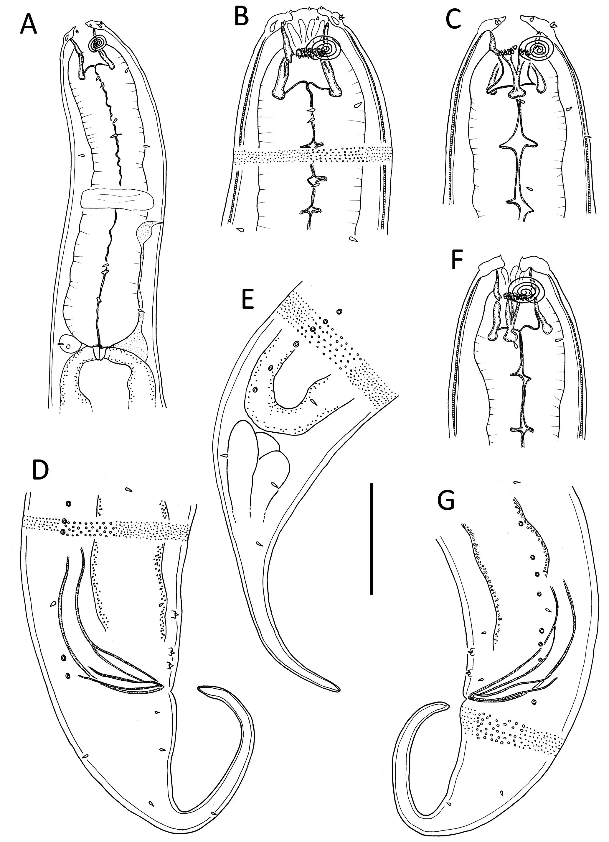
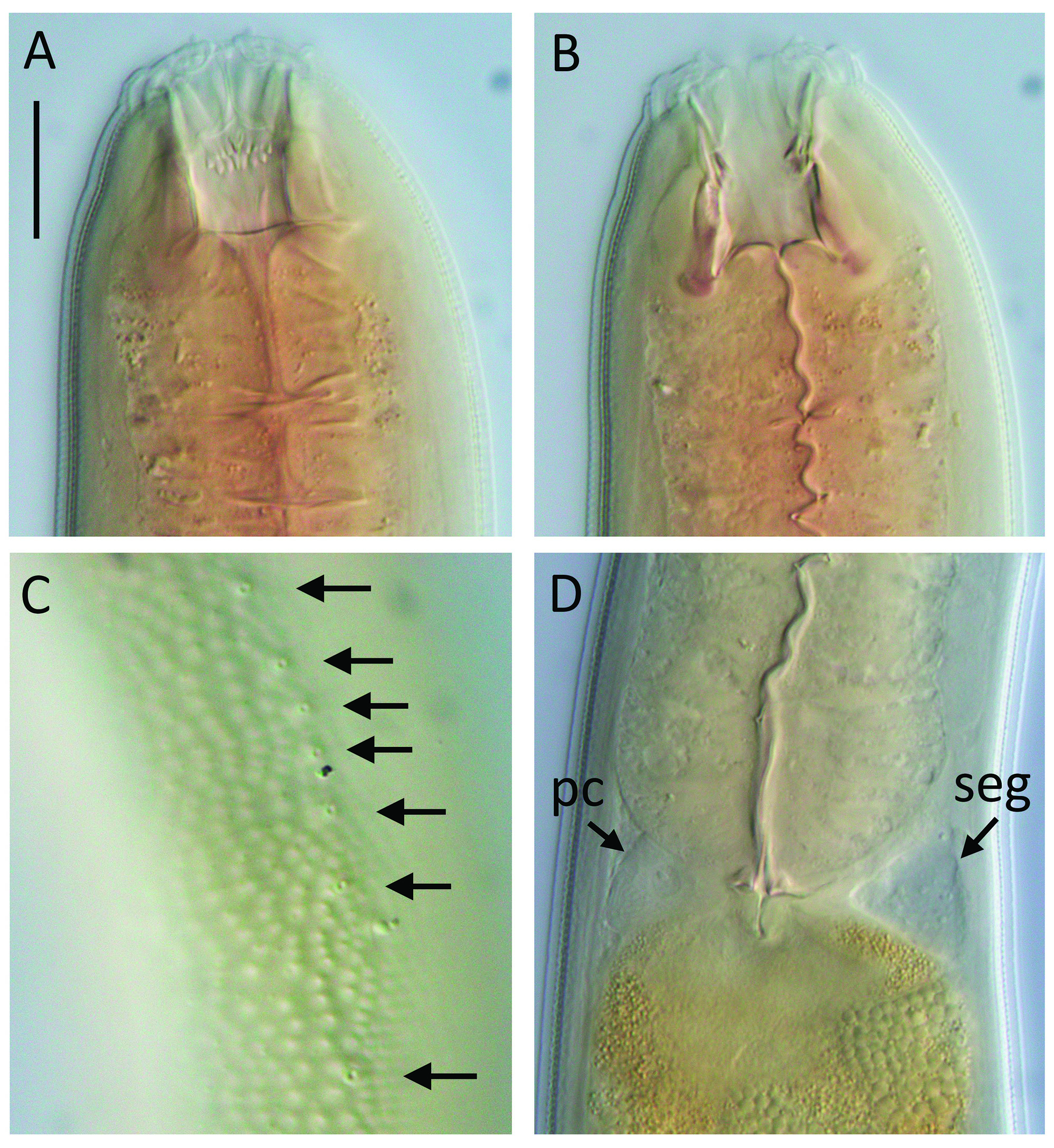
Halichoanolaimus ossilagulus sp. nov. is characterized by a body length of 966–1267 µm, amphideal fovea with 4.5 turns in both males and females, rhabdions of anterior portion of buccal cavity ending in three sets of 10 pairs of denticles with single raised denticle in centre of each set, pharyngeal lumen conspicuously cuticularized, single pseudocoelomocyte associated with SE system and located dorsally opposite renette cell, spicule length 1.6–1.7 body diameters at level of cloacal opening, gubernaculum 24–26 µm long, two or three precloacal supplements and tail 2.4–3.2 cbd long with cylindrical portion comprising about half (or three quarters in one female) of total tail length.
Differential diagnosis
The new species is most similar to H. consimilis Allgén, 1933 in general body dimensions and number of amphideal fovea turns, but differs from the latter in having shorter spicules (56–59 vs 72 µm in H. consimilis ), fewer precloacal supplements (2–3 vs 4 supplements), and shorter tail (males: 2.9–3.2 vs 3.7 cbd in H. consimilis ; females: 2.4–3.2 vs 5.2 cbd in H. consimilis ) (note that the tail dimensions for H. consimilis were derived from Allgén’s drawings using the blind end of the intestine as starting point for the tail region in females). In addition to the morphological differences noted above, the arrangement of the precloacal supplements differs between the two species: in H. consimilis the four precloacal supplements are equidistant, whereas in H. ossilagulus sp. nov. the distance between the second and third (when present) anterior-most supplements is much greater than the distance between the two posterior-most supplements.
Etymology
The species name is derived from the Latin ossilago ('bony hardness') and gula ('gullet', 'throat') and refers to the cuticularized pharyngeal lumen of this species.
Material examined
Holotype
NEW ZEALAND • 1 ♂; Southern flank of Chatham Rise, 1241 m water depth (44.4853° S, 177.1410° E, voyage TAN0705, station 45), sandy mud (83% silt/clay, 17% sand); NIWA 139246 View Materials .
GoogleMapsParatypes GoogleMaps
NEW ZEALAND • 1 ♂, 2 ♀♀; same location as for holotype; 6 April 2007; NIWA 139247 View Materials .
Type habitat and locality
Southern flank of Chatham Rise , 1241 m water depth (44.4853° S, 177.1410° E, voyage TAN0705, station 45), sandy mud (83% silt/clay, 17% sand). GoogleMaps
Description
Males
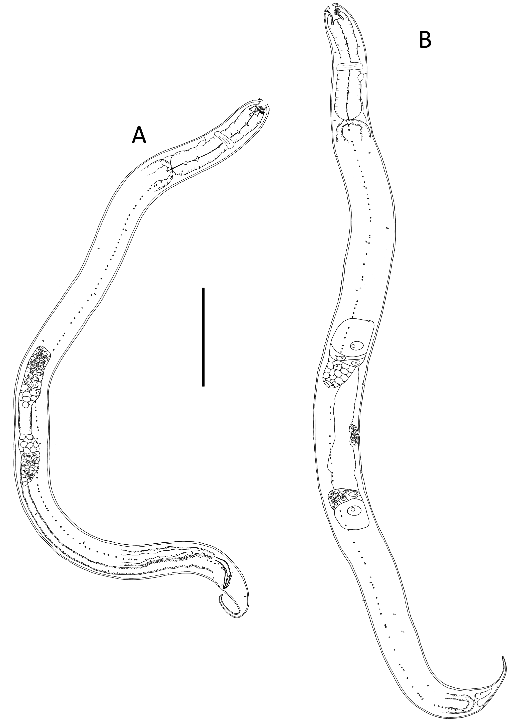
BODY. Cylindrical, tapering slightly towards both extremities.Cuticle with transverse rows of punctations; lateral differentiation consisting of larger, more widely spaced punctations. Two dorsosublateral rows of pore complexes extending from posterior end of pharynx to cloacal region, more or less equally distributed along each row; each pore complex ca 1.5 µm in diameter. Somatic setae short, 1–2 µm long, sparse, irregularly arranged along body. Cephalic region slightly rounded, lip region raised and offset. Six inner labial papillae; six short outer labial papillae, 1–2 µm long, at same level as four cephalic papillae of same length. Amphideal fovea multispiral with 4.5 turns, situated <0.3 cbd from anterior end. Buccal cavity (pharyngostome) large, ca 20 µm deep, divided into anterior (gymnostome) and posterior portions (stegostome). Anterior portion of buccal cavity cup-shaped, with three sets of three cuticularized rhabdions, 8–10 µm long, terminating in three sets of 10 pairs of teeth (denticles), with raised central denticle located in middle of each set; posterior portion of buccal cavity narrower, cylindrical, surrounded by three Y-shaped pairs of cuticularized rhabdions with swollen bases, 13 µm long. Pharynx cylindrical, muscular, without anterior or posterior bulb; pharyngeal lumen conspicuously cuticularized. Nerve ring at 55–60% of pharynx length from anterior. Secretory-excretory system present. Renette cell 13–15 µm long, 7–14 µm wide, situated at level of cardia; single nucleated pseudocoelomocyte present dorsally and opposite renette cell, ampulla small, pore situated slightly posterior to nerve ring. Cardia small, surrounded by intestine. Posterior extremity of intestine blind, rectum absent.
REPRODUCTIVE SYSTEM. Diorchic with short outstretched testes. Anterior testis to the left of intestine, posterior testis to the right of intestine. Sperm cells globular, 7–8 × 8–10 µm. Spicules paired, curved, tapering distally, length 1.6˗˗1.7 body diameters at level of cloacal opening; gubernaculum consisting of two detached lateral pieces (crurae) tapering distally, median portion of gubernaculum (corpus and cuneus) not visible. Two or three precloacal supplements present, consisting of conical papillae set on cylindrical cuticular elevations. When present, third anterior-most supplement conspicuously further away from other two supplements (12 µm) than two posterior-most supplements are from each other (4–6 µm). Tail conicocylindrical, cylindrical portion ca half of total tail length; a few short and sparse somatic setae present. Caudal glands not observed.
Females
Similar to males; one female with cylindrical portion of tail comprising ca three quarters of total tail length. Reproductive system didelphic-amphidelphic, with reflexed ovaries. Anterior ovary to the left of intestine and posterior ovary to the right of intestine. Vulva situated near mid-body. Proximal portion of vagina surrounded by constrictor muscle, two pairs of vaginal glands present. Intestine blind, anus not observed.
No known copyright restrictions apply. See Agosti, D., Egloff, W., 2009. Taxonomic information exchange and copyright: the Plazi approach. BMC Research Notes 2009, 2:53 for further explanation.
|
Kingdom |
|
|
Phylum |
|
|
Class |
|
|
Order |
|
|
Family |
|
|
Genus |