
Halichoanolaimus funestus, Leduc, 2020
|
publication ID |
https://doi.org/ 10.5852/ejt.2020.726.1175 |
|
publication LSID |
lsid:zoobank.org:pub:3099C8E5-38D0-4985-90AE-B8AD4CB66D98 |
|
DOI |
https://doi.org/10.5281/zenodo.4328605 |
|
persistent identifier |
https://treatment.plazi.org/id/92FE70A3-3C0B-434F-94ED-71931889DC40 |
|
taxon LSID |
lsid:zoobank.org:act:92FE70A3-3C0B-434F-94ED-71931889DC40 |
|
treatment provided by |
Plazi |
|
scientific name |
Halichoanolaimus funestus |
| status |
sp. nov. |
Halichoanolaimus funestus sp. nov.
urn:lsid:zoobank.org:act:92FE70A3-3C0B-434F-94ED-71931889DC40
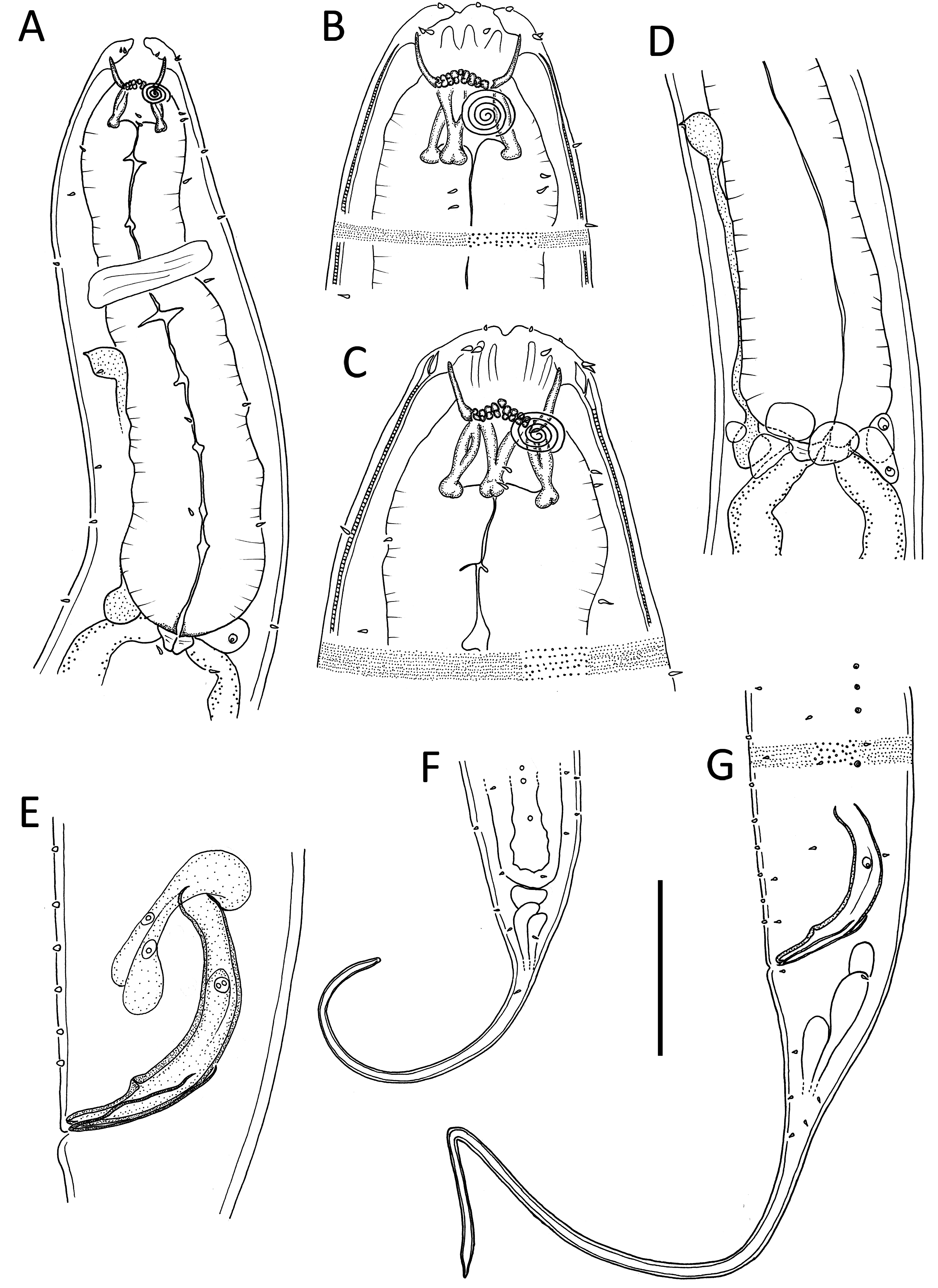
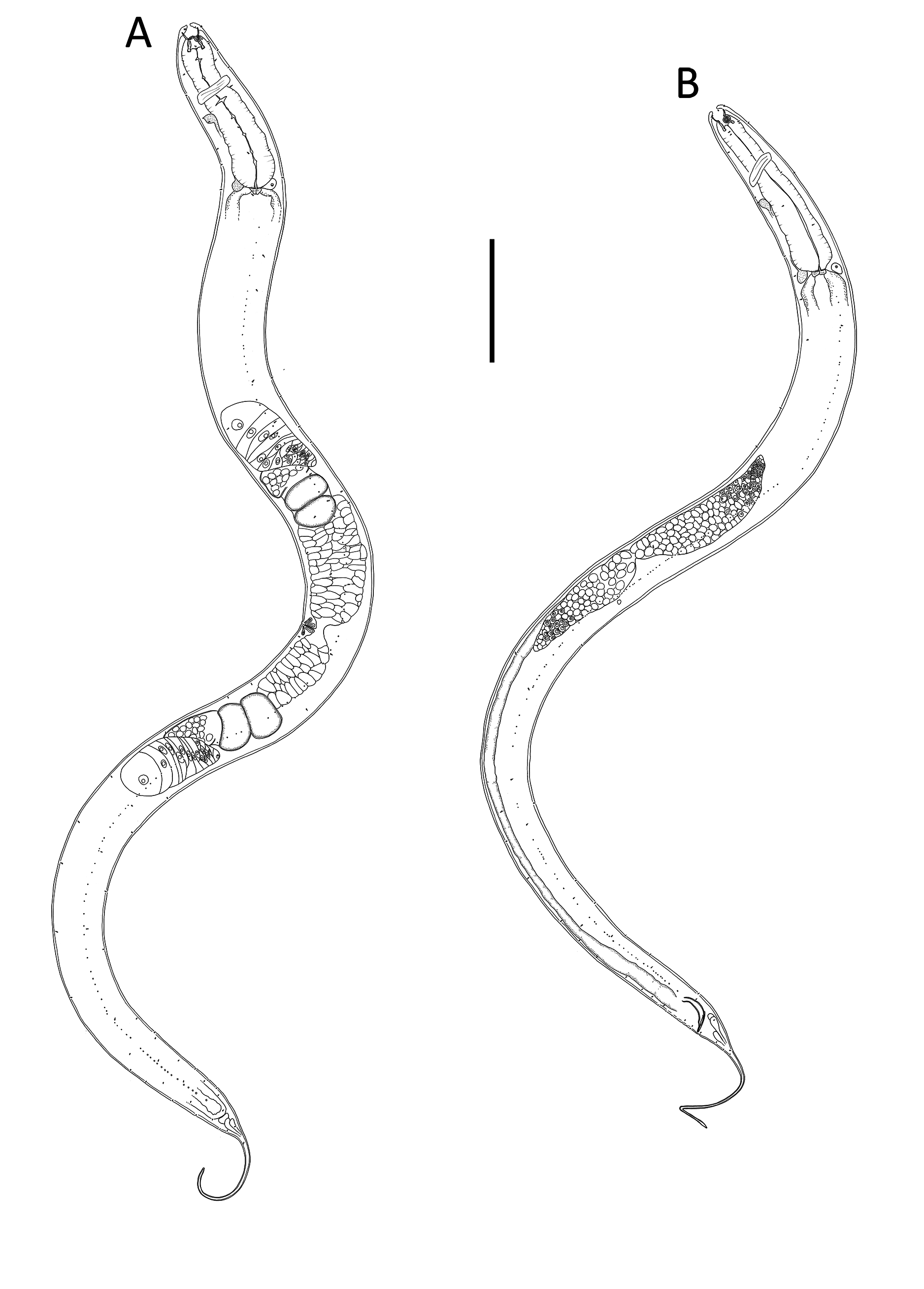
Table 1 View Table 1 , Figs 7–9 View Fig View Fig View Fig
Diagnosis
Halichoanolaimus funestus sp. nov. is characterized by relatively large body, 2.7–3.0 mm long, amphideal fovea spiral with 4.5–5.0 turns, rhabdions of anterior portion of buccal cavity ending in three sets of 10 pairs of denticles with single raised denticle in centre of each set, several pseudocoelomocytes associated with SE system surrounding base of pharynx, spicule length 1.8 body diameters at level of cloacal opening with ventral protrusion near distal tip, gubernaculum 47 µm long, seven inconspicuous papillose precloacal supplements and tail 5.1–6.3 cbd long with cylindrical portion comprising slightly more than three quarters of total tail length.
Differential diagnosis
The new species is most similar to H. dolichurus Ssaweljev, 1912 in body length, shape of the copulatory apparatus, number of precloacal supplements and tail length, but can be differentiated from the latter by the lower ‘a’ ratio (20–23 vs 30–40 in H. dolichurus ) and number of amphideal fovea turns (4.5–5.0 vs 3.75–4.0 turns in H. dolichurus ). The new species is also similar to H. ovalis in the number of amphideal fovea turns, structure of the spicular apparatus and number of precloacal supplements, but can be differentiated from the latter by the longer body (2.7–3.0 vs 1.3–1.8 mm in H. ovalis ), higher ‘a’ ratio (20–23 vs 17–18 in H. ovalis ), longer tail (in males, 5.7–6.3 vs 3.6 cbd in H. ovalis ) and longer spicules (97 vs 60 µm in H. ovalis ).
Etymology
The species name is derived from the Latin funestus ('causing death', 'calamity, deadly') and refers to the ability of this species to feed on relatively large nematodes owing to its voluminous armed buccal cavity and large body size.
Material examined
Holotype
NEW ZEALAND • 1 ♂; Main axis of Kaikōura Canyon , 1061 m water depth (42.5082° S, 173. 6325° E, voyage TAN1006, station 7, site K4); NIWA 139248 View Materials .
GoogleMapsParatypes GoogleMaps
NEW ZEALAND • 2 ♀♀; same location as for holotype; 3 May 2010; NIWA 139249 View Materials .
Type habitat and locality
Main axis of Kaikōura Canyon , 1061 m water depth (42.5082° S, 173. 6325° E, voyage TAN1006, station 7, site K4). GoogleMaps
Description
Male
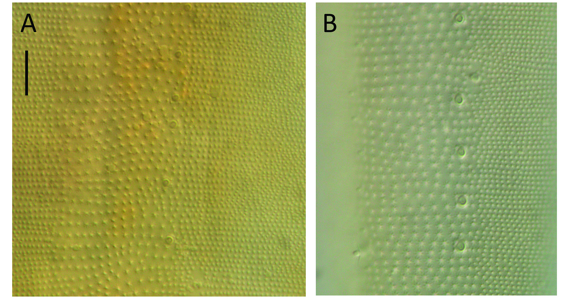
BODY. Cylindrical, tapering slightly towards both extremities.Cuticle with transverse rows of punctations; lateral differentiation consisting of larger, more widely spaced punctations. Two dorsosublateral rows of pore complexes extending from posterior end of pharynx to cloacal region, becoming more closely spaced posteriorly; each pore complex ca 2 µm in diameter in middle body region and ca 3 µm in diameter near cloacal region. Somatic setae 2–5 µm long, sparse, arranged in eight longitudinal rows along body. Cephalic region slightly rounded, lip region slightly offset. Six inner labial papillae; six short outer labial setae, 3–4 µm long, at same level as four cephalic papillae of same length. Amphideal fovea multispiral with 5.0 turns, situated 0.4–0.5 cbd from anterior end. Buccal cavity (pharyngostome) large, ca 45–50 µm deep, divided into anterior (gymnostome) and posterior portions (stegostome). Anterior portion of buccal cavity cup-shaped, with three sets of three cuticularized rhabdions, 17–25 µm long, terminating in three sets of 10 pairs of teeth (denticles), with raised central denticle located in middle of each set; posterior portion of buccal cavity narrower, cylindrical, surrounded by three Y-shaped pairs of cuticularized rhabdions with swollen bases, 24–27 µm long. Pharynx cylindrical, muscular, without anterior or posterior bulb; pharyngeal lumen not cuticularized. Nerve ring at 40–50% of pharynx length from anterior. Secretory-excretory system present. Renette cell 24–32 µm long, 21–33 µm wide, situated at level of cardia; several nucleated pseudocoelomocytes surrounding base of pharynx, ampulla small, pore situated slightly posterior to nerve ring. Cardia small, surrounded by intestine. Posterior extremity of intestine blind, rectum absent.
REPRODUCTIVE SYSTEM. Diorchic with outstretched testes. Anterior testis to the left of intestine, posterior testis to the right of intestine. Sperm cells globular, 21–22 × 22–25 µm. Spicules paired, curved, tapering distally, length 1.8 body diameters at level of cloacal opening, with ventral protrusion near distal tip. Gubernaculum consisting of two detached lateral pieces (crurae) tapering distally, median portion of gubernaculum (corpus and cuneus) not visible. Seven inconspicuous papillose precloacal supplements present; posteriormost four supplements 10–13 M m apart, anteriormost three supplements 16–21 M m apart. Tail long, conicocylindrical, cylindrical portion slightly more than three quarters of total tail length; a few short and sparse somatic setae present. Three caudal glands present.
Females
Similar to males, but with slightly lower ‘a’ ratio and slightly fewer (4.5–4.75) amphideal fovea turns. Reproductive system didelphic-amphidelphic, with reflexed ovaries. Anterior ovary to the left of intestine and posterior ovary to the right of intestine. Mature eggs 95–99 × 48–53 µm. Vulva situated slightly pre-median. Proximal portion of vagina surrounded by constrictor muscle, two small vaginal glands present. Intestine blind, anus not observed.
Remarks
A nematode belonging to the genus Parodontophora Timm, 1963 was observed in the intestine of the holotype, and a nematode of the genus Sabatieria Rouville, 1903 was observed in the intestine of one of the female paratypes.
No known copyright restrictions apply. See Agosti, D., Egloff, W., 2009. Taxonomic information exchange and copyright: the Plazi approach. BMC Research Notes 2009, 2:53 for further explanation.
|
Kingdom |
|
|
Phylum |
|
|
Class |
|
|
Order |
|
|
Family |
|
|
Genus |