
Sufetula carbonalis Hayden, 2013
|
publication ID |
https://doi.org/ 10.5281/zenodo.5175913 |
|
publication LSID |
lsid:zoobank.org:pub:0986651C-DD2A-41B4-A937-563B5E366536 |
|
persistent identifier |
https://treatment.plazi.org/id/E72B87B2-C03F-512E-53CC-F34BF41F0C83 |
|
treatment provided by |
Felipe |
|
scientific name |
Sufetula carbonalis Hayden |
| status |
sp. nov. |
Sufetula carbonalis Hayden , new species
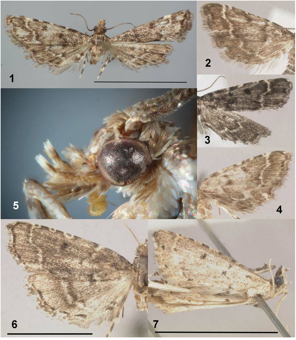
( Fig. 1–5 View Figures 1–7 , 17–19 View Figures 17–22 , 23–25 View Figures 23–29 )
S. [ Sufetula ] sp. Kimball, 1965: 200 (McDunnough No. 5352,2).
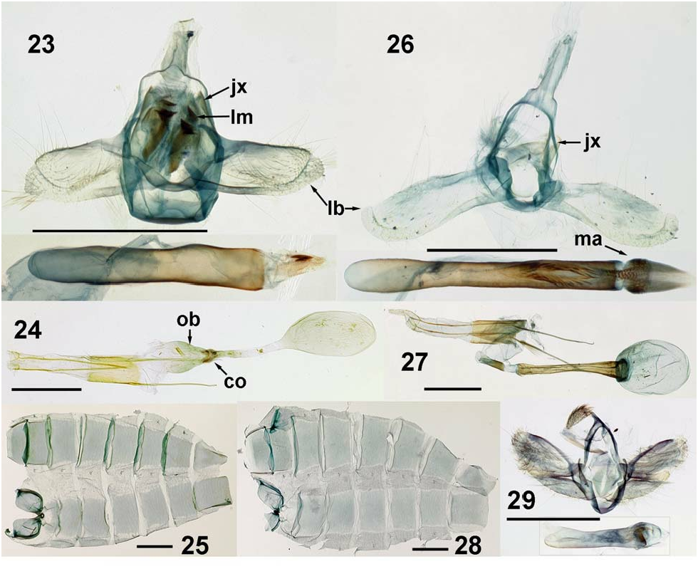
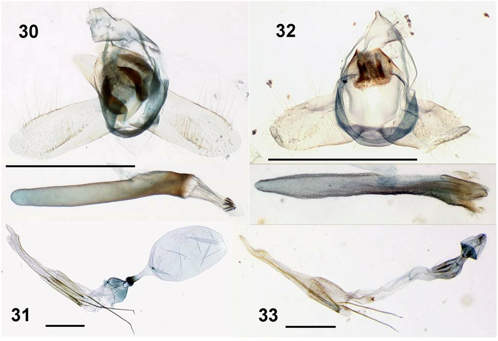
Diagnosis. Sufetula carbonalis is both smaller and darker in color than known congeners. Adults of species that are similarly small in size are paler in color. The second meron of the labial palpi is ventrally tufted ( Fig. 5 View Figures 1–7 ). The anterior half of the forewing PM line runs nearly perpendicularly from the costa before turning at a 45° angle ( Fig. 1–4 View Figures 1–7 ). Rs 2 and Rs 3 in the forewing are entirely fused ( Fig. 17 View Figures 17–22 ). The manica of the male genitalia has two pairs of lamellae that are flat, elongate and distally recurved and bifid, like crowbars ( Fig. 23 View Figures 23–29 : lm). The phallus has two small distal cornuti ( Fig. 23 View Figures 23–29 ). In other species of Sufetula , the manica is densely spiculose ( Fig. 26 View Figures 23–29 : ma) but without flat plates (although the dense granules may resemble plates when the manica remains with the annellus, e.g. Fig. 32 View Figures 30–33 ), and the other species lack cornuti or have more than two. The female genitalia of S. carbonalis have no sclerotizations of the ductus bursae or corpus bursae; the ostium bursae is wide ( Fig. 24 View Figures 23–29 : ob), the membrane at the junction of the ostium and ductus bursae is thickened, and a rudimentary colliculum is visible as two transverse sclerotizations ( Fig. 24 View Figures 23–29 : co).
Similar species. The sympatric S. diminutalis is larger in size and lighter in color. The labial palpi do not have a ventral tuft. The PM line slants slightly distad from the costa to the medial fold, where it sharply turns basad at an angle more nearly parallel to the wing’s long axis. Females of S. diminutalis have two frenular bristles, a well-developed colliculum and an elongate ductus bursae that is completely and evenly sclerotized along its entire length.
Sufetula carbonalis is closely related to S. grumalis Schaus and possibly S. sacchari (Seín) . Sufetula grumalis , described from Cuba, is larger in size, but it has very similar dark gray maculation and a PM line of identical curvature ( Fig. 6 View Figures 1–7 ). The AM line differs in that it extends distad from the costa to the Cu vein, then jaggedly basad on the anal fold, then distad on the anal area. Like S. carbonalis , S. grumalis has labial palpi with a ventral tuft, the ocelli are absent, and the female has one frenular bristle. The manica has one pair of curved, tridentate sclerites and eight small cornuti ( Fig. 30 View Figures 30–33 ). The colliculum in S. grumalis ( Fig. 31 View Figures 30–33 ) is better defined than in S. carbonalis , and the ductus bursae is shorter. Sufetula sacchari , described from Puerto Rico, is similarly small in size ( Fig. 7 View Figures 1–7 ). The labial palpi are ventrally tufted, and the forewing PM line has similar curvature. It differs from S. carbonalis in its pale gray color, conspicuous ocelli, elongate costal lunules that equal the gray interstices in length, manica without large, discrete, elongate lamellae ( Fig. 32 View Figures 30–33 ), and membranous ductus bursae without sclerotization ( Fig. 33 View Figures 30–33 ).
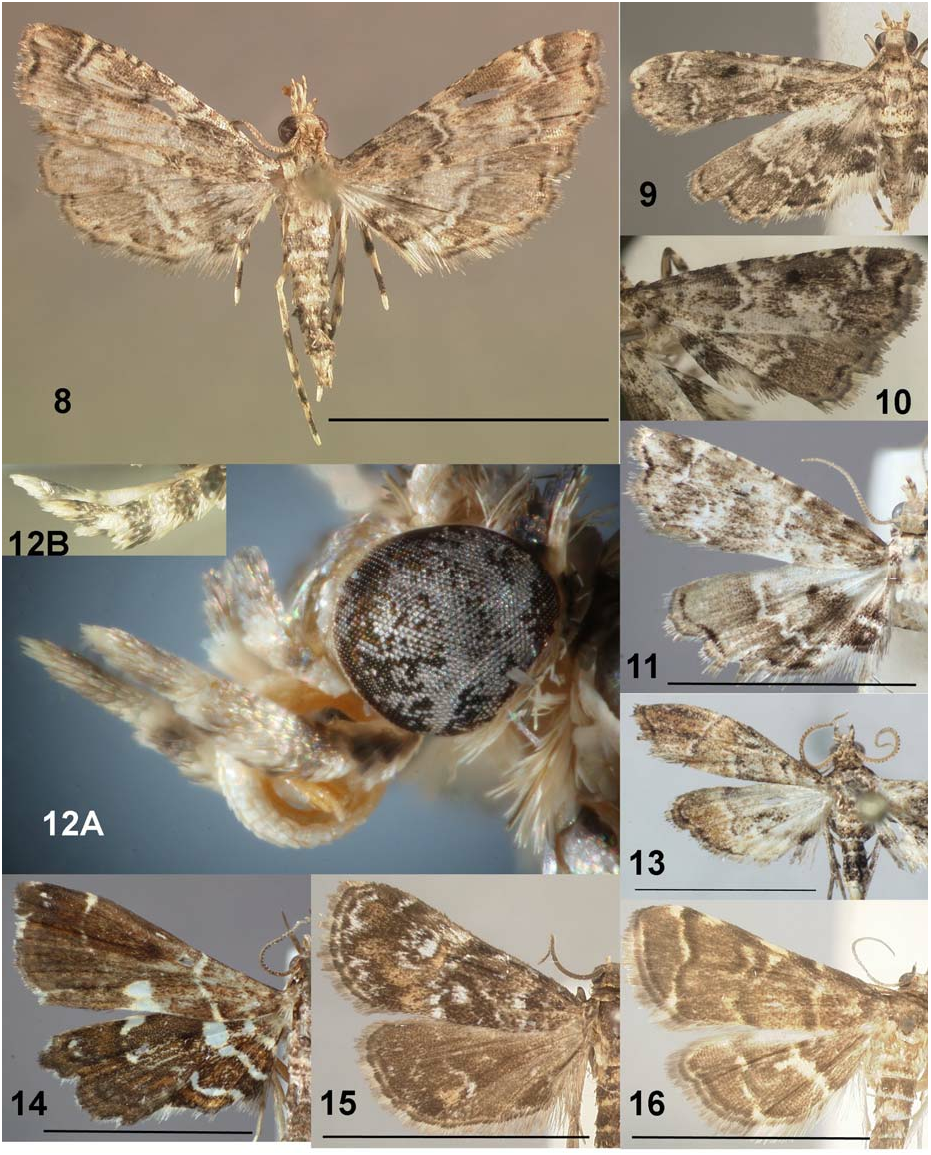
Among relatively common species in Florida, the most superficially similar are the acentropines Elophila (Synclita) obliteralis (Walker) ( Fig. 15 View Figures 8–16 ) and E. tinealis (Munroe) . Females have one frenular bristle, like S. carbonalis , but differ in having evenly colored palpi and large ocelli and chaetosemata that are situated on a swollen vertex. Elophila obliteralis has a white discal spot. Females are larger and paler than S. carbonalis . Males are the same size, and although darker than the female, they have forewings with orange scales on the AM and PM areas and also on the hind wing around the anal fold. In E. tinealis , the males are even smaller, but females are the same size. The wings have a few irregular white scales, and the ventral body is distinctly white.
Microphysetica hermeasalis (Spilomelinae) is Caribbean in distribution, including southern Florida. Characters shared with Sufetula include slightly ascendant labial palpi, relatively large maxillary palpi, rather thick antennae and the absence of chaetosemata and of the male frenular hook. The maculation is similar ( Fig. 13 View Figures 8–16 ), but the PM line is angled out at nearly 45° in the apical half, then directly inward and slightly anteriad up into the cell. The male genitalia ( Fig. 29 View Figures 23–29 ) have dorsally angled valvae with a roundended, transverse fibula, a capitate, spinose uncus and a large, curved cornutus.
Description. Head ( Fig. 5 View Figures 1–7 ): frons round with appressed, glossy whitish-gray scales; vertex round with dark gray scales directed forward. Chaetosemata absent. Ocelli rudimentary, barely visible posterior of scale tuft. Labial palpi dark gray with mixed white scales; basal meron short; second meron porrect, extended beyond head, ventral side with forward-directed tuft; third meron ascendant at 45° angle. Haustellum well-developed. Antennal scape and pedicel without modifications; flagellum simply scaled, with two rows of scales per meron, lightly ciliate in female, more strongly so in male, with alternating rows directed outward at angle, exposing surface of meron.
Thorax: dorsal side dark gray with mixed white scales; ventral side similar but predominantly white. Legs: epiphysis present; tibial spurs 0, 2, 4, outer spur half length of inner. Robust chaetae not visible on legs. Mera of legs basally white, distally dark gray. Tibiae of hind legs with row of rough, ascendant scales.
Wings ( Fig. 1–4 View Figures 1–7 , 17 View Figures 17–22 ): forewing length (base to apex) 3.5–5.0 mm, mean 4.4 ± 0.3 mm; width (costa to tornus) 1.2–1.7 mm, mean 1.5 ± 0.1 mm (n = 12). Forewing apex rounded, incised on medial fold between M 2 and M 3. Male without retinacular hook. Retinacular hairs in male and female from membrane posterior of base of Cu vein. Female and male both with one frenular bristle in hind wing. Forewing R 1 free, from cell at 0.85 length; Rs 1 free; Rs 2+3 fused; Rs 4 from anterior corner of cell or very shortly stalked with Rs 2+3; cell closed; discal cross-vein nearly perpendicular to wing axis; M 1 from cross-vein 1/4 distance from anterior corner; M 2 and M 3 short-stalked; CuA 1 and CuA 2 free; CuP present as tubular marginal vein. 1A+2A straight; 3A not tubular, only a faint, straight trace. Hind wing Sc+R1 stalked with Rs 1 1/ 3 distance; cell closed; M 1 present; M 2 and M 3 shortly stalked; CuA 1 and CuA 2 free; CuP present from base but weak; 1A+2A strong; 3A present, weak. Forewing color gray to dark gray. AM and PM lines white edged with dark gray; white basal line sometimes visible. Discal spot round, nearly black, in anterior center of wing. Two white spots on costa basal and distal of discal spot, sometimes appearing as lunules with black centers. AM and PM lines expanded slightly on costa and sometimes appearing as lunules. AM line faint and slightly jagged, nearly perpendicular to long axis of wing from costa to posterior margin. PM line nearly perpendicular from costa, bent basad at 45° angle along M veins, then nearly perpendicular to posterior margin, again slightly curved basad on margin. Ventral side like dorsal side, paler. Hind wing gray to dark gray with round, dark gray discal spot. AM line not visible. PM line white, jagged, complete from costa to posterior margin, meeting posterior margin near base. Ventral side similar in color, paler.
Abdomen: Dorsally gray with white scales on posterior margin of segments, ventrally paler. Praecinctorium simple. Male T8 and S8 not modified, without membranous areas or scale tufts; T8 a blunt triangle, S8 square ( Fig. 25 View Figures 23–29 ).
Tympanal organs ( Fig. 18–19 View Figures 17–22 ): fornix tympani longer than wide, lateral and posterior margins nearly at right angles, slightly protruded over venula prima. Venulae secundae straight, slightly divergent, longer in female than in male. Posterior depressions small, occupying mesal third of width of tympanal organs; in male, round, pit-like, with emargination lateral of venula secunda ( Fig. 18 View Figures 17–22 : pd); in female, depressions shallow, narrow, transverse, slightly extended onto venula secunda ( Fig. 19 View Figures 17–22 : pd).
Male genitalia ( Fig. 23 View Figures 23–29 ): uncus narrow, membranous, without visible setae, connected to anal tube. Subscaphium larger than uncus. Gnathos and transtilla absent. Tegumen without dorsal ridges. Vinculum round, ventrally nearly flat, without saccus or process. Valvae short, length twice width, of even width, with costal and saccular margins parallel; fibulae and other processes absent but fine setae present; costal margin distally with a few fine, distinct setae perpendicular to margin; distal margin membranous, labriform and distinct from main part of valve, bearing deciduous scales at apex. Juxta (jx) represented by pair of narrow, triangular plates with acute apices, extended to upper end of tegumen and laterally framing the manica and phallus. Sclerotization of manica complex, bearing two pairs of overlapping, lanceolate lamellae with distal ends flat and recurved, the complex detaching from phallus in dissection and remaining with genitalic capsule; membrane of manica otherwise spinulose. Phallus straight, length seven times width; caecum penis longer than rest of phallus, inception of ductus ejaculatorius about 1/4 length from apex of phallus; two short, straight cornuti.
Female genitalia ( Fig. 24 View Figures 23–29 ): ovipositor elongate and telescoping; anal papillae fused, narrowly pointed. Apophyses thin; posterior apophyses very long, extended to anterior margin of A8; anterior apophyses moderately long. Ostium bursae (ob) unarmed, funnel-shaped, nearly as wide as long. Colliculum (co) not discretely sclerotized, represented by round, thickened area between antrum and ductus bursae. Ductus bursae twice as long as antrum bursae, straight, not sclerotized. Ductus seminalis from posterior end of ductus bursae just anterior of colliculum. Corpus bursae oval, slightly longer than ductus bursae, twice as long as wide, without signa or appendix.
Types. Holotype: 1F: “ Terhune S. Dickel Coll. ”, “ FLORIDA: Dade County Homestead Fuchs Hammock ”, “ May 12–13 1980 ”, “ Malaise trap ”, (red label) “ HOLOTYPE Sufetula carbonalis Hayden ”, “ J.E. Hayden slide no. 1590 F” ( TSD, to be deposited in FSCA).
Paratypes: USA, Florida: 1M: “ Homestead, Fla. xi-5 1959 D.O. Wolfenbarger ” ( MGCL slide 99) ( FSCA) ; 1M: “ Siesta Key Sarasota Co., Fla. April 8 1953 C.P. Kimball ” ( MGCL genitalia slide 98, wing slide 541) ( FSCA) ; 1F: “ Oneco, Manatee Co., Fla. V-2 1955 Paula Dillman”, “5352.2” (green pen) ( FSCA) ; 1: “ FLORIDA: Highlands Co. Archbold Biol. Station 10 mi. S. Lake Placid 2-V-1975 ”, “at ( UV) blacklight”, “ J.B. Heppner collector” ( FSCA) ; 1F: “FL: Levy Co. Goethe St. For. Gasline & Beehive Rds. 29°09’38"N, 82°35’55"W. 30-VII-2011. Pine flatwoods, MV. J.E. Hayden ”, “J.E. Hayden slide no. 1405 F” ( FSCA) GoogleMaps ; 2M: “ FLORIDA: Volusia Co. Tomoka State Park 22-25-V-2000 J.B. Heppner ” ( FSCA) ; 1F (in alcohol): truck at I-10 Westbound Interdiction Station, Live Oak , Suwannee Co., FL, origin: Gould, FL, on Phoenix roebelenii , 9-IV-2012, D. Russell & K. Collins, E2012-2497 ( MGCL slide 540) ( FSCA) ; 1M: “ FLORIDA: LEVY CO. Goethe State Forest Intersection of North Prong Rd & Middle Rd DEC 9 2012 BAIT Terhune S. Dickel ” ( TSD) ; 1 (sex undet., abdomen lost): “ Homestead, Fla. xi.13 1958 D.O. Wolfenbarger ”, (Munroe’s hand) “5351”, “Database # CNC LEP 00074732”, (blue label) “ Barcode of Life DNA voucher specimen Sample ID: CNCLEP00074732 BOLD Proc. ID: ZYPAN531-10” ( CNC) ; 1F (without head): “ Winter Park Fla. 28-VII-39 H.T. Fernald ”, “Collected at light”, [large label] “This is probably an undescribed sp. = my 5352,1 or 2. CPK.” ( NMNH) ; 1M: “ Siesta Key Sarasota Co., Fla. Nov. 1 1952 C.P. Kimball ”, [Munroe’s hand, pencil] “5352” ( NMNH) ; 1F: “ FLORIDA: Parker Is. , Highlands Co. 4-7 June 1964 R. W. Hodges ” ( NMNH) .
Additional material examined: 1 specimen: Miami-Dade Co., Florida City, at gas station light, 18 Jan. 2013, J. Vargo (J. Vargo Collection).
Distribution. The species is endemic to peninsular Florida, USA, as far north as Volusia and Levy Counties, but most records are from the south and south-central areas of the state.
Phenology. Specimens have been collected in April, May, July, November and December.
Hosts. Larval feeding has not been observed directly and vouchered, but two interceptions by FDACS- DPI agree with the general habit of Sufetula species feeding on palm roots. In April 2012, adults were found swarming around potted Phoenix roebelenii O’Brien in a truck carrying stock from a nursery in southern Florida. There is an unverifiable earlier record (FDACS-DPI Entomology database no. E2009- 1521-1, March 2009) from the same nursery of adults reared from larvae feeding on root balls of Dypsis lutescens (H. Wendl.) Beentje & J. Dransf. , the specimens of which were identified as “ Synclita sp. ” and discarded.
Etymology. Latin carbo, charcoal, in reference to the dark gray maculation.
Remarks. Unlike S. diminutalis , S. carbonalis is not commonly collected at sugar bait. In Goethe State Forest, T. S. Dickel and I have frequently caught S. diminutalis of both sexes but only one male S. carbonalis with this method; the other specimen from the forest flew to mercury vapor light early in the evening. It might be overlooked because of its small size and dark color. The possibility of a different flight time should be considered. The Fuchs Hammock holotype was caught in a malaise trap.
The two (or possibly three) pairs of distally curved, crowbar-shaped lamellae are a typical modification of the manica, the pattern of which, like the vesica, varies greatly among Sufetula species. The manica may be removed with the phallus or stay with the genitalic capsule in different preparations, so a standardized method of dissection should take advantage of the manica’s diagnostic potential. The sclerites should not be confused with the elongate halves of the juxta.

No known copyright restrictions apply. See Agosti, D., Egloff, W., 2009. Taxonomic information exchange and copyright: the Plazi approach. BMC Research Notes 2009, 2:53 for further explanation.
|
Kingdom |
|
|
Phylum |
|
|
Class |
|
|
Order |
|
|
Family |
|
|
Genus |