
Titanokorys gainesi, Caron & Moysiuk, 2021
|
publication ID |
https://doi.org/10.1098/rsos.210664 |
|
DOI |
https://doi.org/10.5281/zenodo.5500180 |
|
persistent identifier |
https://treatment.plazi.org/id/E9525344-3C22-D54E-FF4C-95F5FB3CFC97 |
|
treatment provided by |
Diego |
|
scientific name |
Titanokorys gainesi |
| status |
gen. et sp. nov. |
Titanokorys gainesi gen. et sp. nov.
LSID urn:lsid:zoobank.org:act:
LSID urn:lsid:zoobank.org:act:
3.1.1. Etymology
Genus name from Titans, a group of powerful Greek deities of great sizes, in reference to the large size of the central carapace element and from the Greek word Korys meaning helmet; gainesi , after Robert R. Gaines, Professor of Geology at Pomona College, who first joined the ROM-led field expeditions in 2008 as a research collaborator. Robert Gaines was instrumental in the co-discovery of the Marble Canyon fossil deposit in 2012 [ 26] and several related Burgess Shale outcrops along Tokumm Creek [ 25], including many fossils in this study.
3.1.2. Type material
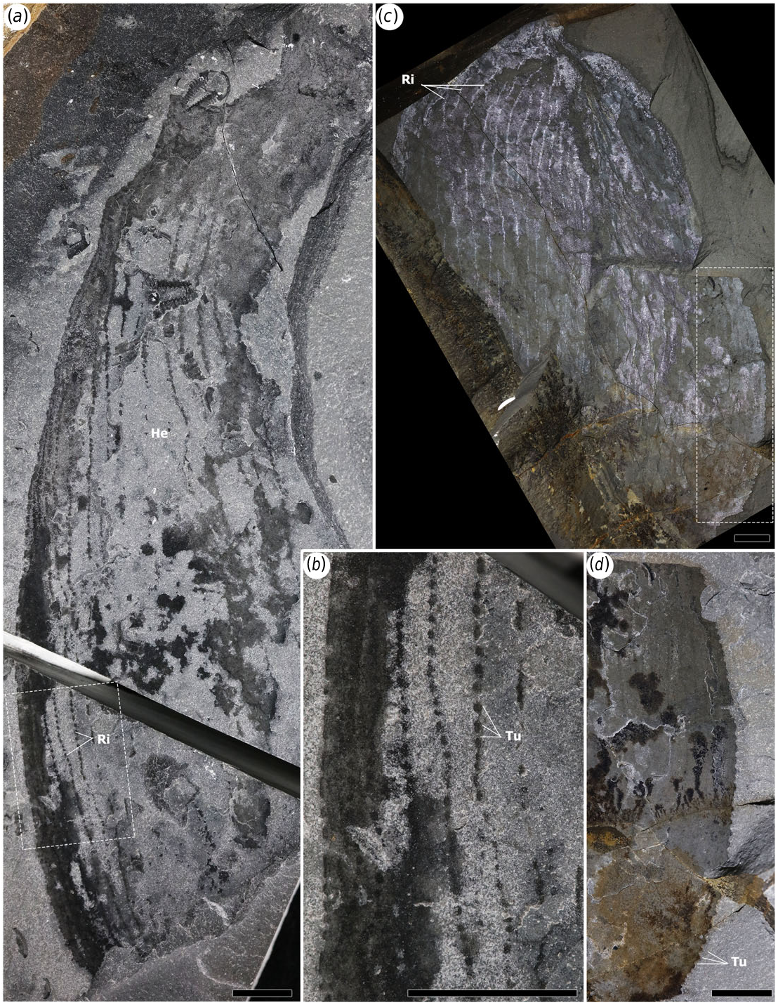
Holotype — ROMIP 65415 , a probable moult assemblage consisting of an H-(central) element, a pair of lateral (P) elements, a pair of frontal appendages, oral cone and probable gill blades ( figure 3 View Figure 3 ). Paratypes — ROMIP 65168 , a complete H-element ( figure 1 View Figure 1 ) ; ROMIP 65741 , an individual H-element with partial endites ( figure 2 View Figure 2 ) ; ROMIP 65748 and ROMIP 65749 , two fragmentary H-elements ( figure 4 View Figure 4 ) ; other materials: seven additional H-elements, one associated with a P-element and poorly preserved appendages, in various states of preservation and completeness (electronic supplementary material, table 2.1) .
3.1.3. Locality and stratigraphy
The upper part of the ‘Thick’ Stephen (Burgess Shale) Formation, Cambrian (Miaolingian Series, Wuliuan Stage), Ehmaniella biozone, from Marble Canyon [26,41] and Tokumm Creek [2,25] localities.
3.1.4. Diagnosis for genus and species
H (central)-element ovoid, bearing a broad anterior sagittal spine ( ca 7% the length of the entire carapace), flanked by a pair of rounded processes pointing forward. Ocular notches moderately incised and positioned at the posterior of the H-element, flanking a short ( ca 10–15% the length of the entire carapace), subtrapezoidal and slightly bilobate axial projection separated by a shallow medial notch. Spinous posterolateral processes are very short, about half the length of the bilobate axial projection. Elongate (sub-equal in length to H-element) and ovoid P (lateral)-element with a very short neck bearing a stout downward-pointing spine. Frontal appendages with elongate spiniform distal endites on podomeres 8–10 and elongate secondary spines on more proximal endites on podomeres 2–6; distal endites and secondary spines at least four times longer than the width of endite 6.
3.1.5. Description: paratype
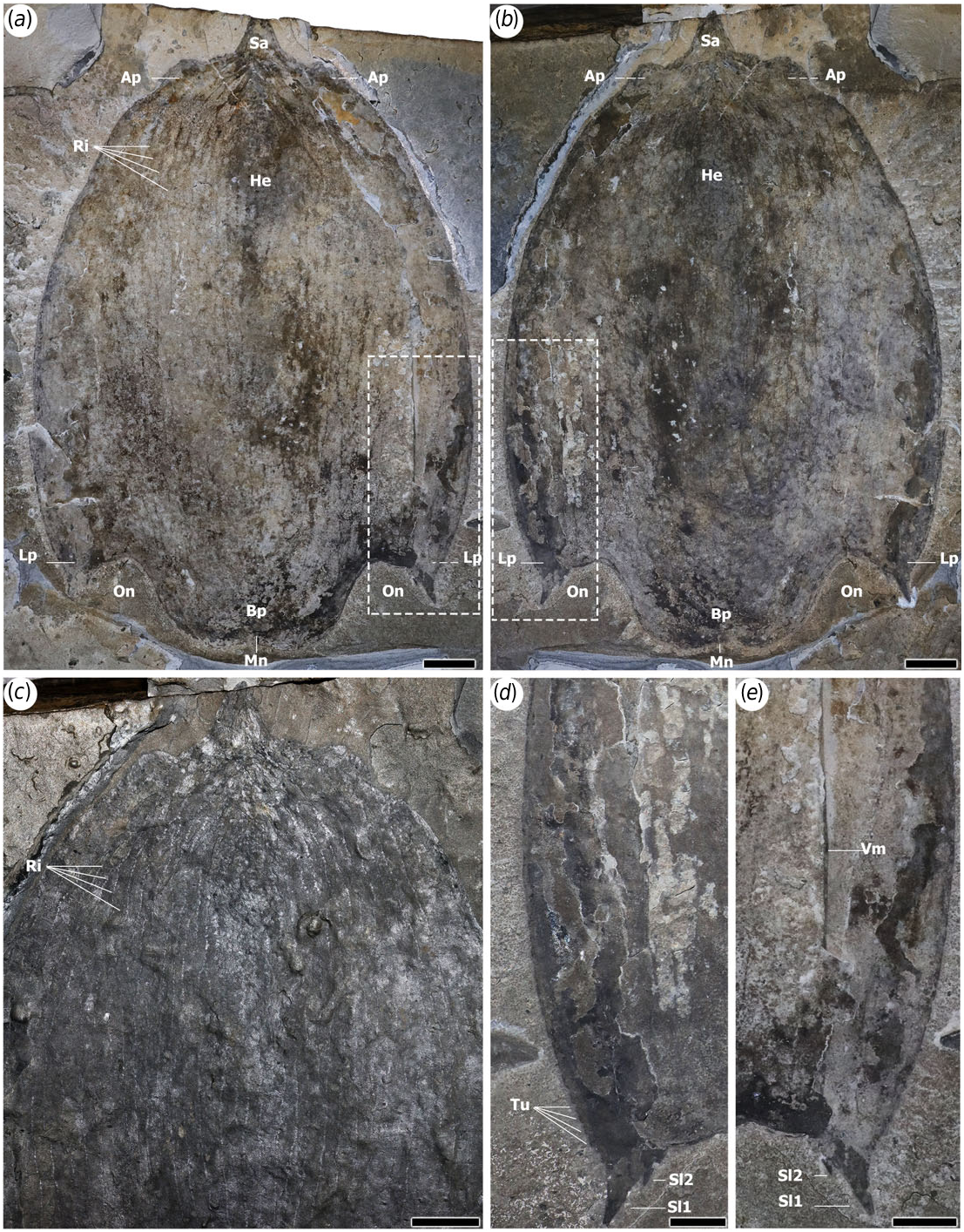
ROMIP 65168 preserves a large (L: 27 cm ; W: 18.5 cm) complete H-element in dorsal view and is the best example of this element for descriptive purposes ( figure 1 View Figure 1 ) . The fact that it appears perfectly bilaterally symmetrical suggests that the carapace was buried and compacted with the dorsoventral axis perpendicular to bedding. The H-element is ovoid with the widest point about 60% from the front and width slightly decreasing along the posterolateral processes. Crescentic to linear compression artefacts, starting from the anterior along the midline and extending towards the lateral areas, suggests that the carapace was convex dorsally, curving down gently from the sagittal plane towards the margins. The front of the carapace yields a sagittal spine with a blunt tip (Sa in figure 1 a–c View Figure 1 ) . This spine is about 7% the total length of the carapace and is roughly the shape of an equilateral triangle. Apair of semicircular to somewhat angular bulges, or anterolateral processes, flank the sagittal spine (Ap in figure 1 a–c View Figure 1 ) . The base of each process is comparable in width to the base of the sagittal spine, but they protrude only a few millimetres forward .
The posterior margin of the H-element projects into a pair of posterolateral processes and a trapezoidal axial projection, separated by ocular notches (On in figures 1 a,b View Figure 1 and 3 a–c View Figure 3 ). The axial projection is subtly bilobate with smooth rounded posterolateral margins separated by a shallow medial notch (Mn in figures 1 a,b View Figure 1 and 3 a–c View Figure 3 ). It represents ca 10–15% the length of the carapace and 60% of its maximal width anteriorly, becoming narrower towards the rear. This implies that the two ocular notches located on either side, are angled ca 20° anterolaterally from the midline. The posterolateral processes in Titanokorys are very short, extending only half as far posteriorly as the axial projection (Lp in figures 1 a,b View Figure 1 and 3 a–c View Figure 3 ). Each process has a smooth ventral margin and terminates in a large spine (Sl 1 in figure 1 d,e View Figure 1 ). Asecond shorter spine, about 25% the dimensions of the large spine, is located along the laterodistal margin of the ocular notch, near the base of the large spine (Sl 2 in figure 1 d,e View Figure 1 ). Both spines are slightly curved such that they point posterolaterally.
The entire H-element is ornamented by a series of longitudinal ridges running more-or-less parallel to each other (Ri in figures 1 a,c View Figure 1 , 2 b View Figure 2 and 4 a–c View Figure 4 ). The ridges occasionally converge, resulting in a reticulated pattern, similar to Cambroraster [ 2] but with particularly elongate and angular cells. The number of ridges probably ranges between 30 and 40 in total across the entire width of the H-element as seen from the top. Each ridge consists of a reflective band dotted with chains of repeated structures, each about 1 mm wide and 2 mm long ( figures 2 d View Figure 2 and 4 a–c View Figure 4 ). Tubercles are present along the margins of the carapace, in particular anteriorly and along the outside margin of the lateral processes (Tu in figures 1 d View Figure 1 , 2 d View Figure 2 , 3 a View Figure 3 and 4 a–c View Figure 4 ). By their comparable sizes and arrangements, these seem to represent the lateral expression of the dorsal repetitive spots suggesting that the carapace is covered by rows of small elongated tubercles. These tubercles increase in size towards the terminal spine of the posterolateral processes ( figures 1 d View Figure 1 and 3 a View Figure 3 ).
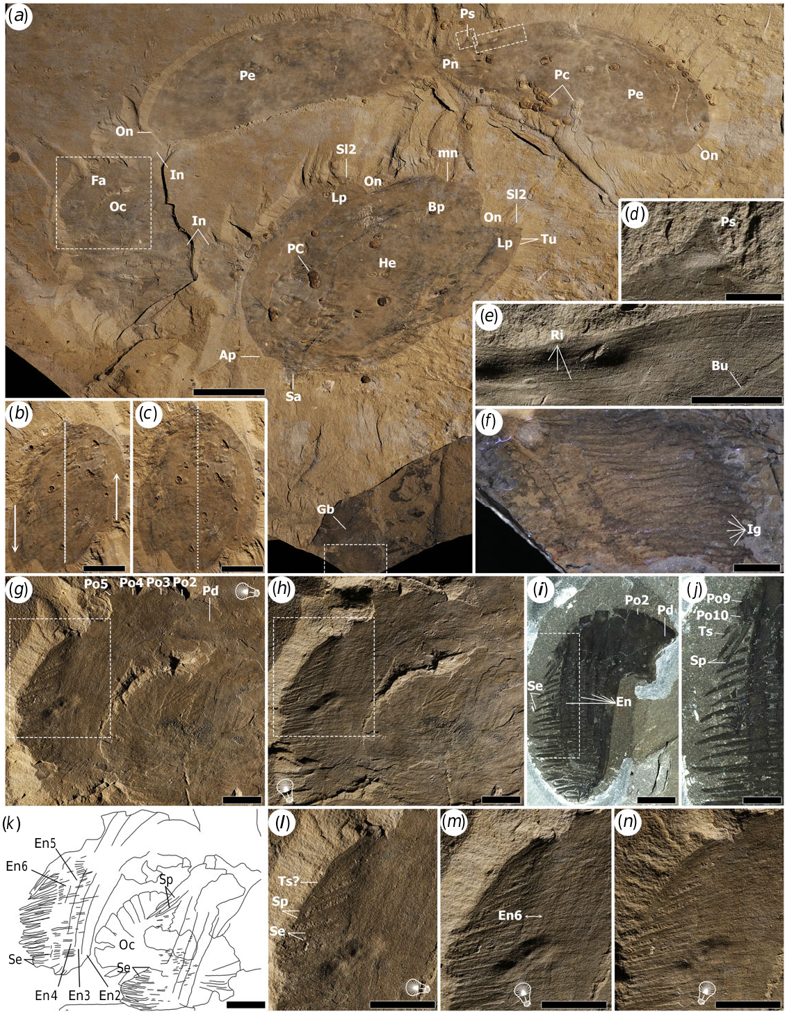
The pair of ventrolateral (P-) elements are best preserved in the holotype ROMIP 65745 ( figure 3 a View Figure 3 ). The P-elements share the same overall dimensions ( ca L = 20 cm and H = 7 cm) are almost perfect mirror images of each other and show only a few marginal compression artefacts suggesting they were buried more-or-less parallel to bedding. This is unlike the associated H-element, which by comparison with the paratype ROMIP 65168 ( figure 1 View Figure 1 ) is not bilaterally symmetrical, suggesting oblique burial. Retrodeformed longitudinally, the H-element, probably would have reached ca 21 cm in length and ca 15 cm in width (assuming a similar L/W ratio as ROMIP 65168), which is subequal to the length of the P-elements ( figure 3 c View Figure 3 ). Each P-element is drop-shaped with a more linear dorsal and truncated posterior margins. The posterodorsal corners of the P-elements are slightly indented, probably representing ocular notches (On in figure 3 a View Figure 3 ). The paired P-elements are connected to each other by a very short anterior neck section (Pn in figure 3 a View Figure 3 ) which bears a single broad-based spine on its curved ventromedial surface (Ps in figure 3 a, d View Figure 3 ). This spine is about a quarter the height of the neck above it. The carapace surface, as in the H-element, bears longitudinal rows of tubercles. In the holotype ( figure 3 e View Figure 3 ), the rows are more visible along the ventromedial surface and appear thinner and with smaller tubercles compared with the H-element in ROMIP 65168 ( figure 1 View Figure 1 ). Based on previous studies [1,2,4], the P-elements would wrap tightly laterally and ventrally along the body, with their anterior attachment point below the base of the H-element sagittal spine ( figure 5 View Figure 5 ).
The holotype also includes a pair of frontal appendages and an oral cone ( figure 3 b View Figure 3 ) as well as a number of indeterminate elements. Smooth plates of the oral cone are faintly visible below or on top of other fossil material (Oc in figure 3 g,h,k View Figure 3 ) and no fine details can be discerned. One appendage, in lateral view, can be distinguished more clearly using different low-angle light orientations ( figure 3 g, h, k–n View Figure 3 ). It preserves the peduncle (Pd) and all the podomeres (Po), with five long endites (numbered according to their respective podomeres, En2 to En6) bearing distally pointing, gradually tapering secondary (or auxiliary) spines (Se). About 35 secondary spines are visible beyond endite 6, but this number includes overlapping spines from more proximal endites as well, so the true number of spines per endite may be closer to 20–25. The secondary spines are also well preserved in ROMIP 65741 ( figure 2 c View Figure 2 ). Each of these spines on that specimen or the holotype is nearly four times longer than the width of endite 6, a third longer compared with equivalent spines in C. falcatus ( figures 2 d View Figure 2 and 3 i,j View Figure 3 ). It is unclear if the distal ends of the secondary spines were hooked like in C. falcatus, although they appear to be curved ( figures 2 c View Figure 2 and 3 l–n View Figure 3 ). The holotype shows evidence of unpaired spines and spiniform endites on more distal podomeres (probably 8, 9 and 10) (Sp in figure 3 g,h,k–n View Figure 3 ). In addition to sclerotized elements, the holotype also preserves bands of lamellae, showing individual gill elements (Gb figure 3 a View Figure 3 ; Ig figure 3 f View Figure 3 ). These are similar to those of other hurdiids (e.g. [1,5]) and were probably associated with the trunk and lateral flaps.
The reconstruction ( figure 5 View Figure 5 ) is based on all of the fossil material available. Details of the trunk, eyes and flaps are not preserved but have been hypothetically reconstructed based on C. falcatus [ 2].


No known copyright restrictions apply. See Agosti, D., Egloff, W., 2009. Taxonomic information exchange and copyright: the Plazi approach. BMC Research Notes 2009, 2:53 for further explanation.
|
Kingdom |
|
|
Phylum |
|
|
Class |
|
|
Order |
|
|
Family |
|
|
Genus |