
Solenofilomorpha pellucida, Kånneby, Tobias & Jondelius, Ulf, 2013
|
publication ID |
https://doi.org/ 10.11646/zootaxa.3736.5.3 |
|
publication LSID |
lsid:zoobank.org:pub:0D412548-2436-434E-93F7-7D2B4EAB53CB |
|
DOI |
https://doi.org/10.5281/zenodo.5658324 |
|
persistent identifier |
https://treatment.plazi.org/id/EB2DA62F-5647-FFBB-37AF-26A8A8E8E6AC |
|
treatment provided by |
Plazi |
|
scientific name |
Solenofilomorpha pellucida |
| status |
sp. nov. |
Solenofilomorpha pellucida View in CoL n. sp. ( Figs. 13–16 View FIGURE 13 View FIGURES 14 – 15. 14 View FIGURE 16 )
Type Material: Holotype—SMNH Type-8472 (longitudinal sections); Paratype 1—SMNH Type-8473 (longitudinal sections).
Type Locality: Marina, El Quisco , Chile (33° 23' 35.664" S, 71° 41' 53.088" W), at 10 m water depth in fine sand.
Other Material Examined: Whole mounts of living specimens.
Etymology: The species epithet pellucida refers to the absence of pigmentation which renders the living specimens translucent.
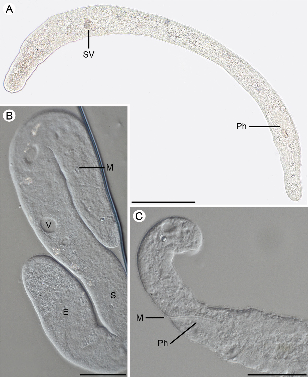
Description: Living specimens up to 1.4 mm in length, width varies between 50–100 µm (L/W ratio 1:18). Fixed specimens approximately 810 µm in length. Body shape variable but usually cylindrical long and slender, slightly tapering towards the rounded anterior and posterior ends ( Figs. 13 View FIGURE 13 A; 15). Anterior end somewhat narrower than posterior end. Body transparent in transmitted light.
Epidermis approximately 5 µm in height and completely ciliated, cilia 3–4 µm in length. Body wall musculature weakly developed. Vacuoles scattered throughout the body but more common in the posterior end ( Fig. 15 View FIGURES 14 – 15. 14 ). They are small, usually less than 15 um in diameter, however a low number of larger vacuoles may also occur ( Figs. 13 View FIGURE 13 B; 15).
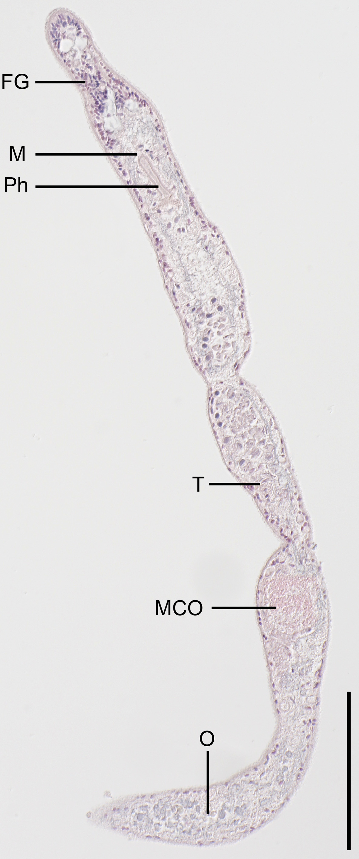
Statocyst 12 µm in diameter and located at U06. Statolith 7 µm. Frontal organ empties through a single terminal pore. The cyanophilic cell bodies of the frontal glands extend from anterior part of the body to the mouth and anterior parts of the pharynx at U15 ( Fig. 14 View FIGURES 14 – 15. 14 ).
Mouth opens on ventral side at U15. Mouth variable in shape depending on degree of contraction of the animal but usually slit-like or wedge-shaped. Mouth slit sometimes as long as 30 µm. Pharynx ciliated, 90 µm in length, widening towards its posterior end ( Figs. 13 View FIGURE 13 C; 15). It extends from U15 to U22. Epithelium of pharynx with few gland cells and covered by cilia approximately 3 µm in length ( Fig. 15 View FIGURES 14 – 15. 14 ). Mouth opening and pharynx well visible in compressed living specimens ( Fig. 13 View FIGURE 13 B). Digestive central syncytium diffuse.
Ovaries unpaired, situated posterior to male copulatory organ, extending from U82 to posterior-most part of body ( Figs. 13 View FIGURE 13 B; 14; 15). Seminal bursa absent. Testis unpaired, dorsal, and separate from ovary ( Figs. 14 View FIGURES 14 – 15. 14 ; 15). Testis extends from U56 to the anterior part of seminal vesicle at U72. Male gonopore located in posterior third of body at approximately U73. Antrum absent. Spherical seminal vesicle, 30–35 µm in diameter located at U73 ( Fig. 15 View FIGURES 14 – 15. 14 ).
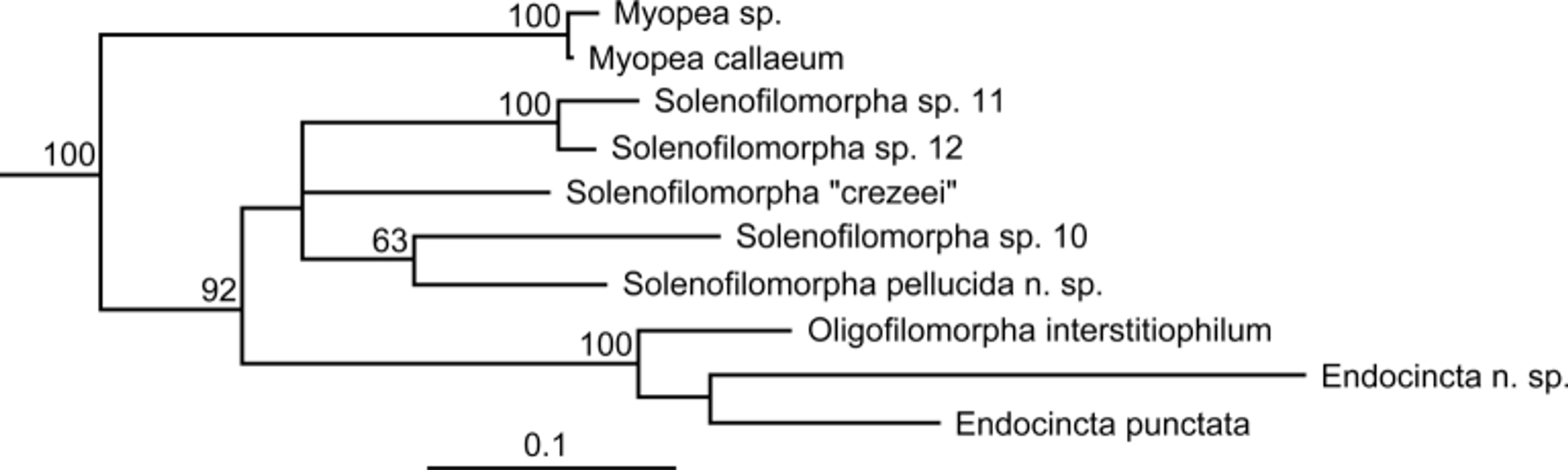
Diagnosis and taxonomic remarks: Solenofilomorpha pellucida n. sp. can be separated from all other species within the genus Solenofilomorpha based on the combination of the following characters: (i) very long and slender body (L/W ratio of 1:18), (ii) ovaries extend to the posterior-most end of the body and (ii) absence of an antrum. An overview of characters of species of Solenofilomorpha is given in table 2. The presence of a ciliated pharynx, 15 the single testis, and the position of the ovaries posterior to the male copulatory organ supports the position within Solenofilomorphidae . The lack of rhammites and a penis further supports the position within Solenofilomorpha according to the diagnoses in Dörjes (1968) and Crezee (1975). The maximum likelihood analysis of the concatenated 18S, 28S and COI sequences provides high support for a clade containing Solenofilomorpha , Endocincta and Oligofilomorpha corresponding to the family Solenofilomorphidae . The individual monophyly of these genera based on molecular data has not yet been addressed. Hence, classification of the new species within Solenofilomorpha is reasonable since this is the type genus for Solenofilomorphidae ( Fig. 16 View FIGURE 16 ).
S. pellucida n. S. funilis S. guaymensis Crezee, S. justinei Nilsson et al. S. longissima sp. Crezee, 1975 1975 2011 Dörjes, 1968
Length/width-ratio 1:18 1:14 1:14 1:5 1:19 Financial support from the European Community - Research Infrastructure Action under the FP7 "Capacities" Specific Programme ASSEMBLE grant agreement no. 227799 is gratefully acknowledged. Grants from The Swedish Research Council (2012-3913) and from the Swedish Taxonomy Initiative (project no Dha 149/10 1.4) are gratefully acknowledged. We wish to thank the director and staff of ECIM. Thanks are also extended to Keyvan Mirbakhsh for producing the sequence data.
No known copyright restrictions apply. See Agosti, D., Egloff, W., 2009. Taxonomic information exchange and copyright: the Plazi approach. BMC Research Notes 2009, 2:53 for further explanation.