
Pseudaphanostoma hyalinorhabdoida, Kånneby, Tobias & Jondelius, Ulf, 2013
|
publication ID |
https://doi.org/ 10.11646/zootaxa.3736.5.3 |
|
publication LSID |
lsid:zoobank.org:pub:0D412548-2436-434E-93F7-7D2B4EAB53CB |
|
DOI |
https://doi.org/10.5281/zenodo.5658316 |
|
persistent identifier |
https://treatment.plazi.org/id/EB2DA62F-564B-FFBC-37AF-2461AB0CE52B |
|
treatment provided by |
Plazi |
|
scientific name |
Pseudaphanostoma hyalinorhabdoida |
| status |
sp. nov. |
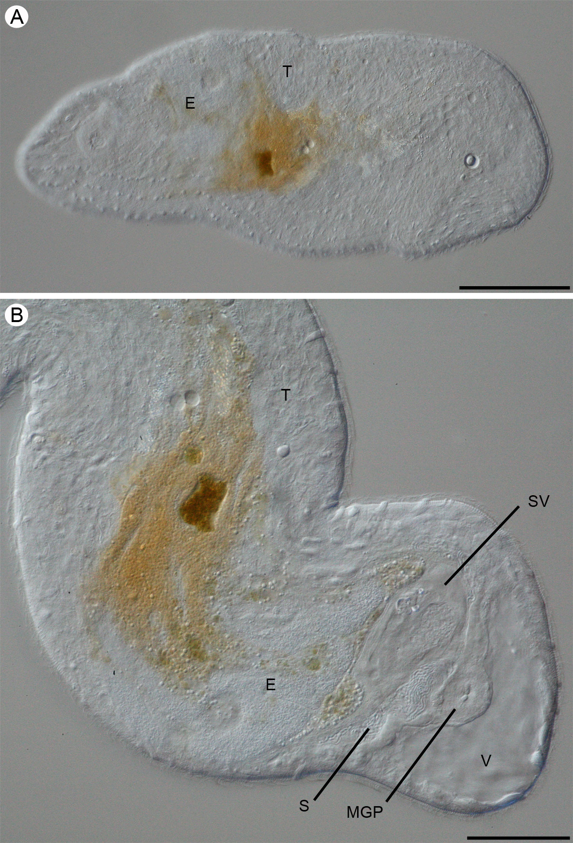
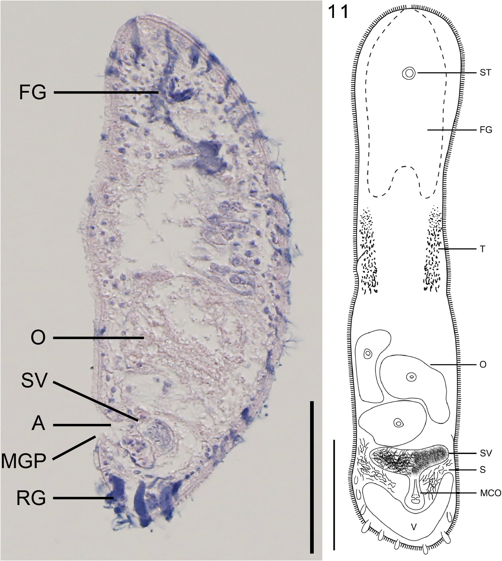
Pseudaphanostoma hyalinorhabdoida View in CoL n. sp. ( Figs. 9–12 View FIGURE 9 View FIGURES 10 – 11. 10 View FIGURE 12 )
Type Material: Holotype—SMNH Type-8468 (sagittal sections); Paratype 1—SMNH Type 8469 (oblique sections); Paratype 2—SMNH Type 8470 (longitudinal sections); Paratype 3—SMNH Type 8471 (transversal sections).
Type Locality: Marina, El Quisco , Chile (33° 23' 35.664" S, 71° 41' 53.088" W), at 10 m water depth in fine sand. Other Material Examined: Whole mounts of living specimens.
Etymology: The species epithet hyalinorhabdoida refers to the unpigmented rhabdoid glands in this species.
Description: Living specimens 400–750 µm in total body length. Body width varies between 100–200 µm. Fixed specimens approximately 270 µm in total body length. Body shape oval, tapering towards the rounded anterior and posterior ends ( Fig. 9 View FIGURE 9 A) Posterior end somewhat more pointed than the usually blunt anterior end. Cyanophilic rhabdoid glands are more numerous dorsally and in the anterior and posterior parts of the body ( Fig. 10 View FIGURES 10 – 11. 10 ). They are well developed, unpigmented, increasing in size towards the posterior end and distributed in longitudinal rows. In cross-section they penetrate the epidermis ( Fig. 10 View FIGURES 10 – 11. 10 ).
Epidermis completely ciliated, approximately 3–4 µm in sagittal sections. One large vacuole, 90 µm in width, present in the posterior end ( Figs. 9 View FIGURE 9 B; 11). A few smaller vacuoles may also be present in the anterior end and along the lateral anterior body sides.
Statocyst 15 µm in diameter and located approximately 75 µm from the anterior end at U10-15, depending on degree of contraction of the animal. Statolith 9 µm in diameter. Frontal organ well developed, consisting of cyanophilic glands and extending from the anterior end of the body to U36.
Mouth opening could not be observed. Central syncytium red and yellow, extending from U32–84. It is most compact between U46–76.
Ovaries unpaired, extending from U62 to the anterior end of the male copulatory organ at U77. Seminal bursa absent. Testes paired extending from U42–U59. Antrum unciliated, opening through male gonopore located at U83 just in front of the large posterior vacuole ( Fig. 10 View FIGURES 10 – 11. 10 ). Spherical seminal vesicle, 60–70 µm in width and 30 µm in length, extending from U78–82 ( Figs. 9 View FIGURE 9 B; 11).
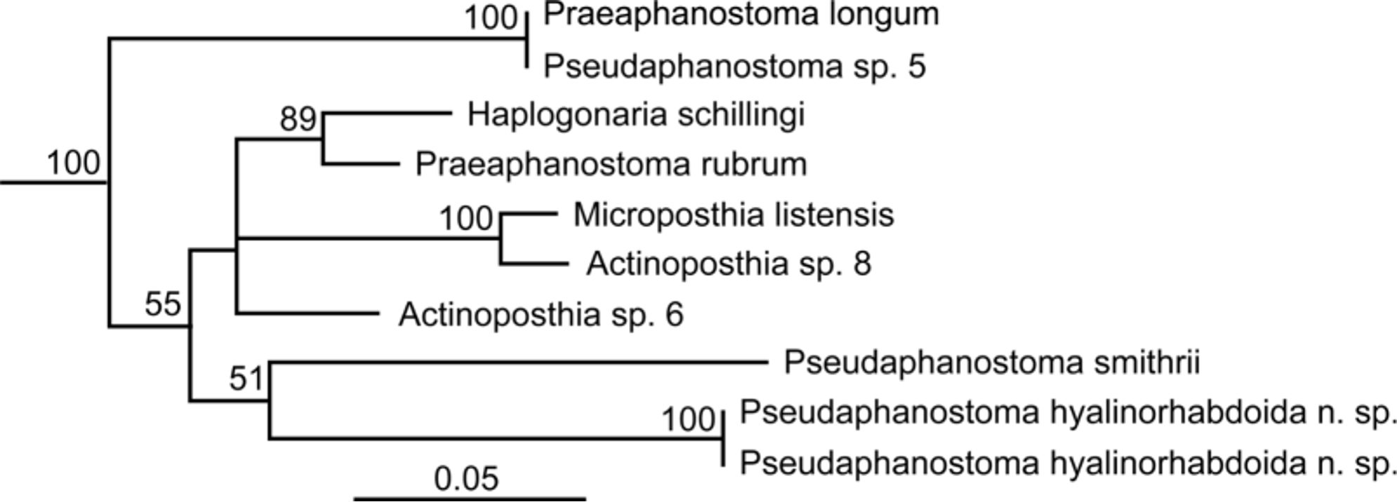
Diagnosis and taxonomic remarks: Pseudaphanostoma hyalinorhabdoida n. sp. can be separated from all other species within the genus based on the well-developed unpigmented rhabdoid glands, which are more numerous dorsally than ventrally. The following morphological characters identify this new species as a Pseudaphanostoma : (i) pigmentation absent; (ii) male gonopore located subterminally in the posterior end; (iii) frontal glands and rhabdoid glands present; (iv) female accessory organs absent and (v) male copulatory organ distinct (Dörjes 1968). The maximum likelihood analysis of the concatenated 18S, 28S and COI sequences indicates a sister group relation of P. hyalinorhabdoida n. sp. with Pseudaphanostoma smithrii Hooge & Tyler, 2003 ( Fig. 12 View FIGURE 12 ). However, P. smithrii is the only other species of Pseudaphanostoma for which sequence data is available.
P. hyalinorhabdoida n. sp. is morphologically similar to Pseudaphanostoma psammophilum Dörjes, 1968 . Both species possess a distinct male copulatory organ with a subterminal gonopore located anterior to a single posterior vacuole.
No known copyright restrictions apply. See Agosti, D., Egloff, W., 2009. Taxonomic information exchange and copyright: the Plazi approach. BMC Research Notes 2009, 2:53 for further explanation.