
Didogobius janetarum, Schliewen & Wirtz & Kovačić, 2018
|
publication ID |
https://doi.org/ 10.11646/zootaxa.4438.2.12 |
|
publication LSID |
lsid:zoobank.org:pub:673356D1-F47F-4CB2-AEFA-3ACF3DB1A3C8 |
|
DOI |
https://doi.org/10.5281/zenodo.5658365 |
|
persistent identifier |
https://treatment.plazi.org/id/345D7297-D9F9-48FF-BBA7-D17F01A1743A |
|
taxon LSID |
lsid:zoobank.org:act:345D7297-D9F9-48FF-BBA7-D17F01A1743A |
|
treatment provided by |
Plazi |
|
scientific name |
Didogobius janetarum |
| status |
sp. nov. |
Didogobius janetarum , spec. nov.
( Figs. 1a, b, c View FIGURE 1 ; 2, 3, 4)
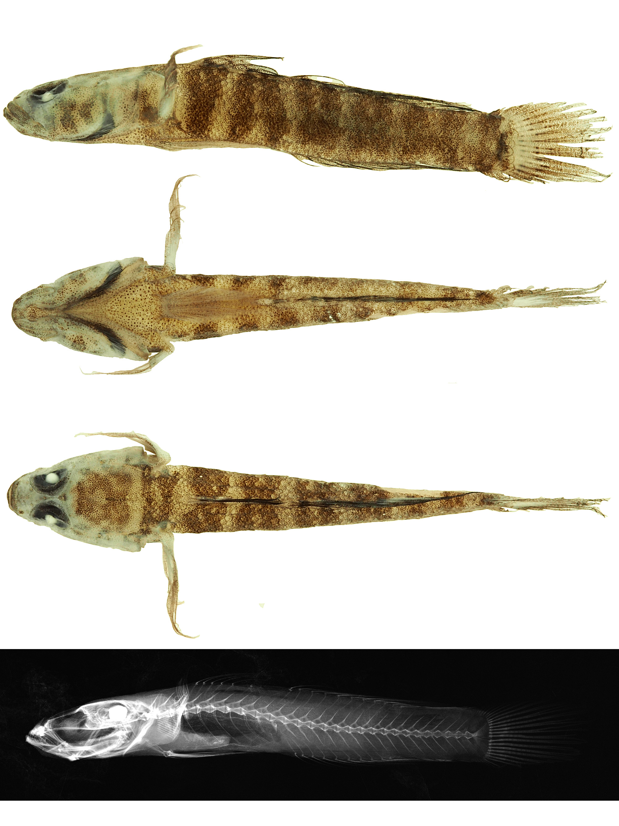
Holotype. ZSM 45303, male, 27.3 + 6.2 mm, Republic of Cabo Verde, Santiago Island, King Bay at Tarrafal (15.275° - 23.758°), from a horizontal rock crevice in approx. 12 m depth, May 2016, collected with a handnet by P. Wirtz.
Paratypes. ZSM 45302, juvenile, 18.2 + 4.9 mm, Republic of Cabo Verde, Santiago Island, cliff approx. 2.5 km west of Cidade Velha from a small cave in 14 m depth rock crevice (14.916° - 23.626°), May 2016, collected with a handnet P. Wirtz ; ZSM 40136, female, 24.7 + 5.7 mm, Republic of Cabo Verde, Santiago Island, SSW of Tarrafal (14.916081° - 23.626626°), dive site “ Danger ” (15.255° - 23.754°) in approx . 20 m depth, March 19th 2010, collected with a handnet by P. Wirtz; PMR VP4134 (ex ZSM-PIS-GO1804); female, 22.8 + 5.5 mm, same collection data as holotype ZSM 45303.
Additional material. ZSM-PIS-GO 1803, juvenile, 12.2 + 2.3 mm, same collection data as paratype ZSM 45302.
Comparative material examined. Didogobius kochi Van Tassell, 1988 : male, 29.1 + 7 mm (Sao Vicente Island, Republic of Cabo Verde ); ZSM 43050, female, 36.6 + 8 mm and male, 25.6 + 6 mm , ZSM 35462 (Sal Island, Republic of Cabo Verde) .
Diagnosis. Didogobius janetarum spec. nov. differs from all currently described congeners by the combination of the following characters: (1) 27 vertebrae, (2) D 2 I + 10, (3) posterior quarter of predorsal region in front of D1 origin scaled, with several rows of cycloid scales, (4) body squamation cycloid anteriorly and ctenoid posteriorly, (5) scales in the lateral series 30–32, (6) anterior oculoscapular canal present, (7) preopercular head canal absent, (8) suborbital row 7 each a single papilla, (9) suborbital rows 2 and 4 close to orbit, (10) branchiostegal membranes uniformely black below preopercle, forming V-mark.
Description. General morphology. Body proportions and meristics of the holotype and four paratypes are given in Table I. For a general view see Figs 1a, 1b and 1c View FIGURE 1 . Body moderately elongate and laterally compressed, head depressed; interorbital space narrow (approx. 20.0–23.5% of eye diameter), dorsolaterally positioned eyes. Mouth oblique (~35° from horizontal), lower jaw slightly projecting, posterior angle of jaws below center of pupil. Snout shorter than eye and rounded in dorsal view. Anterior nostril tubular (longer than diameter), without process from rim, barely reaching but not overlapping the upper lip; posterior nostril with slightly erected rim, but not tubular. Upper lips anteriorly slightly thinner than laterally. Branchiostegal membrane attached along entire lateral margin; posterior margin of operculum approximately reaching frontal edge of pectoral fin base. Cranial roof covered by dorsal axial musculature. Pectoral girdle without dermal flaps on anterior edge.
Fins. D 1 VI; D 2 I/10 (last bifid); A I/9; P 16–17 (counted only on left side); V (left/right) I/5 + 5/I; C (branched/segmented rays) 14–15/17. Fin lengths and proportions are given in Table I. First D1 slightly longer (males) or almost as long (females) as second spine, third to sixth spines becoming progressively shorter; interdorsal space distinct and without fin membrane connection between D1 and D2; longest D2 rays reaching base of uppermost caudal fin rays. A originates slightly posterior of vertical through D2 origin; C rounded, shorter than head length; uppermost rays of P not free of membrane, P reaching D2; V complete and rounded with ray 4 as long as ray 5, and a well-developed anterior pelvic membrane (frenum), its height in midline approx. 1/3 of V spine length and at its lateral margins approx. 2/3 of V spine length.
Scales. Body covered anteriorly with cycloid and posteriorly with ctenoid scales; ctenoid scales commence midlaterally from the vertical below base of the fourth spine of D1, dorsally and ventrally further caudally from approximately the vertical below the base of 2nd ray of D2 and third ray of A. Head area naked, predorsal area with up to six rows (holotype) or fewer (paratypes) cycloid scales in front of D1 origin, covering at maximum the posterior quarter of the predorsal region and extending more anteriorly along the middorsal line than laterally, i.e. with more middorsal predorsal scale rows than dorsolateral ones (see Fig. 1c View FIGURE 1 ), prepectoral area naked, breast with few cycloid scales posteriorly; belly completely scaled with cycloid scales. One row of slightly ctenoid scales on caudal fin base. LL 30–32, TR 12, CP 12–14.
Teeth. Teeth in lower jaw in two rows. Outer row with five large-sized teeth frontally on each side, caniniform, pointing slightly backwards. Inner frontal teeth small and conical, numerous, more or less in one row in frontal position. Inner posterior lateral teeth few and large, caniniform. Teeth in upper jaw in two rows. Outer row with 12 (left side) or 11 (right side) teeth, the frontal three ones on each side large and caniniform, the posteriorlateral ones medium to small, decreasing in size posteriorly; inner row with one (right side) or three (left side) large frontal teeth, caniniform.
Osteology. Vertebral column and pterygiophore insertion pattern (pty) ( Fig. 1d View FIGURE 1 ). 9 precaudal and 18 caudal vertebrae (including urostyle); total count: 27. Pty 3-22110; two pterygiophores anterior to the first haemal spine. One epural. Number of C rays inserting in the hypural 5: 2, 3 + 4 (fused): 6, hypural 1 + 2 (fused): 5 and parhypural: 1, total number of C rays inserting in hypurals, and parhypural: 14; fused hypural 1 + 2 and 3 + 4 separated by a large gap, which is not inserted by a branched caudal ray.
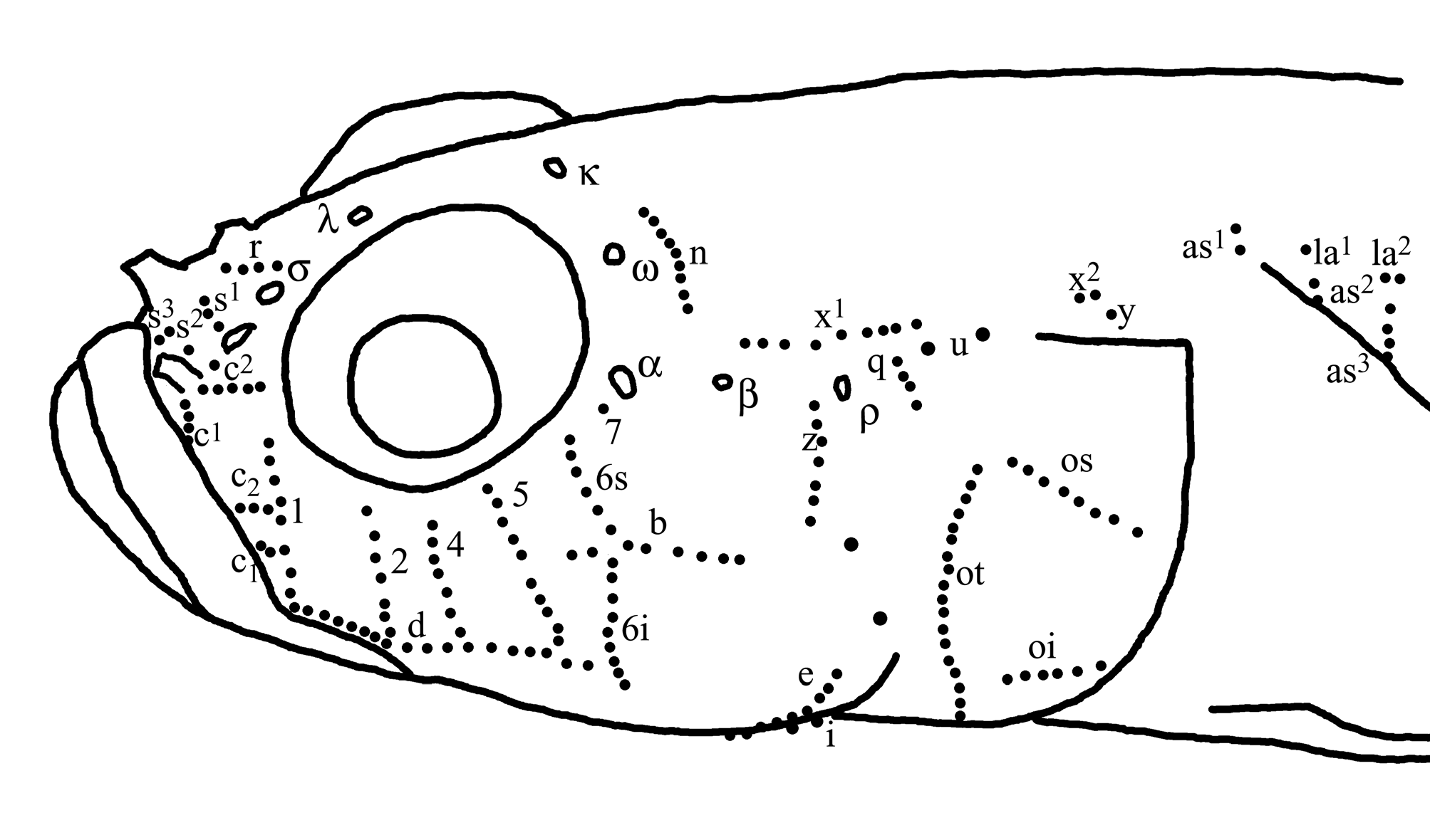
Lateral line system ( Fig. 2 View FIGURE 2 ). Head with anterior oculoscapular canal with pores σ, λ, Κ, ω, α, β, ρ, paratype ZSM 45302 with open furrow between left σ and λ. Posterior oculoscapular canal and preopercular canal absent. Rows and number of sensory papillae as follows, counted on left side of holotype ZSM 45303 (first value) and of paratype PMR VP4134 (second value): (I) preorbital: snout with four rows in median preorbital series. Row r (4, 4) median to pore σ. Upper row s1 (3, 3) transversal near posterior nostril, lower s2 (1, 1) near anterior nostril, and s3 longitudinal above upper lip (2, 3). Lateral series c in four parts: superior c2 between posterior and anterior nostrils (1 + 5, 2 + 4) as two rows; middle transversal c1 (4, 3) below anterior nostril; inferior upper c2 (3, 2) and lower c1 (2, 2) as two rows between lips and row 1. (II) suborbital: six transverse and two longitudinal rows of sensory papillae on cheek. Rows 1-5 before longitudinal row b; row 6 divided by b in superior (6s) and inferior sections (6i); row 7 near pore α (1: 8, 6, 2: 7, 5, 4: 7, 6, 5: 10, 6, 6s: 6, 3, 6i: 9, 10, 7: 1, 1). Row 1 slightly in front of eye orbit. Rows 2 and 4 close to orbit. Row 6i ending ventrally below level of row d. Longitudinal row b (8, 6) starting anteriorly behind row 5 (holotype ZSM 45303) or at row 6 (paratype PMR VP4134), ending posteriorly far behind rear border of eye. Longitudinal row d (18, 15) continuous. (III) preoperculo- mandibular: external row e and internal row i divided into anterior (e: 21, 18, i: 7, 8), and posterior sections (e: 20, 16, i: 7, 7); row f longitudinal (4, 6). (IV) oculoscapular: anterior longitudinal row x1 (9, 7) from behind pore β to behind pore ρ; posterior longitudinal row x2 (2, 2) above transversal row y (1, 1); transversal row z (7, 7) below pore ρ; transversal row q (4, 5) behind pore ρ; row u as two large longitudinally arranged papillae behind row q on the place of absent posterior oculoscapular canal; transversal axillary rows as1 (2, /), as2 (3, 4), as3 (6, /) present and longitudinal rows la1 (1, 1) and la2 (2, 1) above as2 and as3; as1 and as3 not visible in paratype PMR VP4134. (V) opercular: transverse row ot (21, 16); superior longitudinal row os (8, 7); and inferior longitudinal row oi (6, 3); two large papillae present on the place of absent preopercular canal. (VI) anterior dorsal: transversal row n behind pore ω (9, 8); transversal rows o (/, 2) divided from each other; longitudinal row g (/, 4) ends posteriorly to front row o, longitudinal row m (/, 2) behind and below of row g; longitudinal row h continuous (/, 6); rows o, g, m, h not visible in holotype ZSM 45303.
Coloration. In life (based on photograph of holotype ( Fig. 3 View FIGURE 3 ; right side) shortly after capture. Seven dark orange-brown vertical bands extending on flanks from dorsal midline: the most posterior stripe on caudal peduncle and two anterior stripes below D1 and predorsal area strait vertical, the middle four stripes broader and connected ventrally forming a zig-zag pattern, i.e. the seven stripes form a pattern similar to the sequence of letters IAAAAII from caudal peduncle to head: the anteriormost originates from the predorsal area anterior to the origin of D1 and splits ventrally with the posterior branch extending behind pectoral fin base, and the anterior extending obliquely to meet the posterior border of the opercle; the second from the below anterior D1 base from approx. third spine to approx. fourth spine; the third extending from posterior base of the D1, from approx. fifth to sixth spine, the fourth and fifth from anterior and central base of D2, from approx. second to third and sixth to seventh ray, respectively, the sixth from the posterior base of D2 (from approx. tenth ray) and the anterior caudal peduncle, and the seventh extending at middle caudal peduncle. Posterior end of caudal peduncle and caudal fin base brown. White areas of the IAAAAII pattern are two poorly visible whitish vertical stripes on caudal peduncle and two clear stripes anteriorly below D1 and predorsal area; in between three white dorsal saddles are present below D2, seven white triangles along the lower flanks (the three posteriormost on caudal peduncle poorly visible), and several small whitish marks inside orange-brown vertical bands. Nape anterior to first orange-brown band with a narrow white band followed anteriorly by an orange-brown area with embedded whitish marks and with two pairs of orangebrown lateral “legs” extending from that area laterally, followed behind eye by another narrow white band. One orange-brown oblique stripe extends ventrally from orbit slightly backwards ending at about center of the cheek, another one from orbit to mouth; below those two marks are present, one on posterior angle of mouth and one on lower cheek behind the first one. Preopercle and operculum with three irregular oblique orange bands (forming a “/ I/”-pattern) connected dorsally to form a distinctive orange brown pattern. Lower part of anterior pectoral fin base with a prominent white area followed anteriorly by an orange brown band or blotch; anterior parts of branchiostegal membranes, i.e. ventrally below preopercle blackish-grey, forming an intensively pigmented black spot at each of the upper lateral sides. Ventral body coloration varies from white to grey. Pectoral and ventral fins with white rays and clear membranes, anal fin rays white with a membranes having a dusky hue; D1 whitish with a white posterior margin, and four to three brown oblique bands, the anteriormost being darker and broader than the posterior ones; first D1 spin with contrasting dark-brown and white bands partially corresponding with the D1 bands. D2 white with seven to six oblique orange brown series of brown dots on fin rays, the anteriormost being darker and broader than the posterior ones; C base with a narrow brown bar embedded in a white field, the remaining C area whitish to clear with eight or fewer pale rows of brown dots on C rays. Variation (based on color photographs of three additional specimens (not shown)): smaller individual may have less ill-defined and more contrasting lateral bands and orange head coloration pattern may be slightly different in size and distribution of colored elements.
Preserved color. Body bars and blotches on flanks and operculum the same as in life, but orange-brown live colors have changed after preservation to dark brown, and previously white areas have become whitish-beige, the pattern being less distinctive than in living specimens; head, nape, cheek and snout bands and blotches also less distinct; branchiostegal membranes below preopercle uniformely black (without a clear dark spot on the upper part) forming V mark from ventral view, otherwise ventral whitish-grey; narrow bar on caudal fin base black but ill defined, white area on C fin base prominent and larger than in living specimens; D1 and D2 whitish-translucent with blackish-brown bands; ventrals and pectorals dusky; anal fin with blackish-brown rays and whitishtranslucent membranes. Variation (based on all five specimens): smaller individual may have less ill-defined and more contrasting lateral bands and head coloration pattern may be slightly different in size and distribution of elements – analogous to variation in life coloration (see above).
Etymology. The species name is dedicated to Mrs. Janet Camp and Ms. Janet Eyre, who generously supported our goby research. A noun in feminine genitive (plural).
Distribution and habitat. Didogobius janetarum sp. nov. is only known from two locations of Santiago Island, Republic of Cabo Verde. It was collected exclusively from small caves and rock crevices between 12 and 20 m depth. One specimen was observed lurking out of a small cave before it was collected, whereas the other four known specimens were retrieved after spraying a fish anaesthetic (clove oil) into rock crevices on vertical walls covered with the coral Tubastrea caboverdiana .

| ZSM |
Bavarian State Collection of Zoology |
No known copyright restrictions apply. See Agosti, D., Egloff, W., 2009. Taxonomic information exchange and copyright: the Plazi approach. BMC Research Notes 2009, 2:53 for further explanation.
|
Kingdom |
|
|
Phylum |
|
|
Class |
|
|
Order |
|
|
Family |
|
|
Genus |