
Pseudophacopteron caffrariense, Capener, 1973
|
publication ID |
https://doi.org/ 10.11646/zootaxa.2086.1.1 |
|
persistent identifier |
https://treatment.plazi.org/id/EC223817-FFA8-FFD5-FF22-FA8BFA84FCF3 |
|
treatment provided by |
Felipe |
|
scientific name |
Pseudophacopteron caffrariense |
| status |
|
Pseudophacopteron caffrariense View in CoL -group
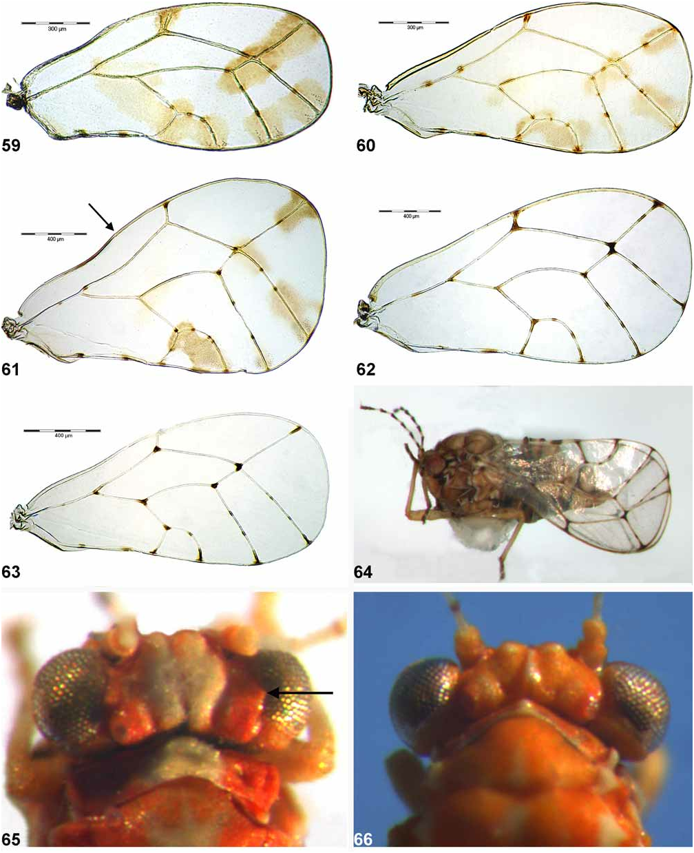
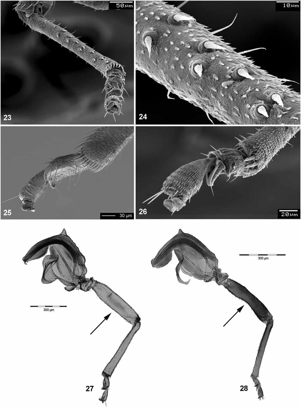
Head, in dorsal view, slightly wider than mesonotum. Vertex relatively flat (Figs. 14, 65) or with weakly raised midline ( Fig. 66 View FIGURES 59–66 ) but lacking pronounced median ridge; lateral ocelli lying on small tubercles above the plane of vertex or lying in it; two large tubercles, each on either side of midline, present on anterior vertex margin, divided by median epicranial suture (Figs. 13–14, 65–66) or vertex is regularly rounded down in front (Figs. 15–16); median epicranial suture usually distinct basally (near posterior vertex margin) and apically (above frons), reduced in the middle; vertex with a small lateral tubercle adjacent to eyes present (Figs. 16, 65) or indistinct (Figs. 14, 66). Occiput and preoccipital sclerite narrow; eyes not stalked, subglobular. Genae relatively large, swollen and slightly produced anterio-laterally; tubercle below torulus blunt (Figs. 15–16) or pointed with apex forming a right angle (Fig. 13). Frons narrowly pentagonal, slightly wider anteriorly (Figs. 13, 15). Clypeus subglobular. Antenna with a large, widely elliptic rhinarium bordered with a wreath of cuticular spines near apex of each of segments 4–9, associated with another smaller elongate cavity with supplementary sensorium situated more basally and lacking cuticular spines on at least some of segments 4–8 ( Figs. 19 View FIGURES 17–22 , 225 View FIGURES 216–229 ), or with multiple rhinaria on segments 3–8 lacking cuticular spines ( Figs. 21–22 View FIGURES 17–22 ). Fore wing relatively broad, pyriform, apex truncate or broadly rounded; veins with distinct dark spots or streaks ( Figs. 59–63 View FIGURES 59–66 ); radular spinules present, forming small triangular patches in both apical corners of cells m 1 and m 2 and proximal apical corners of r 2 and cu 1 ( Figs. 80–82 View FIGURES 77–82 ). Mesotibia with a comb of 4–12 densely arranged stout setae on outer margin subapically (reduced in P. tamessei ). Metacoxa with right-angled posterior margin and small, acute, thorn-like meracanthus; metafemur parallel-sided, not constricted medially ( Fig. 27 View FIGURES 23–28 ). Metatibia bearing an open crown of long, slender, unsclerotised apical spurs ( Fig. 25 View FIGURES 23–28 ), lacking similar spurs laterally. Metabasitarsus relatively long, cylindrical, bearing two black lateral sclerotised spurs apically ( Fig. 25 View FIGURES 23–28 ) or short, lacking distinct spurs (Fig. 169). Male subgenital plate with dorsal margin nearly straight (Figs. 160, 166) or more or less angular (Figs. 154, 157, 163). Paramere asymmetrical (Figs. 155, 158, 161, 164, 167). Distal segment of aedeagus with elongate, narrow and apically rounded apical dilation; sclerotized end tube of ductus ejaculatorius short, sinuate (Figs. 156, 159, 162, 165, 168). Female circumanal ring with two rows of pores, pores of outer row contiguous. Dorsal and ventral valvulae lacking distinct lateral teeth ( Figs. 217, 219, 221–222 View FIGURES 216–229 ).
Fifth instar larva (known only in P. cuniculus ) with long limbs, broad tarsal arolium with indistinct petiole, slender truncate lanceolate setae, and large and strongly sinuate circumanal pore ring, widely extending on both dorsal and ventral surfaces of caudal plate ( Fig. 262 View FIGURES 259–262 ).
Comments. The group of five Afrotropical species is defined by the presence of supplementary rhinaria on the antennae. The ultrastructure and position of rhinaria is similar in P. caffrariense , P. hollisi , P. serrifer and P. tamessei . P. cuniculus is unique in having multiple rhinaria that lack a wreath of cuticular spines. These multiple rhinaria are present also on the segment 3. As P. cuniculus is similar to P. caffrariense and P. hollisi in several characters (e.g. anterior tubercles on vertex, the form of the male and female terminalia, fore wing pattern), the multiple rhinaria of P. cuniculus are interpreted here as the homologous, derived state.


No known copyright restrictions apply. See Agosti, D., Egloff, W., 2009. Taxonomic information exchange and copyright: the Plazi approach. BMC Research Notes 2009, 2:53 for further explanation.
|
Kingdom |
|
|
Phylum |
|
|
Class |
|
|
Order |
|
|
Family |
|
|
Genus |