
Cobbionema brevispicula, Ahmed & Boström & Holovachov, 2020
|
publication ID |
https://doi.org/ 10.5852/ejt.2020.702 |
|
publication LSID |
lsid:zoobank.org:pub:B4DDC9C7-69F4-40D1-A424-27D04331D1F8 |
|
DOI |
https://doi.org/10.5281/zenodo.4328589 |
|
persistent identifier |
https://treatment.plazi.org/id/E6F3CD60-DCE1-4F96-BB28-410199A02705 |
|
taxon LSID |
lsid:zoobank.org:act:E6F3CD60-DCE1-4F96-BB28-410199A02705 |
|
treatment provided by |
Valdenar |
|
scientific name |
Cobbionema brevispicula |
| status |
sp. nov. |
Cobbionema brevispicula sp. nov.
urn:lsid:zoobank.org:act:E6F3CD60-DCE1-4F96-BB28-410199A02705
Figs 6–7 View Fig View Fig , Table 2
Diagnosis
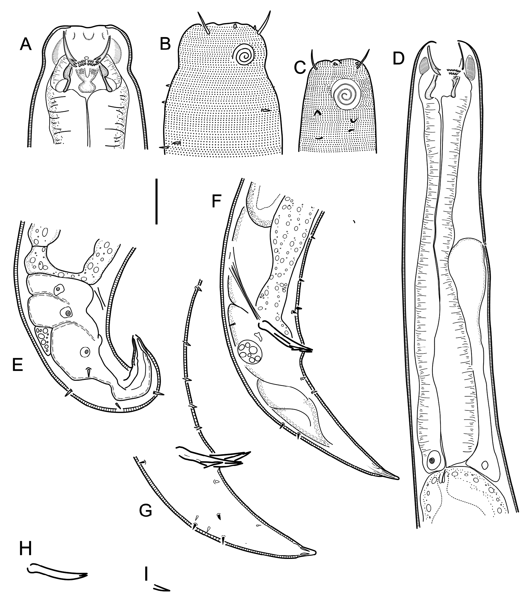
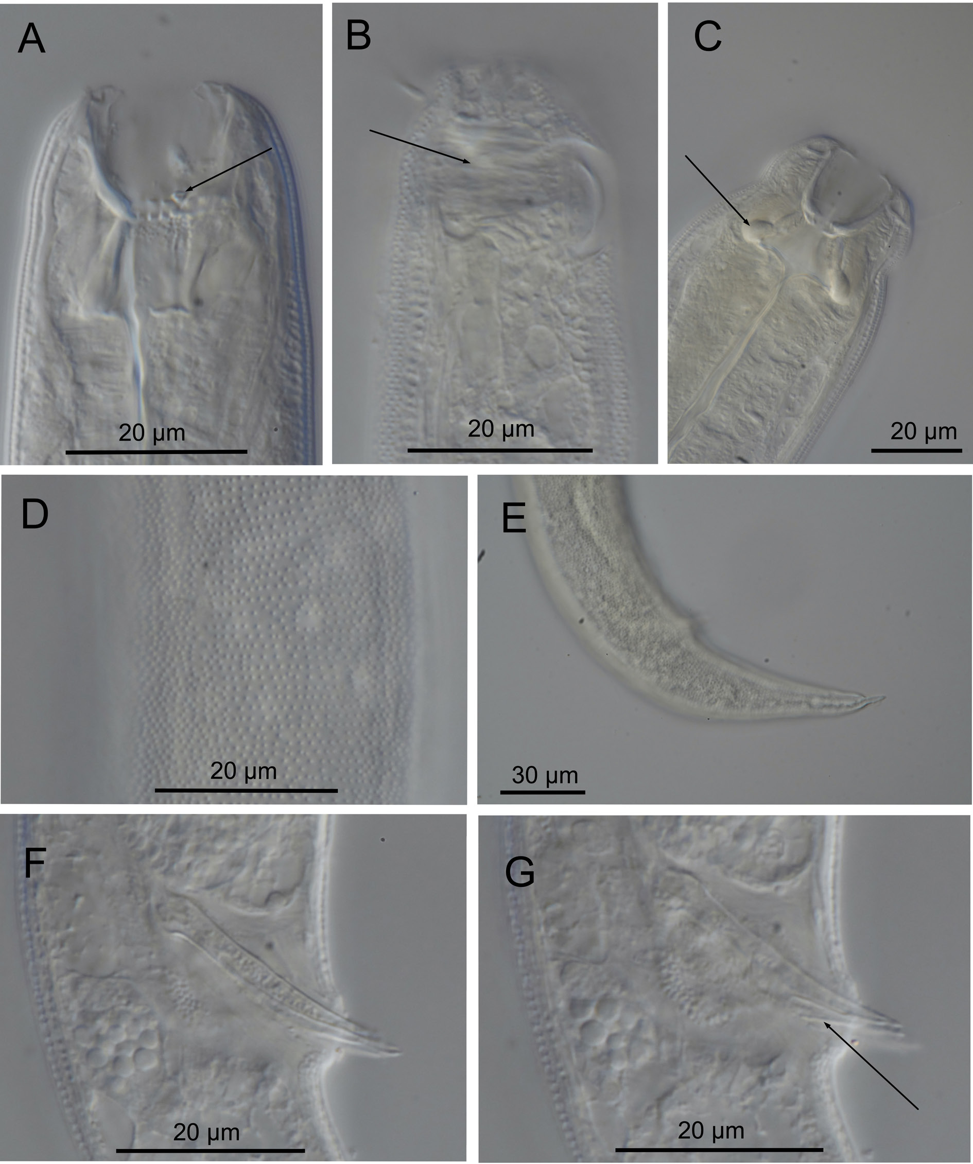
Cobbionema brevispicula sp. nov. is characterised by 1.08–1.37 mm long body; lateral alae absent; body pores present; cephalic sensilla equal to 0.3–0.5 labial region diameters in length; amphideal fovea sexually dimorphic, with 3.5–3.75 turns; two rows of pointed projections and a third row with three prominent sharply pointed tines in the stoma, rows of projections close to one another longitudinally; spicules elongate, nearly straight, with rounded manubrium, subcylindrical shaft and bifid tips, 29–32 µm long. Three or five midventral precloacal papilliform sensilla; tail curved ventrally and conoid in shape.
Etymology
The species name refers to relatively short spicule in the species, compared to other congeners.
Material examined
Holotype
SWEDEN • ♂; Skagerrak ; 58º22′14″ N, 11º05′00″ E; 30–70 m depth; 9 Aug. 2014; O. Holovachov leg.; gravel, mud and algae; SMNH Type-9219 . GoogleMaps
Paratypes
SWEDEN • 2 ♂; same collection data as for holotype; SMNH Type-9220 . GoogleMaps Additional non-type material
SWEDEN • 1 ♀; Skagerrak ; 58º17′32″ N, 11º11′24″ E; 45–55 m depth; 10 Sep. 2012; O. Holovachov leg.; coarse sediment with algae; SMNH-182041 GoogleMaps .
Description
Adult characters
Body cylindrical, tapering slightly towards both extremities. Cuticle with transverse rows of dots, without lateral differentiation. Lateral alae in the shape of a smooth band of cuticle absent. Body pores present (fewer and less obvious in males), starting at level with anterior part of pharynx and extending to tail region, located along either side of the lateral alae, irregularly spaced. Somatic setae scattered along the body. Labial region bluntly-rounded, not set-off. Cephalic region of one male and a female demarcated by a broad and shallow constriction at level with amphid base ( Fig. 6 View Fig A–B); cephalic region of other two males subcylindrical ( Fig. 6 View Fig C–D); this is likely determined by the level of constriction of circum-stomatal sphincter. Six equal lips surrounding mouth opening. Inner labial sensilla indistinct. Outer labial sensilla small papilliform, located on the periphery of labial region. Cephalic sensilla setiform, equal to 0.3–0.5 labial region diameters in length, located at the same level as outer labial sensilla. Amphideal fovea ventrosublateral, large, sexually dimorphic in size, multispiral with 3.5–3.75 turns, with circular outline, equal to about 0.4–0.5 of the corresponding body diameter in male and 0.3 in female. Three to four pairs of cervical setae present, located posterior to amphid in ventrosublateral (1–2 pairs) and dorsosublateral (2 pairs) positions. Buccal cavity voluminous, divided into anterior and posterior chambers. Anterior chamber of buccal cavity barrel-shaped, with 12 cuticularized rhabdions, each with two pairs of large, pointed projections at posterior extremity. These projections are arranged in two horizontal rows, appearing from an en face view as two circles of denticles separating stoma into anterior and posterior chamber. Three (one middorsal and two ventrosublateral) prominent and hollow tines located a short distance in front of anterior circle of denticles. Posterior buccal chamber cone-shaped or cylindrical, widest at (posterior or anterior) extremity (depending on the state of stomatal musculature), with three strongly cuticularized Y-shaped mandibles; anterior branches of each mandible with a field of distinct denticles/knobs on its inner surface; posterior ‘stem’ of each mandible with strongly developed basal (submedian) knobs. Anterior buccal chamber surrounded by strongly developed sphincter muscle. Each mandible is also supported by a strong longitudinal and tangential musculature, forming a muscular anterior pharyngeal bulb. Pharynx muscularised, with developed glandular tissue throughout its entire length; the anterior pharyngeal bulb surrounds the base of buccal cavity; pharynx widens posteriorly but not forming true posterior bulb. Cardia small, with cuticularized lumen. Secretory-excretory system present, renette body located at level with pharyngo-intestinal junction, secretory-excretory pore located at level with nerve ring. Tail curved ventrally, conoid in shape. Caudal glands present, opening via a common spinneret, caudal gland cells/bodies incaudal in the majority of examined specimens, at level with posterior part of intestine in one male.
Male
Reproductive system diorchic, with outstretched anterior testis and reflexed posterior testis, always on opposite sides of the intestine. Anterior testis on either left (holotype) or right (paratype) side of intestine, posterior testis on either right (holotype) or left (paratype) side of intestine. Spicules paired and symmetrical, elongate, nearly straight, with rounded manubrium, subcylindrical shaft and bifid distal tips, equal to 0.9–1.0 anal body diameters in length ( Figs 6H View Fig , 7F View Fig ). Gubernaculum reduced to sublateral crura, conoid in shape, flanking distal part of spicules. Three or five midventral precloacal papilliform sensilla located in a row, starting 20–22 µm in front of cloacal opening. Further papilliform sensilla arranged as follows: one subventral pair located half-way between posteriormost midventral papilliform sensillum and cloacal opening; several subventral and several subdorsal pairs along the tail in a variable manner ( Fig. 6 View Fig F–G).
Female
Reproductive system didelphic, amphidelphic, ovary branches reflexed antidromously. Anterior ovary on right side of intestine, posterior ovary on left side of intestine. Other details of female reproductive system morphology cannot be observed due to suboptimal preservation of the specimen. Vulva located
just posterior to midbody. Intra-uterine egg not seen. Rectum reduced, a string of undifferentiated tissue stretching from posterior blunt end of intestine towards ventral body side is visible.
Differential diagnosis
Both males and females of C. brevispicula sp. nov. can be distinguished from all previously described species of Cobbionema by their conical tail (vs conoid proximal portion and digitate distal section). Males of C. brevispicula sp. nov. are also different from all other previously described species in the unique shape and size of their spicules (nearly straight, with bifid tips and measuring 0.9–1.0 anal body diameters long vs two-sectioned spicules consisting of subcylindrical proximal part and a conoid/ fusiform distal section and measuring 1.9–2.8 anal body diameters long).
Biology
Gut content included undigested remains (spicules and gubernacula) of a variety of nematode species, including a possible Cyatholaimidae .
No known copyright restrictions apply. See Agosti, D., Egloff, W., 2009. Taxonomic information exchange and copyright: the Plazi approach. BMC Research Notes 2009, 2:53 for further explanation.
|
Kingdom |
|
|
Phylum |
|
|
Class |
|
|
Order |
|
|
Family |
|
|
Genus |