
Cobbionema acrocerca Filipjev, 1922
|
publication ID |
https://doi.org/ 10.5852/ejt.2020.702 |
|
publication LSID |
lsid:zoobank.org:pub:B4DDC9C7-69F4-40D1-A424-27D04331D1F8 |
|
DOI |
https://doi.org/10.5281/zenodo.4328591 |
|
persistent identifier |
https://treatment.plazi.org/id/EC598796-8571-D438-FD9B-FD6D549CFAB1 |
|
treatment provided by |
Valdenar |
|
scientific name |
Cobbionema acrocerca Filipjev, 1922 |
| status |
|
Cobbionema acrocerca Filipjev, 1922
Figs 2–3 View Fig View Fig , Table 2
Diagnosis
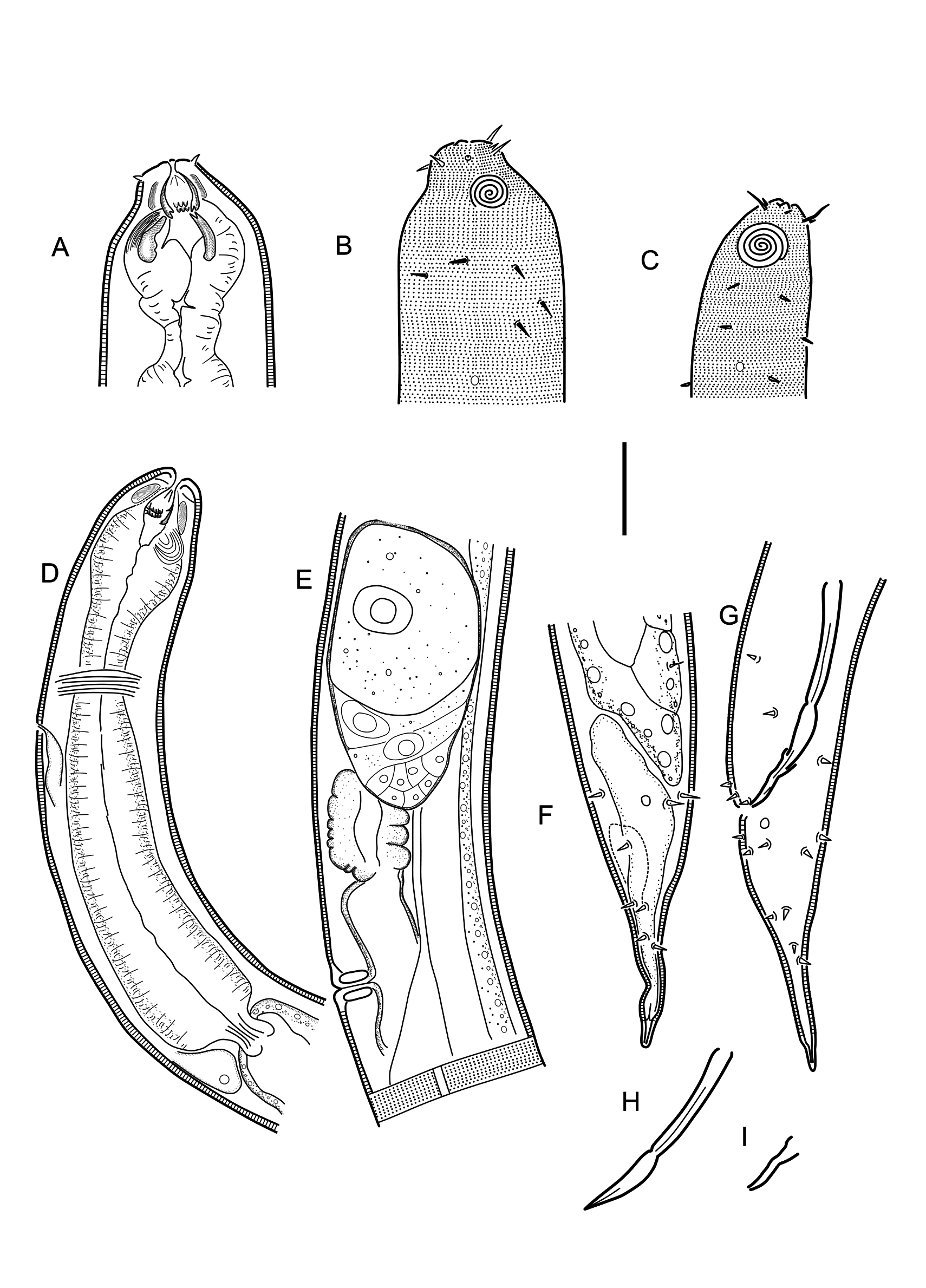
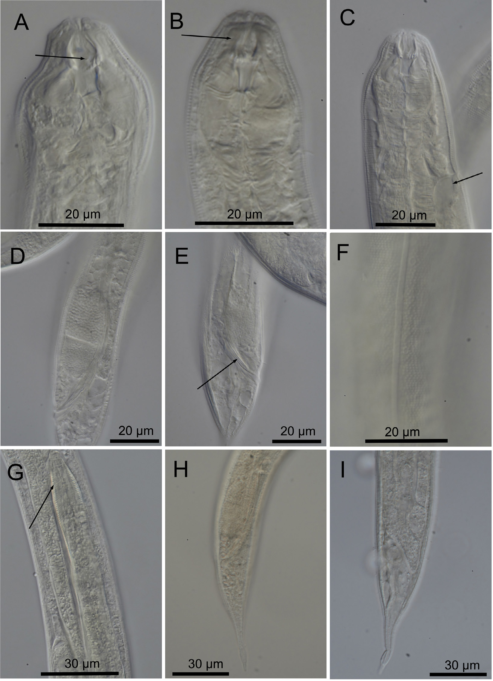
Cobbionema acrocerca is particularly characterised by 0.59–0.86 mm long body; lateral alae present; body pores arranged on either sides of lateral alae; cephalic sensilla equal to 0.3–0.6 labial region diameters in length; amphideal fovea sexually dimorphic, with 4.25–5.25 turns in males and 3.25–3.75 turns in females; two rows of pointed projections and a third row with three prominent sharply pointed tines in the stoma, rows of projections close to one another longitudinally; spicules consisting of two sections, 50–58 µm long; single precloacal papilliform sensillum; tail with conoid proximal and digitate distal section.
Material examined
SWEDEN • 1 ♀; Skagerrak ; 58º20′15.5″–15.2″ N, 10º40′06.9″–25.8″ E; 194–201 m depth; 10 Sep. 2012; “Inventering Bratten” leg.; soft bottom; SMNH-179216 • 1 ♂; Skagerrak ; 58º19′15.6″–20.9″ N, 10º29′ 33.5″–34.0″ E; 352–374 m depth; 10 Sep. 2012; “ Inventering Bratten ” leg.; soft bottom; SMNH- 179217 • 2 ♂♂; Skagerrak ; 58º23′00.8″–22′00.8″ N, 10º20′28.8″–38.3″ E; 390–428 m depth; 10 Sep. 2012; “ Inventering Bratten ” leg.; soft bottom; SMNH-179218 • 2 ♂♂; Skagerrak ; 58º22′17.8″–19.4″ N, 10º23′50.8″–24′03.2″ E; 351–387 m depth; 10 Sep. 2012; “ Inventering Bratten ” leg.; soft bottom; SMNH-179219 • 1 ♂; Skagerrak ; 58º28′28.0″–31.1″ N, 10º33′19.1″–23.8″ E; 221–260 m depth; 11 Sep. 2012; “ Inventering Bratten ” leg.; soft bottom; SMNH-179220 • 3 ♂♂; Skagerrak ; 58º28′12.1″–19.5″ N, 10º37′01.1″–07.1″ E; 180–216 m depth; 11 Sep. 2012; “ Inventering Bratten ” leg.; soft bottom; SMNH- 179221 , SMNH179222 View Materials • 1 ♂, 2 ♀♀; Skagerrak ; 58º34′21.3″–16.6″ N, 10º38′11.2″–29.4″ E; 139– 153 m depth; 12 Sep. 2012; “ Inventering Bratten ” leg.; soft bottom; SMNH-179223 • 1 ♀; Skagerrak ; 58º27′36.7″–43.3″ N, 10º32′52.0″–59.4″ E; 232–240 m depth; 12 Sep. 2012; “ Inventering Bratten ” leg.; soft bottom; SMNH-179224 .
Description
Adult characters
Body cylindrical, tapering slightly towards both extremities. Cuticle with transverse rows of dots, no lateral differentiation. Lateral alae present, single band of smooth cuticle ( Fig. 3F View Fig ) starting at level with basal pharyngeal bulb and ending anterior to anus/cloaca. Body pores present, starting at level with anterior part of pharynx and extending to tail region, located sublaterally on either dorsal or ventral side of lateral alae, irregularly spaced. Somatic setae scattered along the body. Labial region bluntly-rounded, not set-off. Cephalic region of males and females narrower from the level of the base of the anterior chamber of the stoma. Six equal lips surrounding mouth opening. Inner labial sensilla indistinct. Outer labial sensilla small papilliform, located on the periphery of labial region. Cephalic sensilla setiform, equal to 0.3–0.6 labial region diameters in length, located at the same level as outer labial sensilla. Amphideal fovea ventrosublateral, large, sexually dimorphic in size, multispiral with 4.25–5.25 turns in males and 3.25–3.75 turns in females, with circular outline, equal to about 0.5–0.6 of the corresponding body diameter in males and 0.3 in females. Five to seven pairs of cervical setae present, located posterior to amphid in subventral, ventrosublateral, subdorsal and dorsosublateral positions (cervical setae can be distinguished from somatic sensilla in having larger size and regular arrangement in pairs between amphid and nerve ring level). Buccal cavity voluminous, divided into anterior and posterior chambers. Anterior chamber of buccal cavity barrel-shaped, with 12 cuticularized rhabdions, each with two pairs of large, pointed projections at its posterior extremity. These projections are arranged in two horizontal rows, appearing from an en face view as two circles of denticles separating the stoma into anterior and posterior chamber. Three (one middorsal and two ventrosublateral) prominent and sharply pointed tines located a short distance in front of anterior circle of denticles. Posterior buccal chamber coneshaped, widest at (posterior or anterior) extremity (depending on the state of stomatal musculature), with three strongly cuticularized Y-shaped mandibles; anterior branches of each mandible rather short and acute; posterior “stem” of each mandible enlarged but without basal (submedian) knobs ( Fig. 3 View Fig A–B). Anterior buccal chamber surrounded by strongly developed sphincter muscle. Each mandible is also supported by a strong longitudinal and tangential musculature, altogether appearing as a muscular bulb. Pharynx muscularised, with developed glandular tissue throughout its entire length; with conspicuous anterior swelling surrounding the base of buccal cavity; pharynx widens posteriorly but not forming true posterior bulb. Cardia small, with cuticularized lumen. Secretory-excretory system present, renette body located at level with pharyngo-intestinal junction, secretory-excretory pore located just posterior to nerve ring. Tail straight or ventrally curved, distinctly divided into conoid proximal and digitate distal sections. Caudal glands present, opening via a common spinneret, caudal gland cells/bodies may extend anterior to the posteriormost end of intestine.
Male
Reproductive system diorchic, with outstretched anterior testis and reflexed posterior testis, always on opposite sides of the intestine. Anterior testis on either left (n = 6) or right (n = 1) side of intestine, posterior testis on right (n = 6) or left (n = 1) side of intestine. Spicules paired and symmetrical, elongate, consisting of two sections, subcylindrical proximal section and conoid/fusiform distal section ( Fig. 2H View Fig ), equal to 1.9–2.8 anal body diameters in length. Gubernaculum reduced to sublateral crura, arcuate conoid in shape, with central bend ( Figs 2I View Fig , 3E View Fig ), flanking distal part of spicules. Single midventral precloacal papilliform sensillum located 6 µm from the cloacal opening. Further papilliform sensilla arranged in pairs subventrally and subdorsally anterior to cloaca and along tail.
Female
Reproductive system didelphic, amphidelphic, ovary branches reflexed antidromously. Anterior ovary on either left (n = 2) or right (n = 2) side of intestine, posterior ovary on either right (n = 2) or left (n = 2) side of intestine. Oviducts with large cylindrical cells. Uterus thin-walled, filled with round spermatozoa in fertilized females. Vagina straight, without cuticularization, surrounded by single sphincter muscle. Vulva midventral, a transverse slit. Intra-uterine egg not seen. Rectum absent.
Differential diagnosis
The original description of Cobbionema acrocerca by Filipjev (1922) was based on several female specimens of which only one was measured. The Swedish population examined here resembles the type specimen in terms of body size and a number of body measurements ( Table 2), having unremarkable mandibles in the posterior stoma and proximal part of the tail being conical in shape. Cobbionema acrocerca differs from C. cylindrolaimoides in having a smaller body (588–860 µm vs 1360– 1755 µm in C. cylindrolaimoides ), smaller spicules (50–58 µm vs 89–98 µm in C. cylindrolaimoides ), weakly cuticularized mandibles (vs strongly cuticularised), and different tail shape in females (with conoid vs dome-shaped proximal section).
Cobbionema acrocerca differs from all other species of Cobbionema , except C. cylindrolaimodes , in the morphology of the cuticle (lateral alae present in C. acrocerca vs absent in all except C. cylindrolaimoides ). Cobbionema acrocerca can also be distinguished from holotype male of C. trigamma in having smaller body size (592–666 µm vs 1305 µm) and smaller spicules. In comparison with paratypes of C. trigamma , however, C. acrocerca only differs from C. trigamma in the morphology of the cuticle (lateral alae present in C. acrocerca vs absent in C. trigamma ).
Cobbionema acrocerca can be distinguished from C. capense in the relative positions of cephalic and outer labial sensilla (outer labial sensilla more posterior to cephalic sensilla in C. capense vs cephalic sensilla at the same level as outer labial sensilla) as well as the cuticle morphology (lateral alae present in C. acrocerca vs absent in C. capense ).
Biology
Gut content included undigested remains (spicules and gubernacula) or partially digested entire bodies of a variety of nematode species ( Fig. 3G View Fig ), including Chromadoridae Filipjev, 1917 .
No known copyright restrictions apply. See Agosti, D., Egloff, W., 2009. Taxonomic information exchange and copyright: the Plazi approach. BMC Research Notes 2009, 2:53 for further explanation.
|
Kingdom |
|
|
Phylum |
|
|
Class |
|
|
Order |
|
|
Family |
|
|
Genus |