
Cobbionema Filipjev, 1922
|
publication ID |
https://doi.org/ 10.5852/ejt.2020.702 |
|
publication LSID |
lsid:zoobank.org:pub:B4DDC9C7-69F4-40D1-A424-27D04331D1F8 |
|
DOI |
https://doi.org/10.5281/zenodo.4328587 |
|
persistent identifier |
https://treatment.plazi.org/id/EC598796-8573-D435-FDED-FBED5262FD9D |
|
treatment provided by |
Valdenar |
|
scientific name |
Cobbionema Filipjev, 1922 |
| status |
|
Genus Cobbionema Filipjev, 1922
Type species
Cobbionema acrocerca Filipjev, 1922
Diagnosis
Adult characters
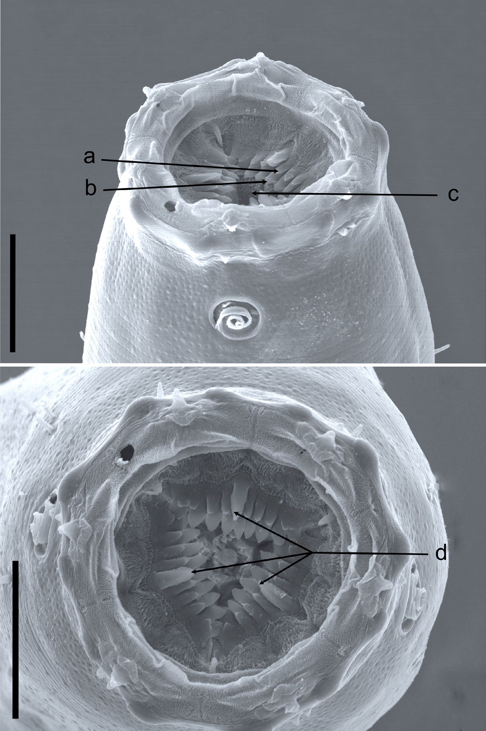
Body cylindrical, tapering slightly towards both extremities. Cuticle with fine transverse striation, with transverse rows of subcuticular dots, one row per annule. Lateral differentiation where punctations are irregularly spaced and further apart (cf. Bendiella Leduc, 2013 ) is absent. Lateral alae present in some species, shaped as single band of raised smooth cuticle. Somatic setae scattered along the body. Cephalic region narrower from the level of the base of the anterior chamber of the stoma. Inner labial sensilla indistinct. Outer labial sensilla small papilliform, located on the periphery of labial region. Cephalic sensilla setiform. Amphideal fovea lateral or ventrosublateral, large, sexually dimorphic in size, multispiral with circular outline. Anterior chamber of buccal cavity barrel-shaped or cylindrical in C. capense and C. trigamma , with 12 cuticularized rhabdions or six in C. trigamma , each with two pairs of large, pointed projections at posterior extremity. These projections are arranged in two horizontal rows, appearing from an en face view as two transverse circles of denticles separating the stoma into anterior and posterior chamber. Three (one middorsal and two ventrosublateral) prominent and sharply pointed tines (slender pointed projections, cf. Maggenti 2005) located a short distance in front of the anterior circle of denticles. Posterior buccal chamber cone-shaped, widest at (posterior or anterior) extremity (depending on the state of stomatal musculature), with strongly cuticularized Y-shaped mandibles; anterior branches of each mandible with denticles on its inner surface; posterior ‘stem’ of each mandible with strongly or weakly developed basal (submedian) knobs. The stoma structure of all studied specimens generally resembles that of Halichoanolaimus both in the arrangement of pointed projections in the anterior chamber of the stoma and the shape of the mandibles in the posterior chamber ( Fig. 1 View Fig ). Anterior buccal chamber surrounded by strongly developed sphincter muscle. Pharynx muscularised, with conspicuous anterior swelling surrounding the base of buccal cavity and widens posteriorly but not forming true posterior bulb. Cardia small, with cuticularized lumen. Tail straight or curved ventrally, conical in shape or with distinct conical proximal portion and a digitate (= subcylindrical) distal end. Caudal glands present, opening via a common spinneret, caudal gland cells/bodies at level with posterior part of intestine, anterior to cloaca or extending anterior to the posteriormost end of the intestine.
Male
Reproductive system diorchic, with outstretched anterior testis and reflexed posterior testis. Anterior and posterior testis can lie on either left or right side of intestine. Spicules paired and symmetrical, elongate, consisting of two sections separated by a constriction, nearly straight, with rounded manubrium, subcylindrical shaft, or short with bifid tips. Gubernaculum reduced only to two sublateral crura, arcuate conoid in shape, flanking distal part of spicules. Precloacal supplements setose or papilloid, midventral and range in number from one to five.
Female
Reproductive system didelphic, amphidelphic, ovary branches reflexed antidromously. Anterior ovary on left of intestine, posterior ovary on right side of intestine or vice versa. Vagina straight, without cuticularizations, surrounded by single sphincter muscle. Vulva midventral, a transverse slit. Rectum absent.
Remarks
Gerlach (1964) could not resolve the relationship of this genus with the other members of Selachinematidae due to the limited understanding of its buccal structure at that time. The descriptions of C. capense and C. trigamma have contributed useful insights into the nature of the buccal cavity of this genus. In spite of this, some differences in terminologies used to describe, possibly the same, features in the buccal cavity of these species need clarification. For example, pointed projections in C. trigamma and denticles in C. capense may refer to the same character; which could also be the same feature Schuurmans Stekhoven (1950) described as rows of denticular corpuscles. Also, the three chitinous hooks mentioned in the description of C. acrocerca could be the same as the teeth that were referred to in the description of C. capense . Moreover, an examination of the Swedish populations identified as C. acrocerca and C. cylindrolaimoides revealed that some of the characters described as unique to some species were actually found in others. For example, the Y-shaped rhabdions found in C. trigamma were also observed in all species of Cobbionema that were examined in this study and are very similar to the ones found in most species of Halichoanolaimus . In the descriptions of C. acrocerca and C. capense , a sphincter muscle around the anterior and part of the posterior chambers of the buccal cavity were described. This character has also been observed in all populations of C. acrocerca and C. cylindrolaimoides as well as the two new species described here, and depicted in Leduc (2013: fig. 10). This character has, however, not been observed in other genera of the family. There is therefore a possibility that this feature could be an apomorphic character which separates this genus from other genera within Selachinematidae .
Relationships
Cobbionema differs from all other genera of Selachinematidae in having a sphincter muscle around the anterior buccal chamber. Species of Cobbionema are similar to Halichoanolaimus in the rows of pointed projections at the intersection between the two buccal chambers as well as the three tines (one middorsal and two ventrosublateral) anterior to these two rows of pointed projections. In H. robustus , for example, the tail shape closely resembles that of some Cobbionema species. However, Cobbionema can be distinguished from Halichoanolaimus by the cephalic sensilla being setiform and not papilliform. Cobbionema can also be distinguished from Choniolaimus by the absence of terminal pharyngeal bulb and shape of precloacal supplements. Table 1 View Table 1 summarizes some of the differentiating features between Cobbionema and other genera included in Selachinematidae .
Valid species
Cobbionema acrocerca Filipjev, 1922
Cobbionema acuminata sp. nov.
Cobbionema brevispicula sp. nov.
Cobbionema capense Furstenberg & Heyns, 1987
No known copyright restrictions apply. See Agosti, D., Egloff, W., 2009. Taxonomic information exchange and copyright: the Plazi approach. BMC Research Notes 2009, 2:53 for further explanation.
|
Kingdom |
|
|
Phylum |
|
|
Class |
|
|
Order |
|
|
Family |