
Cobbionema cylindrolaimoides Schuurmans Stekhoven, 1950
|
publication ID |
https://doi.org/ 10.5852/ejt.2020.702 |
|
publication LSID |
lsid:zoobank.org:pub:B4DDC9C7-69F4-40D1-A424-27D04331D1F8 |
|
DOI |
https://doi.org/10.5281/zenodo.4328593 |
|
persistent identifier |
https://treatment.plazi.org/id/EC598796-857C-D422-FE09-FAB05162FB65 |
|
treatment provided by |
Valdenar |
|
scientific name |
Cobbionema cylindrolaimoides Schuurmans Stekhoven, 1950 |
| status |
|
Cobbionema cylindrolaimoides Schuurmans Stekhoven, 1950
Figs 4–5 View Fig View Fig , Table 2
Diagnosis
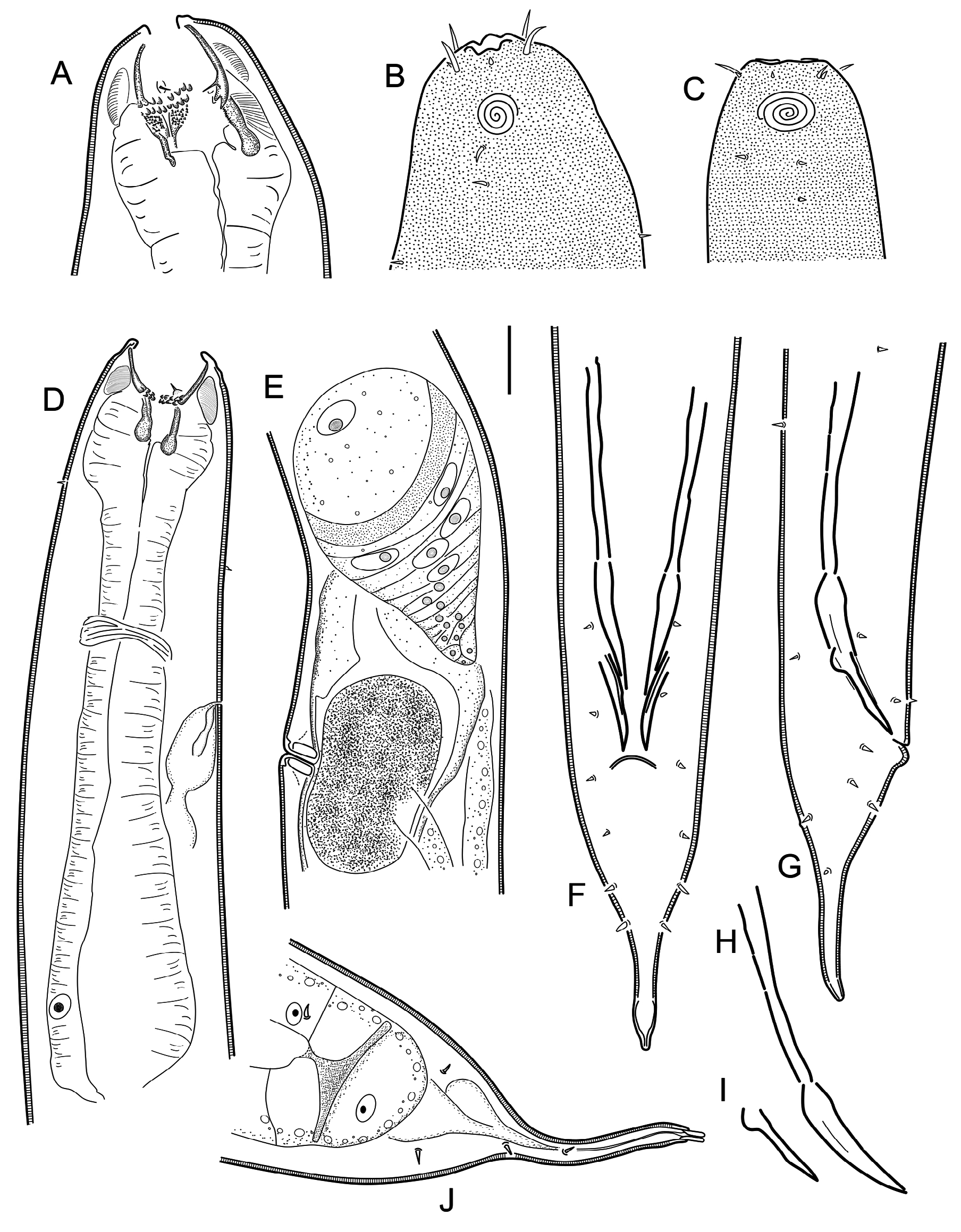
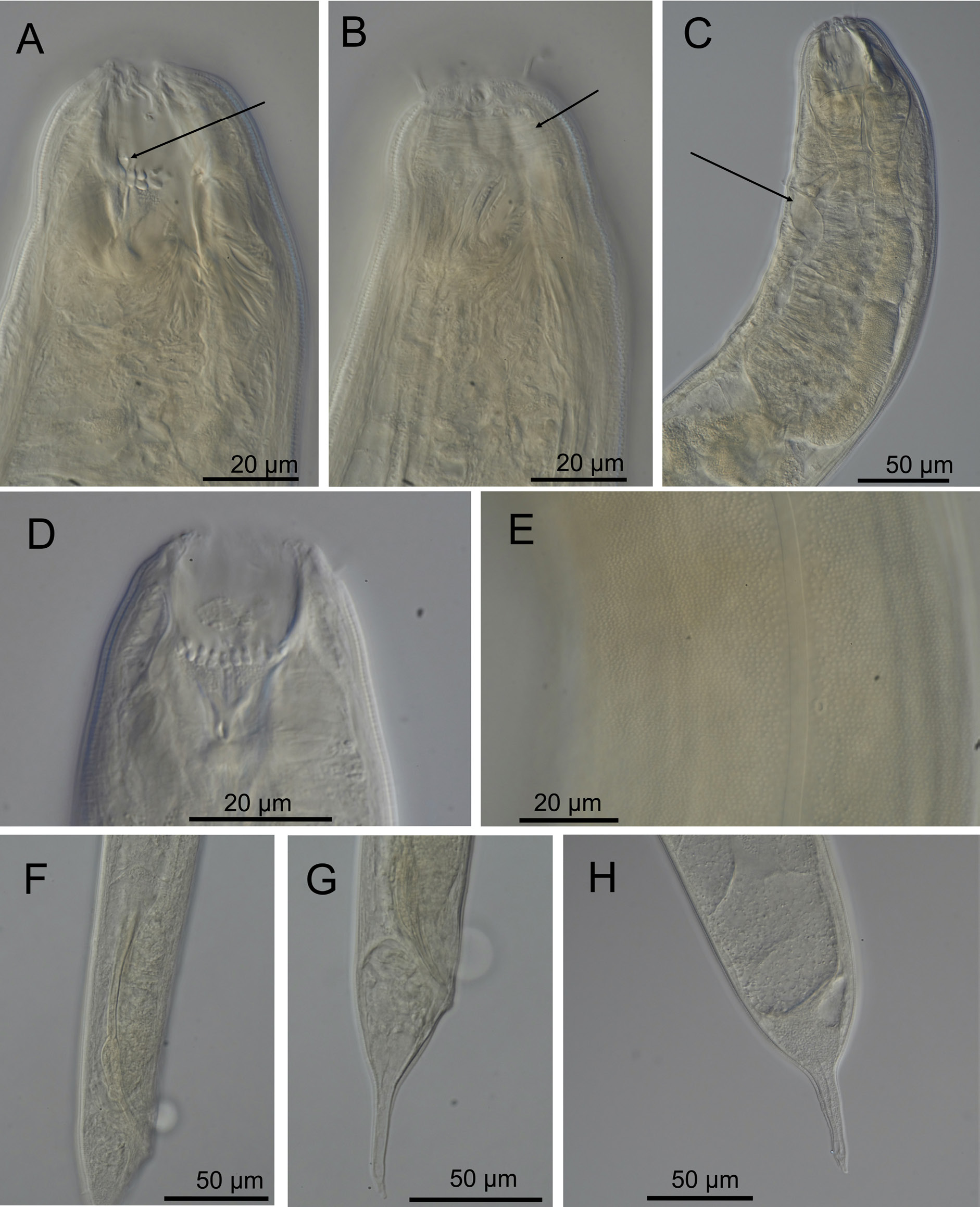
Cobbionema cylindrolaimoides is characterised by 1.36–1.76 mm long body; lateral alae present; body pores arranged on either sides of lateral alae; cephalic sensilla, equal to 0.3–0.5 labial region diameters in length; amphideal fovea sexually dimorphic, with 3.5–4.25 turns in males and 3.25–3.5 turns in females; two rows of pointed projections and three prominent sharply pointed tines in the stoma, rows of projections close to one another longitudinally; spicules consisting of two sections, 89–98 µm long; single midventral precloacal papilliform sensillum; tail with conoid or dome shaped proximal and digitate distal section.
Material examined
SWEDEN • 3 ♀♀; Skagerrak ; 58º22′20″ N, 11°09′26″ E; 25–50 m depth; 9 Aug. 2011; O. Holovachov leg.; muddy sand; SMNH-179211 GoogleMaps • 3 ♀♀, 6 ♂♂; Skagerrak ; 58º19′15.6″–20.9″ N, 10º29′33.5″–34.0″ E; 30 m depth; 11 Aug. 2011; O. Holovachov leg.; soft mud; SMNH-179212 , SMNH-179213 • 1 ♀, 3 ♂♂; Skagerrak ; 58º19′15.6″–20.9″ N, 10º29′33.5″–34.0″ E; 30 m depth; 11 Aug. 2011; O. Holovachov leg.; soft mud; SMNH-179214 , SMNH-179215 .
Description
Adult characters
Body cylindrical, tapering slightly towards both extremities. Cuticle with transverse rows of dots, without lateral differentiation. Lateral alae present, single band of smooth cuticle starting at level with basal pharyngeal bulb and ending anterior to anus/cloaca. Body pores present, starting at level with anterior part of pharynx and extending to tail region, located on either side of the lateral alae, irregularly spaced. Somatic setae scattered along the body. Labial region bluntly rounded, not set off. Depending on which muscles are constricted, cephalic region can be cylindrical or with a broad and shallow constriction at level with amphidial fovea. Six equal lips surrounding mouth opening. Inner labial sensilla indistinct. Outer labial sensilla small papilliform, located on the periphery of labial region. Cephalic sensilla setiform, equal to 0.3–0.5 labial region diameters in length, located at the same level as outer labial sensilla. Amphideal fovea ventrosublateral, large, sexually dimorphic in size, multispiral with 3.5–4.25 turns in males and 3.25–3.5 turns in females, with circular outline, equal to about 0.4– 0.5 of the corresponding body diameter in male and 0.2–0.3 in female. Two to three pairs of cervical setae present, located posterior to amphid in ventrosublateral and dorsosublateral positions. Buccal cavity voluminous, divided into anterior and posterior chambers, partially everted in some specimens. Anterior chamber of buccal cavity barrel-shaped, with 12 cuticularized rhabdions, each with two pairs of large, pointed projections at posterior extremity. These projections are arranged in two horizontal rows, appearing from an en face view as two circles of denticles separating the stoma into anterior and posterior chamber. Three (one middorsal and two ventrosublateral) prominent and sharply pointed, almost setiform tines located a short distance in front of anterior circle of denticles. Posterior buccal chamber cone-shaped, widest at (posterior or anterior) extremity, or cylindrical (depending on the state of stomatal musculature), with three strongly cuticularized Y-shaped mandibles; anterior branches of each mandible broad triangular, with a field of distinct denticles; posterior ‘stem’ of each mandible with strongly developed basal (submedian) knobs ( Figs 4A View Fig , 5A View Fig ). Anterior buccal chamber surrounded by strongly developed sphincter muscle. Each mandible is also supported by a strong longitudinal and tangential musculature, altogether appearing as a muscular bulb. Pharynx muscularised, with developed glandular tissue throughout its entire length; with conspicuous anterior swelling surrounding the base of buccal cavity; pharynx widens posteriorly but not forming true posterior bulb. Cardia small, with cuticularized lumen. Secretory-excretory system present, renette body located at level with pharyngointestinal junction, secretory-excretory pore located at level with nerve ring. Tail straight, or curved either dorsally or ventrally, distinctly divided into conoid (in males and some females) or dome-shaped (in females) proximal and digitate distal sections. Caudal glands present, opening via a common spinneret, caudal gland cells/bodies incaudal.
Male
Reproductive system diorchic, with outstretched anterior testis and reflexed posterior testis, always on opposite sides of the intestine. Anterior testis on right (n = 4) side of intestine, posterior testis on left side (n = 4) of intestine. Spicules paired and symmetrical, elongate, seemingly consisting of two sections, subcylindrical proximal section and conoid/fusiform distal section, equal to 2.2–2.4 anal body diameters in length. Gubernaculum reduced to sublateral crura, subcylindrical to conoid in shape, with proximal swelling in some specimens, flanking distal part of spicules ( Fig. 4I View Fig ). Single midventral precloacal papilliform sensillum located 15 µm from the cloacal opening ( Fig. 4G View Fig ). Further papilliform sensilla arranged in pairs subventrally and subdorsally anterior to cloaca and along tail.
Female
Reproductive system didelphic, amphidelphic, ovary branches reflexed antidromously. Anterior ovary on either right (n = 3) or left (n = 4) side of intestine, posterior ovary on either right (n = 5) or left (n = 2) side of intestine. Sperm present in the uterus. Vulva located at around midbody, a transverse slit.
Differential diagnosis
Swedish specimens of C. cylindrolaimoides resemble the only female specimen used by Schuurmans Stekhoven (1950) for the original description in body size and most body measurements ( Table 2), presence of strongly cuticularized mandibles in the posterior stoma section and sigmoid-conoid shape of the tail. Gerlach (1964) synonymised it with C. acrocerca (and followed by Bezerra et al 2020) but in Gerlach & Riemann (1974) they are treated as separate species.As discussed above, C. cylindrolaimoides differs from C. acrocerca in larger body (1360–1755 µm vs 588–860 µm in C. acrocerca ), longer spicules (89–98 µm vs 50–58 µm in C. acrocerca ) and mandibles with strongly developed basal knobs (vs mandibles without discernible basal knobs) and shape of female tail.
Based on the updated morphological diagnosis, C. cylindrolaimoides most closely resembles the holotype male of C. trigamma in having relatively large body size (1360–1476 µm vs 1305 µm in C. trigamma ) and long spicules (89–98 µm vs 114 µm in C. trigamma ). The only clear morphological difference between C. cylindrolaimoides and C. trigamma is in the morphology of the cuticle (lateral alae present in C. cylindrolaimoides vs absent in C. trigamma ). Paratypes of C. trigamma differ from C. cylindrolaimoides in much smaller body size (1360–1755 µm vs 566–600 µm in both male and female C. trigamma ).
Furthermore, C. cylindrolaimoides differs from C. capense , known from a single female, in body size (1456–1755 µm in females vs 688 µm in C. capense ), cuticle morphology (lateral alae present in C. cylindrolaimoides vs absent in C. capense ) and different morphology of the stoma (see Table 3).
Biology
Gut content included undigested remains (spicules and gubernacula) or even partially digested entire bodies of a variety of nematode species, including Cyatholaimidae Filipjev, 1918 and Axonolaimidae Filipjev, 1918 .
No known copyright restrictions apply. See Agosti, D., Egloff, W., 2009. Taxonomic information exchange and copyright: the Plazi approach. BMC Research Notes 2009, 2:53 for further explanation.
|
Kingdom |
|
|
Phylum |
|
|
Class |
|
|
Order |
|
|
Family |
|
|
Genus |