
Cheilopora elfa, Kuklinski, Piotr, Grischenko, Andrei V. & Jewett, Stephen C., 2015
|
publication ID |
https://doi.org/10.11646/zootaxa.3963.3.7 |
|
publication LSID |
lsid:zoobank.org:pub:BFB9E380-0C93-4EA4-9943-3D07C2ECDF7D |
|
DOI |
https://doi.org/10.5281/zenodo.6100029 |
|
persistent identifier |
https://treatment.plazi.org/id/ED5A878A-FFE4-323A-FF20-C54630275030 |
|
treatment provided by |
Plazi |
|
scientific name |
Cheilopora elfa |
| status |
sp. nov. |
Cheilopora elfa n. sp.
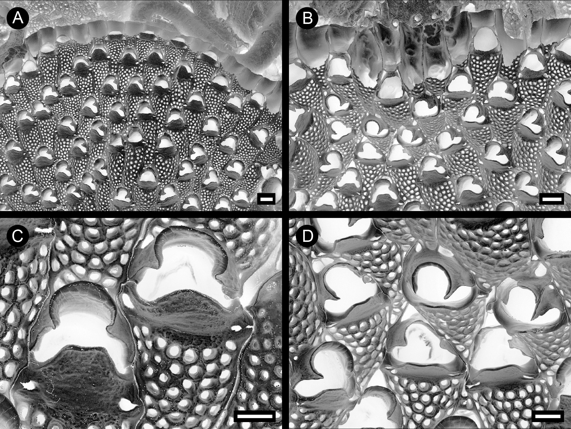
( Figure 2 View FIGURE 2 , A–D)
Material examined. Holotype: NHMUK 2014.12.5.1, colony encrusting oyster shell, RV Norseman, 12 July 2011, Beringian coastal water of Amchitka Island, Rat Islands of the Aleutian Islands, Constantine Harbour, 51°24'31.5" N, 179°17'57.9" E, depth 12 m, SCUBA, coll. P. Kuklinski.
Etymology. The given name, elfa , elf, dwarf, alluding to the very small size of lateral-suboral chambers.
Diagnosis. Colony encrusting, autozooids large, elongated. Frontal shield moderately convex, evenly perforated by large circular pseudopores. Primary orifice deep, partly concealed by trifoliate secondary orifice. Peristome cormidial, distal part formed by proximal edge of daughter zooid, separated by deep pseudosinuses from proximal part with strongly projecting rim angled over orifice at 60–70° to frontal plane. Paired heterozooids at lateral-suboral corners in place of avicularia.
Measurements. ZL 0.67–1.11 (0.87 ± 0.15), ZW, 0.26–0.40 (0.33 ± 0.04). OrL 0.13–0.22 (0.19 ± 0.02), OrW, 0.22–0.26 (0.24 ± 0.02).
Description. Colony encrusting, multiserial, unilaminar, more or less circular. Zooids large, elongate hexagonal, rhombic to pyriform, broadest in distal third, tapering proximally, arranged in divergent rows, demarcated by fine sutures between adjacent vertical walls ( Fig. 2 View FIGURE 2 , A). Frontal shield moderately convex, surface finely granulated, uniformly perforated by numerous large circular and oval pseudopores over entire surface except for suboral peristomial area; pseudopores becoming infundibular and thickening frontal shield acquiring reticulate appearance in ephebic zooids. Primary orifice sunken ( Fig. 2 View FIGURE 2 , D), corresponding to operculum. Secondary orifice trifoliate ( Fig. 2 View FIGURE 2 , C), its distal part comprising the proximal end of the daughter zooid, elevated to form an arcuate rim that curves proximad to form angular projections that mark the edge of the deep pseudosinuses flanking the strongly projecting proximal orifical rim; this rim somewhat trapezoidal to arcuate depending on angle of view, angled at 60–70° to frontal plane and overhanging about 1/4 to 1/3 of proximal area of primary orifice. Paired, rarely single, lateral-suboral heterozooidal chambers, each with an irregular frontal foramen ( Fig. 2 View FIGURE 2 , C, D). Interzooidal communications via multiporous septula. Basal wall fully calcified. Ovicells, ancestrula and early astogeny not observed.
Remarks. In general appearance and particularly in having an elevated cormidial peristome with distal and proximal components separated by deep pseudosinuses, Cheilopora elfa n. sp. is very closely related to C. peristomata n. sp., but can be distinguished from the latter by the following criteria: 1) external surface of peristomial components coarsely granulated in C. peristomata and smooth in C. elfa ; 2) proximal peristomial margin angled at 80–70° to frontal plane and overhanging only about 1/5 of proximal area of primary orifice in C. peristomata , but angled at 60–70° and overhanging about 1/4 to 1/3 of proximal area of primary orifice in C. elfa ; 3) proximal peristomial lip transversely weakly concave and strictly trapezoidal in profile with angled corners in C. peristomata , but transversely straight and either trapezoidal with rounded corners or, more frequently, semicircular in profile in C. elfa ; 4) secondary orifice consistently trifoliate in C. elfa , but variably campanulate in nonovicellate zooids to trifoliate in ovicellate zooids of C. peristomata ; 5) fully developed avicularia in C. peristomata but vestigial structures in C. elfa .
Another congener, C. inermis ( Busk, 1880) resembles C. elfa in the occasional presence of dwarf or incipient avicularia ( Kluge 1962, p. 563). However, the peristome is complete and low in C. inermis , with only an inconspicuous central prominence in the suboral peristomial rim.
The lack of ovicells in the only known specimen of C. elfa n. sp. may indicate that the colony was immature and non-breeding; on the other hand, confamilial Cheiloporina haddoni has polymorphic incubating zooids with internal brood sacs ( Ostrovsky et al. 2009; Ostrovsky 2013). The ooecium is reduced to a vestigial elevation on the proximal part of the distal zooid or, if the latter is missing, is a slightly swollen cap-like structure covering the incubating zooid distally (see Dick et al. 2006). Additional material, especially of obviously brooding colonies, will help to determine the type of incubatory structures in C. elfa .
Distribution. Cheilopora elfa n. sp. is so far known only from the type locality. It thus can be categorized as a Pacific, high-Boreal sublittoral species.
| NHMUK |
Natural History Museum, London |
No known copyright restrictions apply. See Agosti, D., Egloff, W., 2009. Taxonomic information exchange and copyright: the Plazi approach. BMC Research Notes 2009, 2:53 for further explanation.
|
Kingdom |
|
|
Phylum |
|
|
Class |
|
|
Order |
|
|
SubOrder |
Neocheilostomina |
|
Family |
|
|
Genus |