
Paucumara falcata Wang & Li, 2019
|
publication ID |
https://doi.org/10.11646/zootaxa.4568.1.9 |
|
publication LSID |
lsid:zoobank.org:pub:CA1AD232-3D40-4D10-82A6-2A6F61A2730D |
|
DOI |
https://doi.org/10.5281/zenodo.5936975 |
|
persistent identifier |
https://treatment.plazi.org/id/ED5EC61A-9C74-8E27-FF00-FAF811E6FC28 |
|
treatment provided by |
Plazi |
|
scientific name |
Paucumara falcata Wang & Li |
| status |
sp. nov. |
Paucumara falcata Wang & Li , sp. nov.
Material examined. Holotype, PLA-Pa001, eastern beach of Shenzhen City , Guangdong Province, China, 22°28′N, 114°31′E, 29th April 2017, coll. Wei-Xuan Li, sagittal sections on 9 slides. GoogleMaps
Paratypes: PLA-Pa002, ibid., one sagittal sections on 4 slides; PLA-Pa003, ibid., horizontal sections on 5 slides; PLA-Pa004, ibid., whole mount on 1 slide; PLA-Pa005, ibid., whole mount on 1 slide; PLA-Pa006, ibid., whole mount on 1 slide. All specimens are deposited in IZCAS. Observations were made on both live and preserved specimens .
Etymology. The specific epithet is derived from the Latin adjective falcatus, sickle-shaped, and alludes to the curved, chitinized tip of the musculo-parenchymatic organ.
Diagnosis. Paucumara falcata is characterized by: three pale yellow transverse pigmentation bands, one at the anterior body margin, another immediately behind the eyes and a third one across the tail end of the body, while a brown band extends anteriorly from the level of the eyes; 8–11 testicular follicles on either side of the body, extending from immediately behind the ovaries to half-way along the pharyngeal pocket; a musculo-parenchymatic organ provided with a sickle-shaped chitinized, sclerotic tip or stylet.
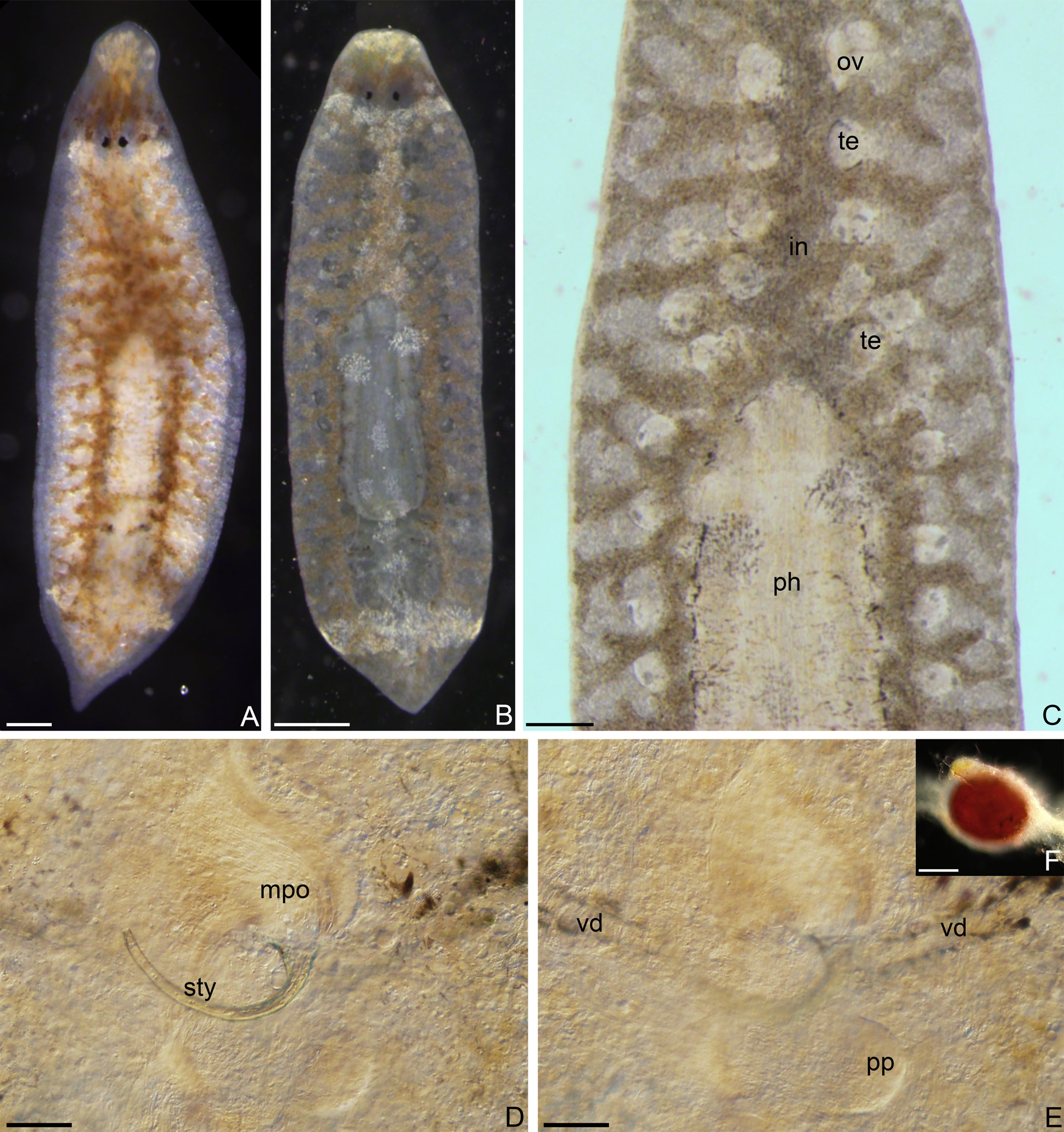
Description. Live specimens with low-triangular head and sharply pointed hind end, their length ranging between 3.8–4.1 mm and their width between 0.48–0.58 mm (n=5) ( Figs. 7 View FIGURE 7 A–B, 8A, 10A). A pair of black, kidney-shaped eyes is located at a position measuring 1/8 of the total body length, as determined from the anterior margin; the eyes are set rather close together, the distance between them ranging between 37–43 µm (n=5). Each eye cup has a large semi-circular lens ( Fig. 8A View FIGURE 8 ). The dorsal body surface shows conspicuous pale yellow stripes as well as a region with brownish pigmentation. Pale-yellow transverse stripes occur at the anterior margin and immediately behind the eyes, and across the tail end of the body ( Fig. 7B View FIGURE 7 ). The transverse band at the anterior margin is 300–330 µm in width, while the stripe behind the eyes measures 100–120 µm in width, while the one on the tail is 140–180 µm wide (n=5). Further there is a pale yellow longitudinal stripe that runs from the transverse pale band behind the eyes to the base of the pharynx that is 600–680 µm long and has a width of 90–100 µm (n=5). In addition to these pale stripes there are snowflake-like pale yellow specks scattered on the dorsal surface. A broad band of brown pigment extends anteriorly from the level of the eyes for a distance of about 180–200 µm ( Figs. 7 View FIGURE 7 A– B).
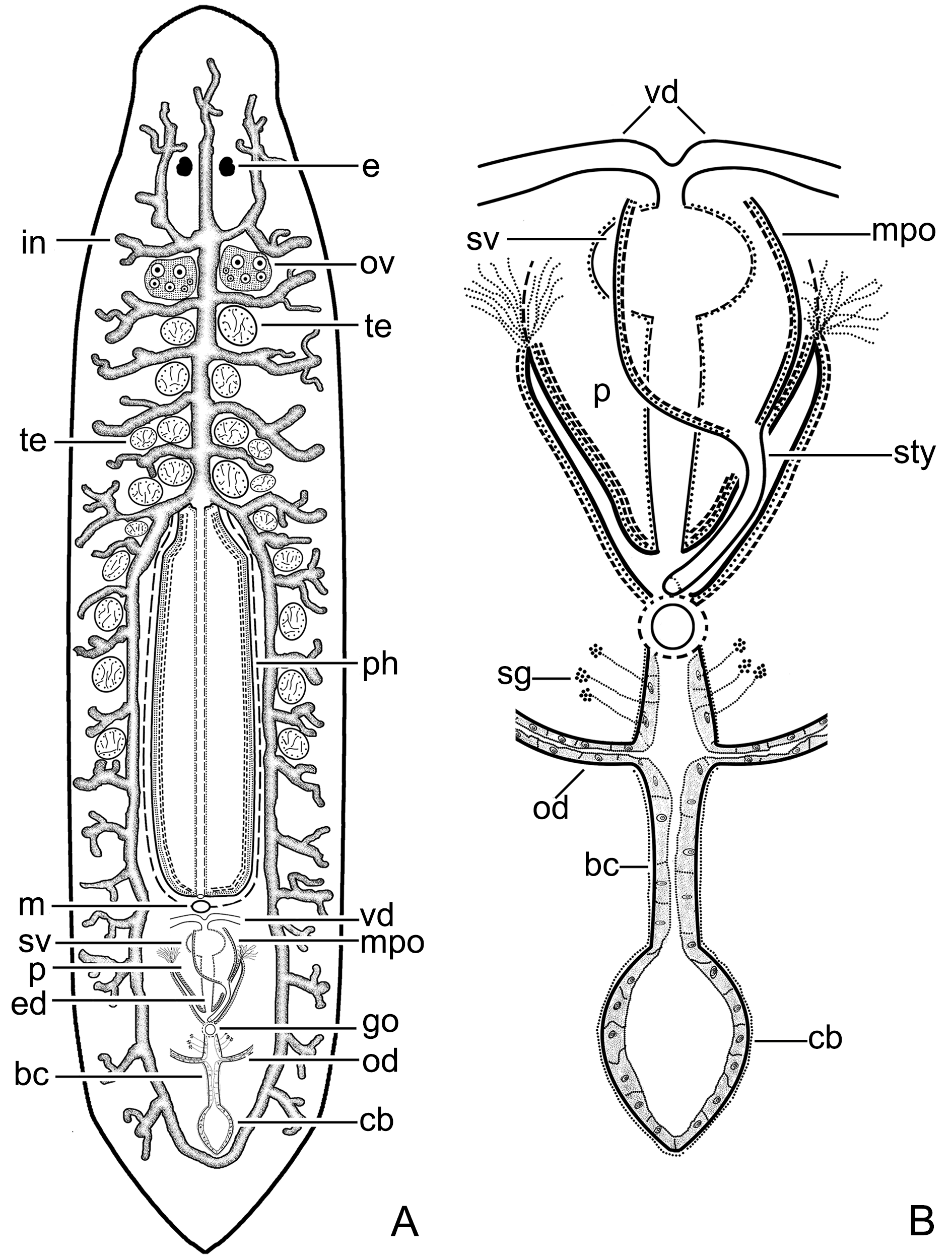
The mouth opening is located at a distance of about 1/3 of the body length, as measured from the posterior body margin; the mouth is situated at the posterior end of the pharyngeal cavity ( Fig. 10 View FIGURE 10 ). The cylindrical pharynx is 840–950 µm in length and 190–220 µm in width (n=5) ( Figs. 7B View FIGURE 7 , 8A View FIGURE 8 , 10A View FIGURE 10 ). The anterior intestinal ramus extends anteriorly to the eyes, while the two posterior gut trunks unite in the hind end of the body ( Figs. 7A View FIGURE 7 , 10A View FIGURE 10 ). The anterior gut trunk gives rise to a pair of lateral intestinal branches that extends anteriorly well beyond the eyes and also gives off 4–6 pairs of short lateral post-ovarial branches ( Fig. 10A View FIGURE 10 ). Each posterior intestinal ramus gives off 10–12 short lateral branches.
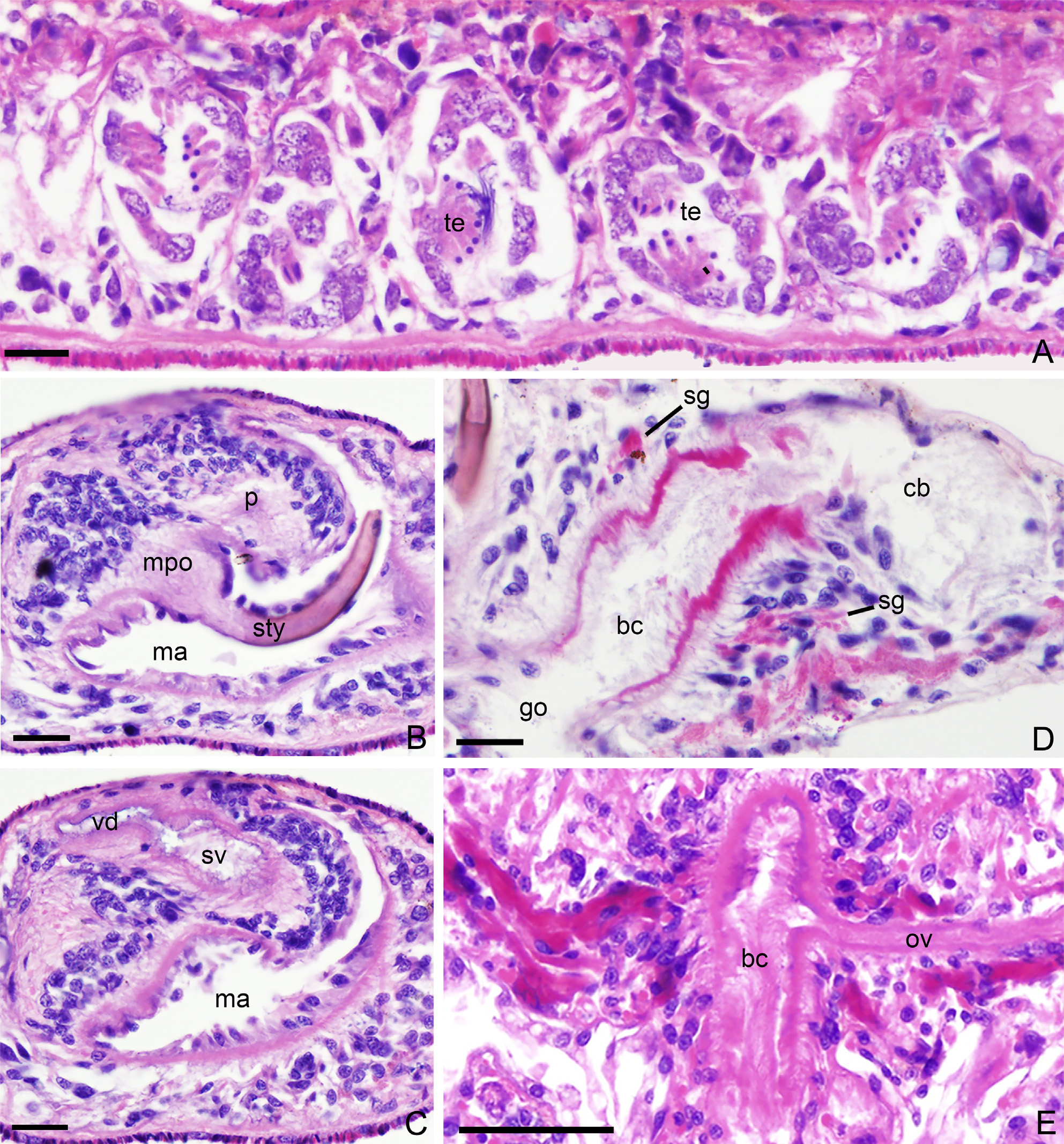
Mature individuals have 8–11 testes on either side of the body, the ventrally located follicles extending from directly behind the ovaries up to about half-way along the pharyngeal cavity ( Figs. 7C View FIGURE 7 , 9A View FIGURE 9 , 10A View FIGURE 10 ). Immediately posterior to the mouth opening the vasa deferentia converge towards the midline of body, meanwhile turning towards the dorsal body surface, after which they separately penetrate the penis and then immediately unite to form a short common vas deferens ( Figs. 7E View FIGURE 7 , 9C View FIGURE 9 , 10 View FIGURE 10 , 11A View FIGURE 11 ). The latter opens into a spacious, oval-shaped seminal vesicle that is situated at the base of the penis papilla and communicates with the ejaculatory duct ( Figs. 9C View FIGURE 9 , 10 View FIGURE 10 ). The seminal vesicle is lined with a simple, cuboidal, nucleated epithelium and covered with a well-developed layer of muscles. The ejaculatory duct is rather broad and lined with a nucleated epithelium and surrounded by a welldeveloped layer of circular muscles; it opens at the tip of the penis papilla. The latter is a sausage-shaped structure that lies dorsally in the male atrium. The penis papilla is lined with a nucleated epithelium that is underlain by circular and longitudinal muscles ( Figs. 9B View FIGURE 9 , 10 View FIGURE 10 , 11A View FIGURE 11 ).
A musculo-parenchymatic organ projects from the anterior wall of the male atrium, ventrally to the root of the penis papilla. The entire organ consist of a broad, muscular base to which is attached a sclerotic, hollow tip or stylet that is curved like a scimitar and that measures 76–80 µm length. The broad base of the musculo-parenchymatic organ consists of a folded epithelium, which is underlain by well-developed circular and longitudinal muscles ( Figs. 9B View FIGURE 9 , 11A View FIGURE 11 ).
A pair of ovaries is situated at a short distance behind the brain ( Figs. 7C View FIGURE 7 , 8B View FIGURE 8 , 10A View FIGURE 10 ). The oviducts arise from the antero-lateral wall of the ovaries. At the level of the gonopore the oviducts turn medially to open separately into the bursal canal ( Figs. 9E View FIGURE 9 , 10 View FIGURE 10 ). The latter is lined with a cuboidal, nucleated epithelium, bearing well-developed cilia, and is surrounded by circular muscle. The bursal canal receives the secretion of shell glands ectally, that is, ventrally to the oviducal openings and is connected with a sac-shaped copulatory bursa, the latter being lined with vacuolated cells ( Figs. 9D View FIGURE 9 , 10B View FIGURE 10 , 11A View FIGURE 11 ).
Feeding and reproduction. Under laboratory conditions, P. falcata seldom moves when starved, and usually assembles in groups that hide from light. When the animals are in search of food, firstly their head touches the food lightly, after which they move their tail end towards the prey item and extend their pharynx, which starts to engulf portions of the prey. After feeding, the animals group together again, away from direct light. We observed that 4 or 5 mature individuals of P. falcata may feed on a prey specimen of Dugesia sinensis of about 10 mm in length. When not starved, copulations may occur. During copulation the two bodies intertwine (either clockwise or counter-clockwise, depending on the copulants), with their heads pointing downwards and the tails pointing upwards ( Fig. 11B View FIGURE 11 ). The process of copulation, during which the tails quiver, lasts for approximately 10 minutes, after which the partners separate.
During cocoon laying, a small amount of mucus is discharged from the common atrium, with which the capsules are being attached tightly to the substrate. The spherical cocoon, which is devoid of a pedicel, is brownred, measuring 340–400 µm in diameter ( Fig. 7F View FIGURE 7 ). From a single cocoon hatches 1–2 young worms (n=5 cocoons observed).
Discussion. When the features exhibited by Paucumara falcata are fed into the key to the genera in Sluys (1989a) the identification leads to the genus Vatapa Marcus, 1948 . This is due to the fact that also Vatapa possesses a musculo-parenchymatic organ (mpo) and the key only offers the choice between mpo present or absent. However, in Vatapa the mpo consists of a large indentation of the posterior wall of the common atrium (see Sluys 1989a) and thus differs greatly from the mpo of P. falcata . Other features also point to the fact that P. falcata belongs to the genus Paucumara and not to Vatapa .
The genus Paucumara was diagnosed as follows: " Ectoplaninae in which the vasa deferentia unite just within the penis bulb to form a more or less curved common vas deferens. Ventral testes, extending to half-way along the pharyngeal pocket or to the level of the mouth. Bursal canal receives the secretion of numerous unicellular glands" ( Sluys 1989a, p.191–192). All of these features apply to P. falcata , while there are a few other characters that point also to a close relationship between P. falcata and P. trigonocephala . Both species exhibit an attenuated head, formed by a narrowing of the body width anterior to the eyes, as well as whitish specks or transverse bands on the dorsal body surface. The eyes are set close together and are provided with a lens, while the posterior gut trunks unite in the hind end of the body.
Paucumara falcata shows one great difference with P. trigonocephala as the latter does not possess the musculo-parenchymatic organ (mpo). However, accessory reproductive structures resembling the musculoparenchymatic organ of P. falcata are present in Pacifides psammophilus Holmquist & Karling, 1972 and P. gladiatoris Sluys, 1989 (see Sluys 1989a). But in the two last-mentioned species these organs concern musculoglandular organs (mgo). In particular the mgo of P. gladiatoris , with its sickle-shaped, sclerotic tip, resembles very much the mpo of P. falcata . Because of this gross resemblance one may initially be inclined to assign P. falcata to the genus Pacifide s. However, upon further reflection it becomes clear that other characters preclude such a taxonomic assignment and point to the fact that P. falcata falls in the genus Paucumara , thus illustrating convergent evolution of such accessory reproductive organs.
Sluys (1989a) synonymized Fovia graciliceps Stimpson, 1857 from Hong Kong, China with P. trigonocephala , albeit with some reservations. Now that we know that another species with an acute triangular head and a pointed tail occurs on Chinese coasts close to Hong Kong, China, i.e., P. falcata , it may be that F. graciliceps actually is a junior synonym of P. falcata . Unfortunately, there is no way to ascertain what actually holds true and therefore the epithet graciliceps should remain suppressed.
Paucumara trigonocephala has been reported from Japan, Australia, the Bismark Archipelago, and South Korea, where it was always collected from low-salinity biotopes, which during ebb tide even may be completely fresh ( Yang et al. 2018). This resembles the habitat of P. falcata , which was collected from 19‰–20‰ salinity biotopes, while in the laboratory animals can also survive in freshwater.


| IZCAS |
Institute of Zoology, Chinese Academy of Sciences |
No known copyright restrictions apply. See Agosti, D., Egloff, W., 2009. Taxonomic information exchange and copyright: the Plazi approach. BMC Research Notes 2009, 2:53 for further explanation.
|
Kingdom |
|
|
Phylum |
|
|
Class |
|
|
Order |
|
|
Family |
|
|
Genus |