
Nerpa fistulata Wang & Chen, 2019
|
publication ID |
https://doi.org/ 10.11646/zootaxa.4568.1.9 |
|
publication LSID |
lsid:zoobank.org:pub:CA1AD232-3D40-4D10-82A6-2A6F61A2730D |
|
DOI |
https://doi.org/10.5281/zenodo.5936973 |
|
persistent identifier |
https://treatment.plazi.org/id/ED5EC61A-9C78-8E2E-FF00-FE081680F82E |
|
treatment provided by |
Plazi |
|
scientific name |
Nerpa fistulata Wang & Chen |
| status |
sp. nov. |
Nerpa fistulata Wang & Chen , sp. nov.
Material examined. Holotype: PLA-N001, eastern beach of Shenzhen City , Guangdong Province, China, 22°28′N, 114°31′E, 29 th April 2017, coll. Jia-Jia Chen, sagittal sections on 2 slides. GoogleMaps
Paratypes: PLA-N002, ibid., sagittal sections on 3 slides; PLA-N003, ibid., sagittal sections on 2 slides; PLA- N004, ibid., horizontal sections on 1 slides; PLA-N005, ibid., whole mount on 1 slide; PLA-N006, ibid., whole mount on 1 slide. All material is deposited in the Institute of Zoology, Chinese Academy of Sciences ( IZCAS) .
Etymology. The specific epithet is derived from the Latin adjective fistulatus, provided with pipes, and alludes to the characteristic intestinal commissures in this species.
Diagnosis. Nerpa fistulata is characterized by: transparent body; semi-circular lens in each eye cup; pentamerous intestine with three distinct commissures, one in the posterior end of the body and the other located between the ovaries and the testes; two very large, prepharyngeal testis follicles; distal end of the penis papilla with a chitinized, pointed stylet; two rounded lateral bursae.
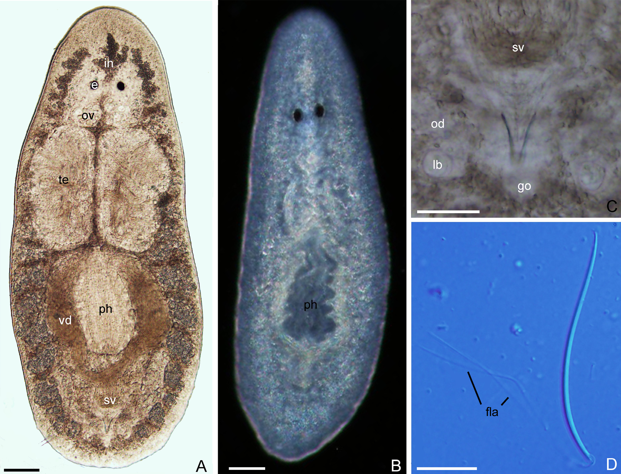
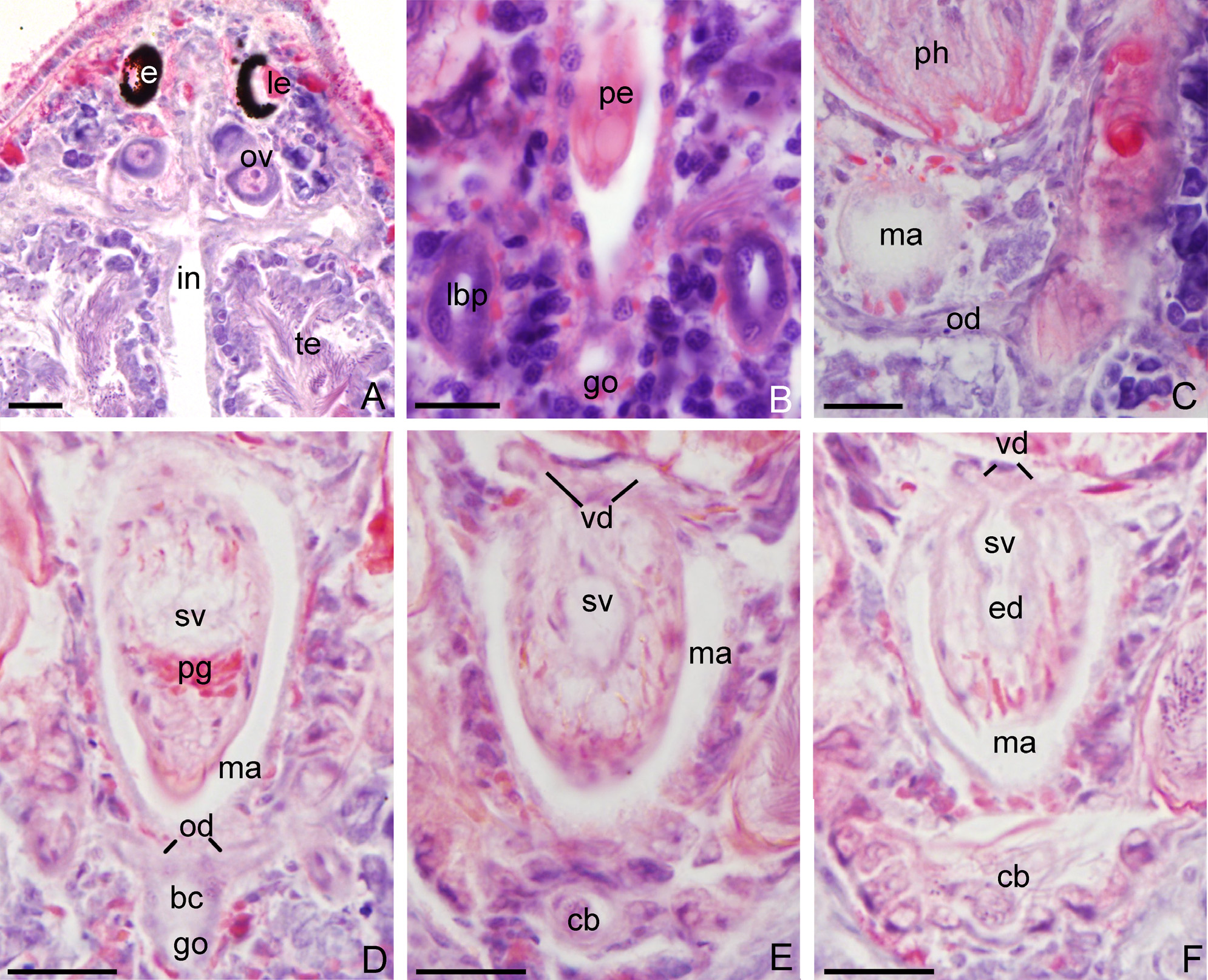
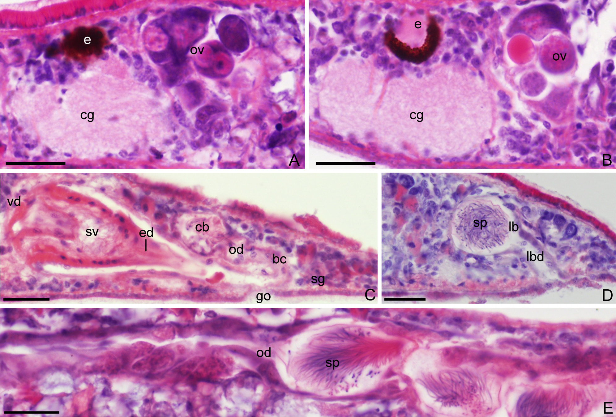
Description. Live animals transparent, without any pigmentation. Body shape varies from elongate to broadly oval-shaped, with a broadly rounded posterior end and a blunt anterior end. Length and width of active specimens varies between 800–1,180 µm (n=5) and 210–435 µm (n=5), respectively ( Figs. 2 View FIGURE 2 A–B, 3A, 6A). A pair of black and elliptical eyes is located at 1/5 of the body length, measured from the anterior margin ( Figs. 2 View FIGURE 2 A–B, 3A, 4A, 6A). The eyes are situated rather close together, the distance between the eye cups being about 25 µm (n=5) and the distance to the body margin being 90–135 µm (n=5). Each eye cup has a large semi-circular lens.
The mouth opening is situated median-ventrally at 1/5 of the body, measured from the posterior body margin; the mouth opening is located at the posterior end of the pharyngeal cavity ( Fig. 6A View FIGURE 6 ). The cylindrical pharynx measures 200–300 µm in length and 130–150 µm in width (n=5) ( Figs. 2 View FIGURE 2 A–B, 3A, 6A).
The principally pentamerous intestine shows three distinct commissures, one uniting the two posterior gut trunks in the hind end of the body, another immediately behind the testes, and a third commissure connecting the central part of the anterior intestinal trunk with the two lateral and forwards directed branches; the latter commissure runs between the ovaries and the testes. This results in the situation that the gut forms three rings, with one pair each encircling the large testes follicles and another, larger ring encircling the pharyngeal pocket and the copulatory apparatus. The median, anterior intestinal trunk extends forwards anteriorly to the eyes and the brain, which holds true also for the forwards directed lateral branches ( Fig. 6A View FIGURE 6 ). Each of the major gut trunks gives rise to 12–16 short lateral diverticula.
A pair of large, ellipsoidal testes, 350–375 µm in length and 180–190 µm in width, are situated immediately in front of the pharynx (n=5) ( Figs. 2A View FIGURE 2 , 3A View FIGURE 3 , 6A View FIGURE 6 ). The follicles are packed with sperm; mature spermatozoa measure 58–60 µm in length and are biflagellate, the flagella being 55–56 µm long (n=5) ( Fig. 2D View FIGURE 2 ). The vasa deferentia originate from the ventro-medial wall of the testis follicles and extend posteriorly alongside the pharynx as greatly swollen spermiducal vesicles, which decrease in diameter before opening separately into the intrapenial seminal vesicle ( Figs. 2A View FIGURE 2 , 4 View FIGURE 4 E–F, 5C, 6A–B). The proximal, anterior section of the seminal vesicle is separated from the smaller posterior section by means of a shallow constriction. The posterior section of the seminal vesicle communicates with the ejaculatory duct, which opens at the tip of penis papilla. Penis glands discharge their secretion into the proximal section of the ejaculatory duct ( Figs. 3C View FIGURE 3 , 5C View FIGURE 5 , 6B View FIGURE 6 ). The tip of the penis papilla is provided with a sclerotic stylet, which is about 50 µm long (n=5) ( Fig. 3C View FIGURE 3 ). The penis papilla is lined with a nucleated epithelium and is surrounded by well-developed layers of circular and longitudinal muscles.
The vitellaria are well developed and lie between the lateral diverticula ( Fig. 3A View FIGURE 3 ). A pair of ovaries is situated immediately behind the brain and the eyes ( Figs. 2A View FIGURE 2 , 3A View FIGURE 3 , 4A View FIGURE 4 , 5 View FIGURE 5 A–B, 6A). The ovaries are oval-shaped and have an oblique orientation, as they tilt forwards. After mating, the anterior end of the oviducts extends to form an ampulla, which houses the allosperm ( Figs. 3B View FIGURE 3 , 5E View FIGURE 5 ). From the ovaries each oviduct runs posteriorly and at the level of the posterior portion of the male atrium it gives rise to two branches, one of which communicates with the lateral bursa, while the other opens into the bursal canal ( Figs. 2C View FIGURE 2 , 3B View FIGURE 3 , 6 View FIGURE 6 A–B). From its point of communication with the atrium, the bursal canal curves anteriad and opens into a rounded copulatory bursa, which lies dorsally to the male atrium. The bursal canal, surrounded by circular muscles, is lined with a nucleated epithelium and receives the openings of shell glands ventrally to the oviducal openings, ( Figs. 5C View FIGURE 5 , 6B View FIGURE 6 ).
Two rounded lateral bursae, about 45 µm in diameter, are located on either side of the body, more or less at the level of the gonopore ( Figs. 2C View FIGURE 2 , 5D View FIGURE 5 , 6 View FIGURE 6 A–B); a duct connects each lateral bursa with the ventrally located lateral gonopore. Ample sperm is present in the lateral bursae ( Fig. 5D View FIGURE 5 ). The bursae and the last-mentioned connecting ducts are lined with a single layer of nucleated epithelium and are surrounded by circular muscle.


| IZCAS |
Institute of Zoology, Chinese Academy of Sciences |
No known copyright restrictions apply. See Agosti, D., Egloff, W., 2009. Taxonomic information exchange and copyright: the Plazi approach. BMC Research Notes 2009, 2:53 for further explanation.
|
Kingdom |
|
|
Phylum |
|
|
Class |
|
|
Order |
|
|
Family |
|
|
Genus |