
Thouarella variabilis Wright and Studer, 1889
|
publication ID |
https://doi.org/10.11646/zootaxa.3602.1.1 |
|
publication LSID |
lsid:zoobank.org:pub:10304FBF-3969-4EFA-83F1-BB8A5E2B37F3 |
|
persistent identifier |
https://treatment.plazi.org/id/EE36E867-FF9C-FFF6-FF0A-AE0CFD580AA2 |
|
treatment provided by |
Felipe |
|
scientific name |
Thouarella variabilis Wright and Studer, 1889 |
| status |
|
2. Thouarella variabilis Wright and Studer, 1889 View in CoL
Figs 6 View FIGURE 6 , 7 View FIGURE 7
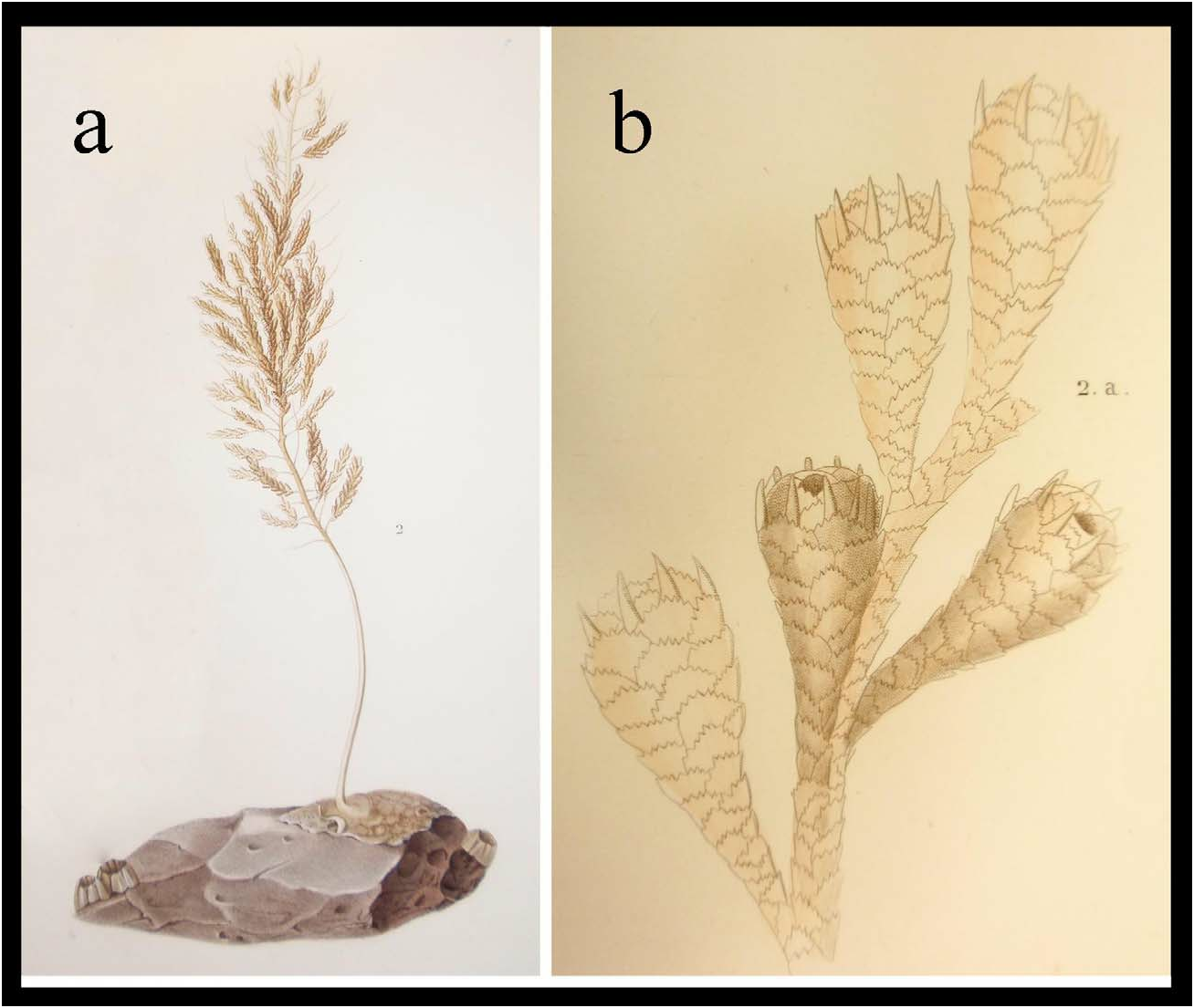
Thouarella variabilis View in CoL var. a Wright & Studer, 1889: 68–69, pl. 21, fig. 1 (incorrectly listed as pl. 14, figs 1–2); Thomson & Henderson 1906: 40 (list)
Thouarella variabilis View in CoL var. c gracilis Wright & Studer, 1889: 70; Thomson & Henderson 1906: 40 (list)
Thouarella variabilis Menneking 1905: 260–262 View in CoL , pl. 9, figs 9, 10, 21, 22 (samples not seen); Versluys 1906: 37–38; Gravier 1914: 56–61, pl. 1 fig. 6, pl. 3 fig. 13–14 (samples not seen); Kükenthal 1915: 150 (key); Molander 1929: 74–5 (samples not seen); Broch 1965: 30–31, pl. 6, figs 17–19; Brito, Tyler & Clarke 1997: 63–69
Not Thouarella variabilis Thomson 1927: 33 View in CoL , pl. 1, fig. 10 (=unknown)
Thouarella (Parathouarella) variabilis Kükenthal 1919: 428 View in CoL , fig. 202 (in text); 1924: 297 (key); Thomson & Rennet 1931: 27–30, pl. 7, fig. 3, pl. 9, figs 4–5 (samples not seen)
Thouarella aff. variabilis Kükenthal 1912: 305–306 View in CoL , figs 9–12 (in text), pl. 20, fig. 2
Thouarella (Thouarella) variabilis typica Cairns & Bayer 2009: 27 View in CoL (listed)
Material examined: Syntype of Thouarella variabilis var. a, NHM 89.5 .27.56, H.M.S. Challenger, sta. 145, SE of Prince Edward Island, 46˚43’S, 38˚4’30”E, 256 m, 27 Dec 1873, 3.5 cm fragment seen; Holotype, T. variabilis var. gracilis, NHM 1889.5.27.55–56, sta. 145, SE of Prince Edward Island , 46˚43’S, 38˚4’30”E, 256 m, 27 Dec 1873, 3.5 cm fragment seen .
Other material: USNM 76897, R/ V Eltanin, Antipodes Island , New Zealand, 49˚51’S, 178˚35’E, 2010–2100 m, 26 Feb 1968 , 1 colony; USNM 98226, R/V Hero , cruise 731, sta. 1842, west of Renaud Island , Biscoe Islands, Antarctic Peninsula, 65˚30’S, 67˚31’W, 180 m, 24 Feb 1973 ; USNM 1130283, R/V Glacier , cruise 1, sta. 5, 76˚00’S, 55˚00’W, 457 m, 9 Feb 1968 , 2 colonies; SMF, EPOS 03 , sta. 281, AGT 21, 402 m, 18 Feb 1989 ; SMF, Am Twist, D. Sudpolar Expedition, 385 m , 1902.
Description of var. a typical
The main stem is simple. Wright and Studer (1889: pl. 21, fig. 1) recorded a specimen 300 mm long (which must have been damaged since as it now stands at 220 mm); its axis is brown-yellow and firm but brittle towards the base and more flexible distally. Branchlets leave the main stem in 3 to 4 directions; the fourth branch in a series is often in line with the first, in a spiral formation. Branchlets occur at intervals of 1.5–2.0 mm, at near right angles to the main stem; branchlets are 50–150 mm long, narrow, flexible, and frequently branched 2 or 3 times (usually dividing close to the stem in the proximal one third).
On the branchlets and stem there are wide, distally flared isolated polyps ( Fig. 6b,d–f View FIGURE 6 ) which are upwardly inclined at 60˚ and arranged in irregular short spirals of 3 or 4 with 5–10 polyps per cm on the branchlets (sometimes more closely spaced at branchlet base). Polyps are 1.50–1.85 mm high (average 1.70 mm) including the long marginal point, with 4–5 scales in the abaxial row and 2–3 in adaxial rows; the number of longitudinal rows reduces quickly from 8 at the marginals to 4 or 5 at the base.
The polyps are sheathed in scales of 4 categories: 8 operculars, 8 marginals, 1 or 2 circlets of pointed submarginals and a variable number of body-wall scales.
The operculars do not form a perfect opercular cone and there are gaps into the opercular cavity ( Fig. 6e View FIGURE 6 ). They are shaped like an isosceles triangle ( Fig. 7a–c View FIGURE 7 ), range in size from 330–650 µm high (average 470 µm) and 150–370 µm wide (average 250 µm), with a H:W of 1.1–2.4 (average 1.9), which is just over half the size of marginals. The proximal half of the inner surface of the operculars are tuberculate; a flat-surfaced, relatively simple keel is present on the inner surface ( Fig. 7c View FIGURE 7 ). The outer surfaces of the scales are covered with granules that extend radially from the central point in the proximal half.
The marginals are long, spinose ( Fig. 7e–g View FIGURE 7 ), 650–960 µm in height (average 800 µm), 430–580 µm wide (average 520 µm), with an average H:W of 1.6, usually splayed out and too long to fold over the operculum neatly. Sometimes the abaxial 2 or 3 marginals are much longer than the adaxial ones ( Fig. 6e View FIGURE 6 ). The outer marginal surface has 2 or 3 longitudinal furrows down the length of the elongated distal point and granules cover the remaining scale area ( Fig. 7e,g View FIGURE 7 ). The keel is channelled and the base of the keel is tuberculate.
The submarginals have a pointed distal edge ( Fig. 7h–k View FIGURE 7 ). They are wider than the marginals, with a width of 440–610 µm (average 525 µm), and a height of 520–600 µm (average 560 µm). They curve away from the polyp body (similar slant to marginals) and are thus visible from an anterior view (see Figs 6e View FIGURE 6 , marked 1, and 6f).
The body-wall scales are round to elliptical in shape ( Fig. 7l–n View FIGURE 7 ), larger and wider towards polyp head (average of 400 µm high, 485 µm wide, H:W 0.9), smaller and round towards polyp base (average of 325 µm high, 455 µm wide, H:W 1.2). The distal edge of the sclerites in all the above categories is finely serrate; proximal edge is irregularly lobate.
The outer layer of coenenchymal scales are elliptical ( Fig. 7o,s,p View FIGURE 7 ), 190–320 µm high (average 240 µm), 590–750 µm wide (average 680 µm) with an average H:W of 0.35. The outer surface of these scales is covered in granules, the inner surface is tuberculate. The inner layer of scales are smaller, thin and roughly circular ( Fig. 7q,r View FIGURE 7 ), with granules on the outer surface and a finely tuberculate inner surface. Coenenchymal scales generally have finely serrated proximal and distal edges.
Distribution
Circum-Antarctic, from 115 to 2100 m depth.
Remarks
Thouarella variabilis View in CoL var. a (hereafter called T. variabilis typica View in CoL ) and T. variabilis var. gracilis Wright & Studer, 1889 View in CoL have identical polyp shape and structure, and sclerite sizes and shapes. Thouarella variabilis var. gracilis View in CoL is described as, what we interpret from Wright and Studer (1889), having more secondary and tertiary ramification than T. variabilis typica View in CoL . However, there are varying degrees of ramification within a single colony and given the similar nature of all sclerites these two varieties are thus proposed to be synonymous with T. variabilis View in CoL .
Within the material examined, we found the branching of T. variabilis to occur in up to four directions (rather than three directions as noted in Wright & Studer 1889). Some juvenile colonies can appear almost pinnate (e.g. SMF, EPOS 03, sta. 281), although on closer inspection branchlets depart in three directions. Also, although there is some regularity with spiral branchlet placement, it is not consistent.
USNM 1130283 and SMF ‘Am Twist’ samples have smaller polyps than the holotype, their maximum length being 1.5 mm rather than 1.5–1.85 mm, but are otherwise identical .
The complex bushy bottlebrush shape of T. variabilis specimens make them ideal habitat for associates and worms, brittlestars, and ascidians were found within their branches.
True to its name, T. variabilis can have polyps that look dissimilar ( Brito 1993); polyps with elongated clawlike marginals are shown in Fig. 6c View FIGURE 6 (some of these polyps were brooding) and open, flared polyps ( Fig. 6b View FIGURE 6 ) with elongated submarginals ( Fig. 6b View FIGURE 6 1 View FIGURE 1 ) are also common.
Comparisons
Thouarella variabilis is unusual within the genus Thouarella in having elongated marginals that do not fold over the operculum neatly, and it is this character that makes this species distinct from all others. The species has a similar number of scales in the abaxial row as T. pendulina , T. hicksoni , T. brucei , and T. striata and comparisons to these species are made here:
Polyps of T. pendulina are much smaller and more clustered (up to 70 per cm) than those of T. variabilis .
Polyps of T. hicksoni are a similar size and have a comparable number of abaxial scales to those of T. variabilis , however, the isosceles triangle-shaped operculars in the latter differ from the operculars of the former which have a blunt, rounded distal edge (and operculars of T. variabilis which consequently form a tighter opercular cone). Also, T. hicksoni lacks elongated submarginals, although they can have a pointed distal edge.
Polyps of T. brucei are a similar size to T. variabilis but have wider triangular operculars.
Opercular, marginal, and submarginal sclerites of T. striata have densely arranged granules on their outer surface that could be confused with those on sclerites of T. variabilis . However, the inner surface of T. striata marginals have sharp striations running perpendicular to the distal edges; these are absent in T. variabilis .
Although now transferred to Plumarella , the species originally described as Thouarella diadema has polyps with 3–5 body-wall scales in the abaxial row and polyps of a similar-size and shape to those of T. variabilis . The major difference between these two species is that marginals of the former species lack a keel (the defining difference between Plumarella and Thouarella ).


| SMF |
Forschungsinstitut und Natur-Museum Senckenberg |
No known copyright restrictions apply. See Agosti, D., Egloff, W., 2009. Taxonomic information exchange and copyright: the Plazi approach. BMC Research Notes 2009, 2:53 for further explanation.
|
Kingdom |
|
|
Phylum |
|
|
Class |
|
|
Order |
|
|
Family |
|
|
Genus |
Thouarella variabilis Wright and Studer, 1889
| TAYLOR, M. L., CAIRNS, S. D., AGNEW, D. J. & ROGERS, A. D. 2013 |
Thouarella (Thouarella) variabilis typica
| Cairns, S. D. & Bayer, F. M. 2009: 27 |
Thouarella variabilis
| Thomson, J. A. 1927: 33 |
Thouarella (Parathouarella) variabilis Kükenthal 1919: 428
| Thomson, J. A. & Rennet, N. I. 1931: 27 |
| Kukenthal, W. 1919: 428 |
Thouarella aff. variabilis Kükenthal 1912: 305–306
| Kukenthal, W. 1912: 306 |
Thouarella variabilis
| Brito, T. A. S. & Tyler, P. A. & Clarke, A. 1997: 63 |
| Broch, H. 1965: 30 |
| Molander, A. R. 1929: 74 |
| Kukenthal, W. 1915: 150 |
| Gravier, C. H. 1914: 56 |
| Versluys, J. 1906: 37 |
| Menneking, F. 1905: 262 |
Thouarella variabilis
| Thomson, J. A. & Henderson, W. D. 1906: 40 |
| Wright, E. P. & Studer, T. 1889: 68 |
Thouarella variabilis
| Thomson, J. A. & Henderson, W. D. 1906: 40 |
| Wright, E. P. & Studer, T. 1889: 70 |