
Eurypedus Gistel
|
publication ID |
https://doi.org/ 10.11646/zootaxa.4161.3.2 |
|
publication LSID |
lsid:zoobank.org:pub:91523D20-348F-40AE-865D-BA49D28E913F |
|
DOI |
https://doi.org/10.5281/zenodo.6057418 |
|
persistent identifier |
https://treatment.plazi.org/id/EE398790-FFBD-FFFC-59DD-FDF3D177D796 |
|
treatment provided by |
Plazi |
|
scientific name |
Eurypedus Gistel |
| status |
|
( Figs 1 View FIGURES 1 – 5 –40)
Eurypedus Gistel 1834: 31 View in CoL [description, type species: Cassida oblonga Sturm in Thon (1827) by monotypy]; Barber 1946: 290 [as valid generic name]; Seeno & Wilcox 1982: 175 [checklist]; Borowiec 1999: 171 [catalogue]; Borowiec & Świętojańska 2014 [online catalogue].
Ischyrosonyx Sturm 1843: 273 [catalogue, type species: Cassida oblonga Sturm in Thon (1827) by monotypy]; Chapuis 1875: 382 [description]; Spaeth 1914: 65 [catalogue]; Hincks 1952: 330 [key to tribes, genera list].
Type species. Cassida oblonga Sturm in Thon (1827) by monotypy (= E. peltoides (Boheman) in this study, see Remarks section of E. peltoides ).
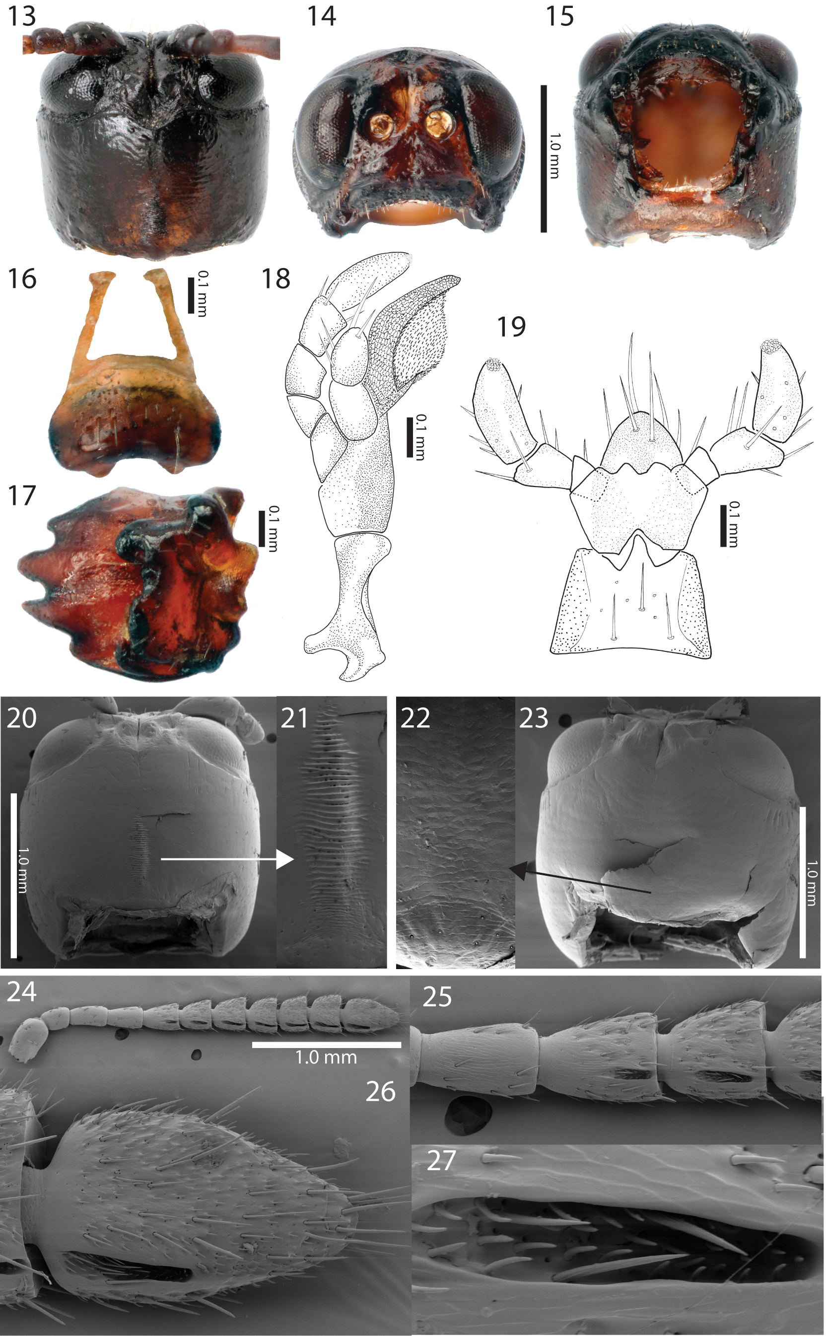
Diagnosis. Eurypedus ( Figs 1 View FIGURES 1 – 5 –12) is distinguished from the other six genera of Ischyrosonychini by its oblong and laterally parallel-sided body, elytral lamella narrower than elytral submesal interval, and prosternal process between procoxae narrower than trochanter ( Borowiec & Świętojańska 2014). It can also be distinguished by newly found diagnostic characters: presence of notches on the ventral surface of antennomeres V–XI (Figs 24–27), and projections on each anterolateral region of the prosternum on the ventral surface of the pronotum ( Figs 29–30 View FIGURES 29 – 30 , marked by black arrow). The detailed morphological features of the stridulatory files on the head can be also used to distinguish Eurypedus from the other six genera of Ischyrosonychini , including the sexual dimorphism with respect to the stridulatory file (Fig. 20–23, see Discussion), the number of ridges on the stridulatory file (48–59, Fig. 21), and the absence of a microtrichial patch on anterior portion of stridulatory file (Fig. 20).
Description. Color ( Figs 1 View FIGURES 1 – 5 –12) mainly red to yellowish brown, occasionally tan, with black marks; pronotal and elytral bases with black spots or marked differently depending on individual; antennae black with brown setae; venter and legs generally shiny, black or blackish red, with basal half of each ventrite often brown to reddish brown; setae on tarsus reddish brown to tan.
Body ( Figs 1 View FIGURES 1 – 5 –12) oblong with anterior margin of pronotum rounded or weakly sinuate, broadest between posterior 1/3 region of pronotum and middle of elytra in dorsal view; profile ovoid with pronotal anterior margin slightly lifted.
Head ( Figs 13–15 View FIGURES 13 – 15 ) completely concealed by pronotum in dorsal view; shape roundly quadrate, broadest at middle, slightly broader than long in dorsal view, with stridulatory file in posteromedial region of vertex. Frons triangular with upper margin slightly projected, sparsely punctuate, medially depressed by medial frontal line; lateral region of anterior margin of frons fitted with depression above condylic projection; clypeus narrow with anterior margin slightly arched; frontoclypeal sulcus indistinct; interantennal region slightly broader than antennal socket. Eyes large, oval, bulging, located on upper anterolateral region of head; interocular area about 1.8 times as broad as eye diameter at broadest point in anterior view, slightly depressed with deep medial sulcus. Antennae (Figs 24–27) reaching elytral base under pronotum; antennomere I oval, with length about 2 times width and about 2 times length of antennomere II; antennomere II length subequal to width; antennomeres III–IV slender, slightly expanded apically; antennomere III shiny, sparsely setose; antennomere IV often more pubescent than antennomere III and less than antennomeres V–XI; antennomeres V–XI each pubescent, with notch on ventral surface; antennomere VII length equal to width; antennomeres VIII–X each with length shorter than width; antennomere XI length 2 times length of antennomere X.
Mouth fossa ( Fig. 13 View FIGURES 13 – 15 ) rounded, subquadrate, with upper half broader and more sclerotized than lower half. Labrum (Fig. 16) with basal half withdrawn under frontoclypeus, sparsely punctate and setose, broadest at middle, with anterior edge well-sclerotized; anterior margin emarginate medially with paired projections. Mandible as in Fig. 17, well-sclerotized, fist-shaped, with 4 teeth; apical half oriented toward mouth fossa; middle region projected, sparsely punctate and setose. Maxilla (Fig. 18) long and slender; cardo distinctly narrower at middle than at base and apex; stipes slightly longer than cardo, narrower at apex than at base; lacinia membranous, oval, greater in length than galea, densely setose; galea 2-segmented with basal segment slightly greater in length than apical segment; apical segment of galea oval, with long setae; palpus 4-segmented with palpifer laterally connected to middle of stipes; palpomere I triangular, slightly shorter in length than palpomere II; palpomere II slightly curved, with length greater than width, slightly expanded apically; palpomere III equal in length to palpomere II, expanded apically, with long setae near apex; palpomere IV elongate oval, with length about 1.5 times that of palpomere III, with sensillae on apex. Labium ( Figs 8 View FIGURES 6 – 8 , 11, 19) with mentum withdrawn into prosternum in ventral view; ligula oval with long setae on apical region; palpus 3-segmented; palpomere I quadrate, slightly expanded apically; palpomere II irregularly quadrate, expanded apically, with outer length about 1.8 times inner length, with long setae in apical region; palpomere III elongate oval, slight curved, sparsely setose, with sensillae on apex.
Pronotum ( Figs 1 View FIGURES 1 – 5 –12) hemispherical in dorsal view, widest near base or occasionally between posterior 1/3 and base; posterior and lateral margins of pronotum forming distinct angle; profile irregularly trapezoidal with rounded dorsum, highest at base; pronotal disc convex, smooth, finely punctate, often with shallow furrow medially and depression in posteromedial region; pronotal lamella separated from disc by depression, except in anterior region; posterolateral region of pronotal base and posterior margin of pronotal hypomeron emarginate; posterior margin of pronotum extended posteriorly.
Prosternum ( Figs 29–30 View FIGURES 29 – 30 ) convex medially, with distinct short collar; anterolateral margin projected onto ventral surface of pronotum ( Figs 29–30 View FIGURES 29 – 30 , marked by black arrow); projections forming upper-lateral part of cervical cavity; tergosternal sulcus distinct; prosternal process smooth with arrow-shaped apex; apex distinctly depressed laterally.
Mesonotum (Fig. 31) pentagonal with anterior apodemes well-developed; mesoscutellum well-sclerotized, triangular, convex with anterior half withdrawn into pronotal base. Mesoventrite (Fig. 32) deeply notched in middle; mesepisternal ridge well defined; process of mesoventrite extended beyond middle of mesocoxal cavity, rigidly connected to metaventrite.
Metanotum (Fig. 33) over 2 times wider than long; scutellar groove and scutoscutellar sulcus distinct. Metaventrite (Fig. 32) flat or slightly convex medially, with distinct longitudinal sulcus medially, slightly broader posteriorly; anterolateral region convex, extended anteriorly, forming mesocoxal cavity.
Elytra ( Figs 1 View FIGURES 1 – 5 –12, 34) oblong, slightly convex, broadest near base, with base of each elytron rounded; anterior margin crenulate; surface shiny, finely punctate; punctures similarly sized; punctures forming striae, occasionally arranged irregularly in lateral region; elytral lamella narrow, distinct from elytral disc, with thickened edge continuous to posterior end; brace (Fig. 34) distinct, with posterior end weakly connected to longitudinal carina, forming an angle.
Legs ( Figs 29–30 View FIGURES 29 – 30 , 32) slender, shiny, extending beyond elytral margin when extended; trochanters elongate, triangular; femoral width subequal to length of trochanter or slightly narrower, broadest at middle, with base much narrower than distal end; mesotibia shorter than mesofemur (protibia and metatibia as long as each respective femur); tibiae coarsely setose apicolaterally, broadened apically, with apicodorsal end notched medially and projected laterally; tarsus dorsally convex, sparsely setose; ventral surface of tarsus densely setose; tarsomere I small, hemispherical; tarsomere II weakly bilobed with apices of lobes pointed, length about 2 times length of tarsomere I; tarsomere III deeply bilobed, with length 3 times length of tarsomere I; tarsomere V as long as tarsomere III, slender, slightly expanded apically, hiding base of pretarsal claws; pretarsal claws robust, evenly curved, tapered.
Abdomen ( Figs 8 View FIGURES 6 – 8 , 11) with outline subparallel, rounded apically; venter slightly convex medially, with surface finely setose; each ventrite well-sclerotized with depressions laterad of meson; ventrite I with greatest length; ventrites II–IV subsequently shorter and narrower; ventrite V with length equal to that of ventrite IV, with more distinct lateral depressions.
Aedeagus ( Figs 35–38 View FIGURES 35 – 38 ) curved in lateral view, slightly broader medially, with aedeagal basal piece oval; apical end pointed; tegmen well-sclerotized, Y-shaped; gastrale speculum ( Fig. 35 View FIGURES 35 – 38 , marked by arrow) U-shaped, with anterior end slightly extended; ejaculatory duct longer than base piece; seminal vesicle thin, slightly shorter than aedeagal base piece, with sclerotized bead ( Fig. 37 View FIGURES 35 – 38 ; marked by arrow) between ejaculatory duct and seminal vesicle.
Spermatheca (Figs 39–40) falcate, short; basal part longer than apical part; receptacle fused to pump, with one opening; spermathecal duct long and coiled.
Remarks. Antennal notches on the ventral surface of antennomeres V–XI (Figs 24–27) are also found in 10 species of Cistudinella — C. apiata (Boheman) , C. foveolata Champion , C. inanis (Boheman) , C. lata Spaeth , C. lateripunctata Spaeth , C. notata (Boheman) , C. obducta (Boheman) , C. parva (Wagener) , C. peruana Spaeth , and C. punctipennis (Boheman) (personal observation). Similar antennal notches were also observed in other tortoise beetles ( Chaboo 2007; Simões & Monné 2014). However, the number of antennomeres with antennal notches varied: seven notched antennomeres (from V to XI) or five notched antennomeres (from VII to XI) (see Chaboo 2007). The projection on each anterolateral portion of the prosternum is found as a unique diagnostic character of Eurypedus ( Figs 29–30 View FIGURES 29 – 30 , marked by black arrow). These projections are weakly connected to the ventral surface of the pronotum.


No known copyright restrictions apply. See Agosti, D., Egloff, W., 2009. Taxonomic information exchange and copyright: the Plazi approach. BMC Research Notes 2009, 2:53 for further explanation.
|
Kingdom |
|
|
Phylum |
|
|
Class |
|
|
Order |
|
|
Family |
Eurypedus Gistel
| Shin, Chulwoo 2016 |
Ischyrosonyx
| Hincks 1952: 330 |
| Spaeth 1914: 65 |
| Chapuis 1875: 382 |
| Sturm 1843: 273 |
Eurypedus
| Borowiec 1999: 171 |
| Seeno 1982: 175 |
| Barber 1946: 290 |
| Gistel 1834: 31 |