
Renicola buchanani (Martin and Gregory)
|
publication ID |
https://doi.org/ 10.11646/zootaxa.4711.3.3 |
|
publication LSID |
lsid:zoobank.org:pub:85D81C2D-0B66-4C0D-B708-AAF1DAD6018B |
|
persistent identifier |
https://treatment.plazi.org/id/EF6AD377-8950-8B31-FF39-FACEFE82FEFD |
|
treatment provided by |
Plazi |
|
scientific name |
Renicola buchanani (Martin and Gregory) |
| status |
|
Renicola buchanani (Martin and Gregory) View in CoL
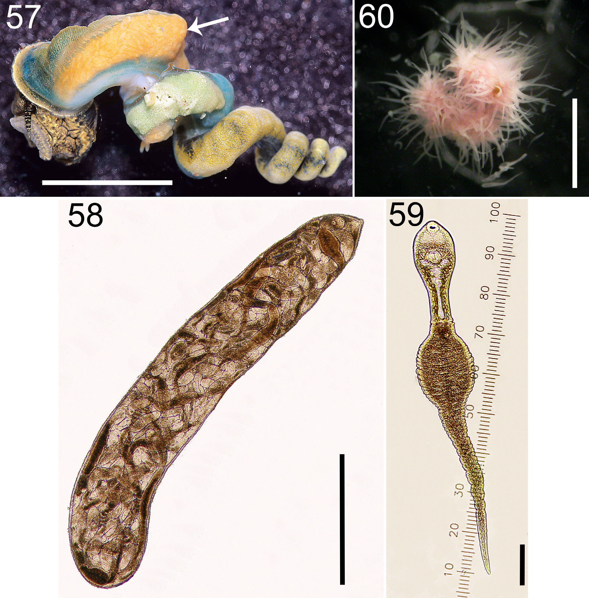
(14. Rebu; Figs. 1 View FIGURE 1 , 57–60 View FIGURES 57–60 )
Diagnosis: Parthenitae. Colony comprised of inactive sporocysts, densely concentrated in snail mantle (in enlarged perirectal sinus). Sporocysts translucent orange, yellow, sometimes white; 1000–2000 µm long, elongate (length: width up to ~6:1), ~sausage-shaped.
Cercaria . Body opaque white; non-oculate; with oral and ventral sucker; with a large Y-shaped excretory blad- der, the arms of which wrap around sides of ventral sucker; body ~ 228 µm long, much shorter than tail (<1/2 length); tail narrow at base, but then with large, sometimes bulbous, proximal portion that narrows distally to a simple tip.
Cercaria behavior: Upon emergence, cercariae either (1) swim solitarily, lashing the distal potion of tail while pressing body adjacent to the greatly inflated, egg-shaped, proximal portion of tail, or (2) form aggregations, where they attach to each other using adhesive proximal-most portions of tail (Martin & Gregory 1951). Cercariae that aggregate together are best referred to as known as “zygocercariae” (see review by Beuret & Pearson 1994). Our unpublished observations clarify that cercariae from the same infection may exhibit these different behaviors (often in different shedding events) (D.C. Metz, unpublished data).
Similar species: The “magnacercous” Rebu is not confusable with any of the other trematodes in this guild.
Remarks: Martin and Gregory (1951) described the sporocysts and cercariae (as Cercaria buchanani ). Based on morphological similarities, Martin (1971) decided that renicolid metacercariae he encountered in estuarine fish livers were the same species, and he assigned the species to Renicola . Hechinger and Miura (2014) provided COI and ITS1 DNA sequence data for this species.
Early Rebu infections can be detected. The sporocysts appear to typically initially form in the basal visceral mass, as generally expected for species that infects the snail with ingested eggs ( Galaktionov & Dobrovolskij 2003) (unpublished observations).
Mature, ripe colonies comprise ~17% the soft-tissue weight of an infected snail (summer-time estimate derived from information in [ Hechinger et al. 2009]).
Nadakal (1960b) presents information on the pigments of the sporocysts and cercariae of this species.
As part of one of the first studies documenting the syncytial nature of trematode integuments, Bils and Martin (1966) examined the fine structure and development of the tegument for the sporocysts and cercariae of this species.
Fingerut et al. (2003a) presents information on the relationship between cercaria emergence and temperature for this species.

No known copyright restrictions apply. See Agosti, D., Egloff, W., 2009. Taxonomic information exchange and copyright: the Plazi approach. BMC Research Notes 2009, 2:53 for further explanation.
|
Kingdom |
|
|
Phylum |
|
|
Class |
|
|
Order |
|
|
Family |
|
|
Genus |