
Myialges anchora Trouessart, 1907
|
publication ID |
https://doi.org/ 10.22073/pja.v8i1.41265 |
|
publication LSID |
lsid:zoobank.org:pub:998D7A66-84BD-4D18-AA66-EEE4C57C513E |
|
DOI |
https://doi.org/10.5281/zenodo.10528613 |
|
persistent identifier |
https://treatment.plazi.org/id/F03E87D2-2574-4846-9FD1-B8BCFCF34F41 |
|
treatment provided by |
Felipe |
|
scientific name |
Myialges anchora Trouessart, 1907 |
| status |
|
Myialges anchora Trouessart, 1907 ( Figs. 5–8 View Figures 5 )
Myialges anchora – Furman and Tarshis (1953), Fain (1965), Macchioni et al. (2005), Mironov et al. (2005), Valim and Gazêta (2007), Marcelino et al. (2009), Bilal (2012).
Type host – Pseudolynchia canariensis ( Diptera : Hippoboscidae ) from domestic pigeons Columba livia .
Material examined
Ten females ( JAZM and ACSIAU) from two locations in Iran, Alborz, Karaj , 35º 48' N, 50º 59' E, alt. 1550 m, collected by O. Joharchi. Six GoogleMaps females ( JAZM and ACSIAU) Italy, Tuscany, Pisa , 43° 42' N, 10° 24' E, alt. 6 m, collected by F. Macchioni GoogleMaps
Redescription
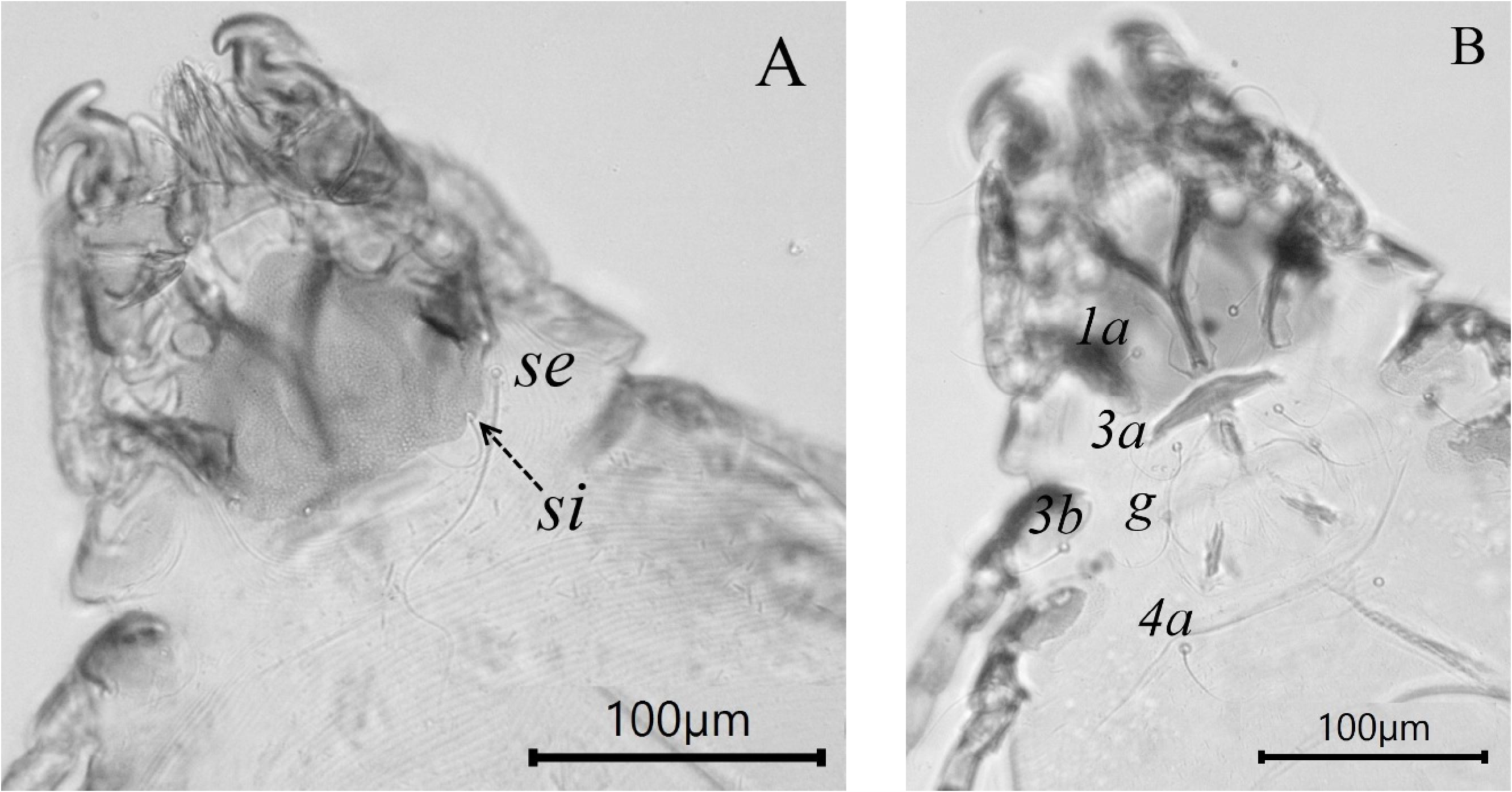
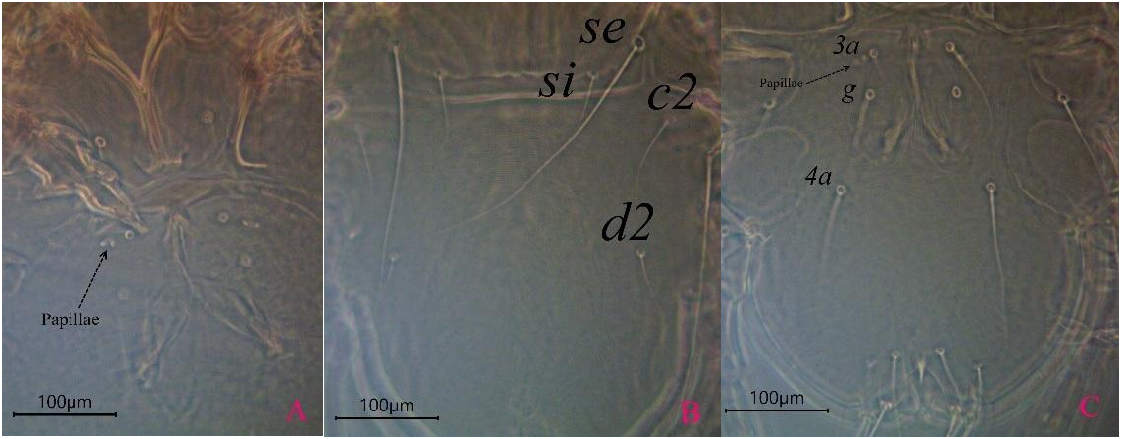
Female ( Figs. 5 View Figures 5 , A, B) (n = 10, all specimens ovigerous) – Idiosomal length × width, 665 × 373 (635–665 × 365–392). Gnathosoma: subcapitulum trapezoidal, strongly attenuated anteriorly, not reach to anterior of apex of Leg I. Length including palps 64 (62–65), width at base 51 (50–58), length/width ratio 1.1–1.2; pseudorutellar membrane not extends laterally; distal part of palps simple; length of chelicerae 75 (58–77). Dorsum ( Fig. 5 View Figures 5 , A): with striated lines. Idiosoma ovoid, without sejugal furrow, dorsal setae smooth, si 41, se 135, c1 35, d2 38, 1a 30, 3a 60, 4b 35, g 47, 4a 48, f2 290, h1 130, h2 62, ps1 52. Prodorsal shields: triangular in shape with compact spot, postero-lateral extensions blunt, not reach to beyond bases of scapular setae se and si or maybe reach to basis of si; antero-lateral curved and extensions to lateral margins of prodorsum and fuse with bases of epimerites Ia; median part of posterior margin slightly concave ( Fig. 8 A View Figure 8 ); greatest length of shield 95 (84–100), width in widest level 126 (120–139). Distance between scapular setae: se: se 103 (103–113), si: si 69 (67–75); setae si postero-mesal to corresponding setae se; distance between transverse levels of these setae 25 (22–26). Scapular shields well developed. Humeral shields present, moderate. Setae c2 on humeral shields. Length of hysterosoma 524 (501–641). Setae h2 located on posterior margin of opisthosma with short extension. Setae h3 ventral, at level of bases of h2. Distance between hysteronotal setae: c2: d2 110 (103–153), d2: e2 152 (147–196), e2: h2 272 (272–318), h2: h2 61 (61– 69). Epimerites I fused in Y-shape, surrounded with narrow sclerotized area; lateral margins of this area surrounded by irregular lines ( Fig. 5 View Figures 5 , 8B View Figure 8 ). Epimerites II slightly curved. Coxal fields II with sclerotized area of triangular form. Coxal fields III and IV ovoid and completely sclerotized. Epimerites IVa absent. Epigynum at level of anterior of epimeites I, thick anchor-shaped, posterior margin with straight line, maximum thickness (at mid-line) 21 (17–23), width 77 (75–85). Genital papillae at level of setae 3a, close to their bases. Folds of ovipore sclerotized in posterior of (g) setae and at mid-line of coxa IV. Setae 3b on sclerotized areas of coxal fields III ( Fig. 8B View Figure 8 ), and with two pairs of genital papillae covered by genital valves and a pair of genital setae ( Fig. 13A View Figure 13 ). Distance between ventral setae: 3a: g 28 (18–28), g: 4a 72 (72–90). Spermathecal duct with the same size than diameter of copulatory opening, spermathecal sac shaped like a bubble. Pretarsi of all legs bilobate. Claw of tarsus I with two arms, anchor shaped. Length of transverse piece of this extension 45 (42– 49) ( Fig. 6). Tibia II sharp, reflexed apophysis; Caruncles bilobed.
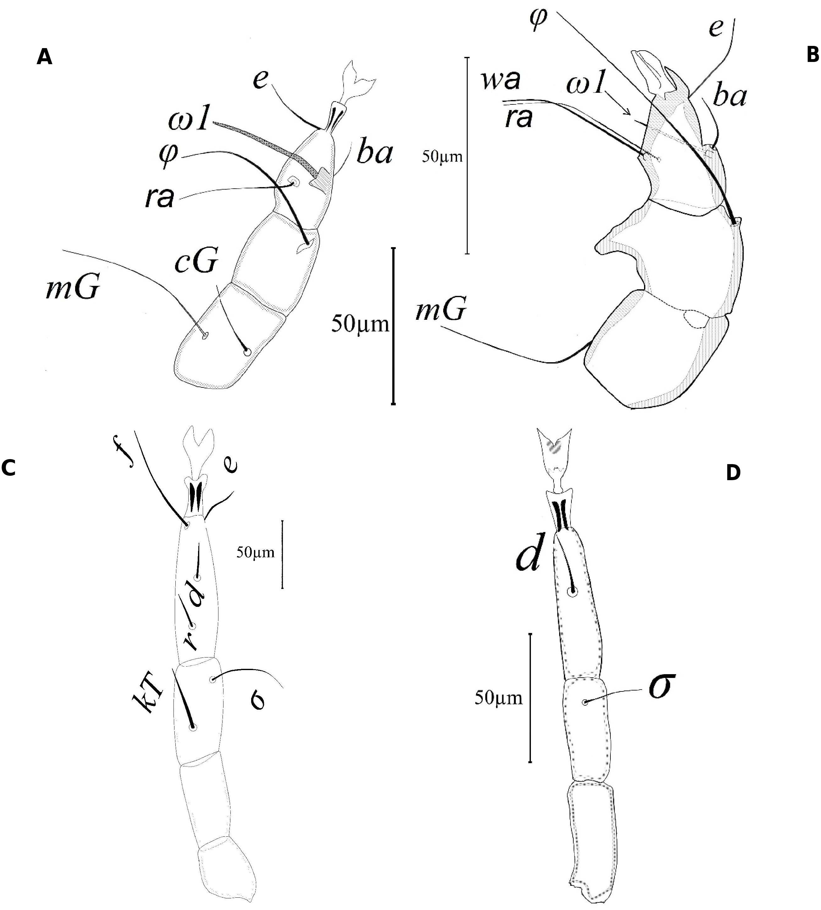
Legs ( Figs. 7 View Figure 7 , A-C) – coxae 1, 0, 1, 0; trochanters 1, 1, 1, 1; femur 1, 1, 0, 0; genua 2, 2 + 1 σ, 0, 0; tibiae 2 + 1 φ, 1 + 1 φ, 1 + 1 φ, 1 + 1 φ; tarsus 5, 7, 5, 4. Claw like structure absent on some legs or smaller and vestigial. Apophyses of tarsi prominent though modified and anchor like on 1st tarsus, claw absent on tarsus II, claw like of tarsus I with two arms, anchor shaped ( Fig. 6), tibia II without apophysis. Legs dark brown pigmented.
Remarks
The two species Myialges anchora Trouessart, 1907 and My. trinotoni Cooreman, 1944 are morphologically most similar to each other, but differ by the shape of tibia II, which bears a reflexed apophysis and bilobed ambulacral disc in M. anchora while in Myialges trinotoni , the tibia II lacks an apophysis and the ambulacral disc is bell-shaped.
Male – not found.
Subfamily Epidermoptinae Trouessart, 1892
Genus Promyialges Fain, 1965
Type species: Microlichus uncus Vitzthum, 1934

No known copyright restrictions apply. See Agosti, D., Egloff, W., 2009. Taxonomic information exchange and copyright: the Plazi approach. BMC Research Notes 2009, 2:53 for further explanation.
|
Kingdom |
|
|
Phylum |
|
|
Class |
|
|
Order |
|
|
Family |
|
|
Genus |
Myialges anchora Trouessart, 1907
| Faradonbeh, Majid Moradi, Ostovan, Hadi, OConnor, Barry M., Gheibi, Mehdi, Joharchi, Omid & Macchioni, Fabio 2019 |
Myialges anchora
| Sergent & Trouessart 1907 |