
Paulita, Guinot, Danièle, 2012
|
publication ID |
https://doi.org/ 10.5281/zenodo.208589 |
|
DOI |
https://doi.org/10.5281/zenodo.6166632 |
|
persistent identifier |
https://treatment.plazi.org/id/F1205E76-056D-FFD4-68CB-FB6B369FF8EB |
|
treatment provided by |
Plazi |
|
scientific name |
Paulita |
| status |
gen. nov. |
Paulita View in CoL n. gen.
Dasygyius Lemos View in CoL de Castro, 1949: 349. Non Dasygyius Rathbun, 1897: 164 . Non Paradasygyius Garth, 1958: 67 View in CoL .
Type species. Dasygyius tuberculatus Lemos de Castro, 1949.
No other species included in the genus.
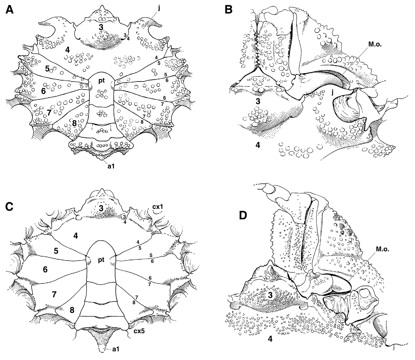
Diagnosis. Carapace ( Figs. 1 View FIGURE 1 , 2 View FIGURE 2 A) urn-shaped, with moderate anterior narrowing, only anterior outline broadly triangular; flattening of the body particularly marked. Dorsal surface ( Figs. 1 View FIGURE 1 , 2 View FIGURE 2 A, B) divided into convex regions delineated by several parallel, deep, broad grooves: cervical groove well marked, with median gastric pits; branchial groove delimitating main part of branchial region; longitudinally, marked branchiocardiac groove. Dorsal surface uniformly covered with prominent tubercules. Lateral margins rounded, unarmed. Pleurites 5–8 ( Fig. 2 View FIGURE 2 A, B) exposed on same level as carapace, calcified, ornamented as carapace; first abdominal somite in prolongation of cephalothorax, dorsally visible, calcified, ornamented; pleurites 5–8, first abdominal somite seemingly parts of carapace; insertion of carapace into setting gutter ( Fig. 2 View FIGURE 2 B); branchiostegite absent. Rostrum ( Figs. 1 View FIGURE 1 , 2 View FIGURE 2 ) simple, broadly triangular, pentagonal. Antennules vertically folded beneath rostrum. Eyestalk narrow. Antenna: articles 1–3 fused; urinal article not sunken in epistome; basal article 2 + 3 large, fused to front, unarmed, granular, outer angle only slightly projecting dorsally; articles 4, 5 free; flagellum short. Supraorbital margin with small median tooth. Exorbital tooth blunt. Pterygostomial lobe ( Fig. 3B View FIGURE 3. A, B ) well marked. Mxp3: ischium with inner margin coarsely dentate, produced into lobe at inner distal angle; merus three-fifths as long as ischium, cordiform; outer angle slightly rounded laterally, excavated at inner subdistal angle for reception of carpus; inner angle produced to prominent, narrow lobe; palp stocky, with carpus enlarged, three-fifths as thick as long. Thoracic sternum ( Fig. 2 View FIGURE 2 A) markedly wide, with conspicuous anterior shield inserted between mxp3; sternite 3 developed, medially inflated; suture 3/4 laterally visible, medially prolonging in deep depression ( Figs. 2 View FIGURE 2 C, D, 3A, B). Sutures 4/5–7/8 all interrupted, with distant interruption points. Sternum/pterygostome junction complete thanks to curved extension of sternite 4. Milne-Edwards openings separated from chelipeds, large, entirely filled by Mxp3 coxa ( Fig. 3B View FIGURE 3. A, B ). Wide sternal extensions joining exposed pleurites (sternum/pleurites connections) between P1 and P2, P2 and P3, P3 and P4, P4 and P5 ( Figs. 2 View FIGURE 2 C, 3A). Sternal device for abdominal-locking system represented by 2 or several granules disposed between sutures 4/5, 5/6; abdominal edge of somites 4–6 markedly thickened, ending in deep socket. Male, female abdomens with first somite developed, entirely dorsally visible in both sexes, ornamented like carapace, not produced into spine. Male abdomen ( Figs. 2 View FIGURE 2 C, 3A) with all somites free, except for somite 6 fused to telson (pleotelson); somite 1 narrow but high; somites 2, 3 widened, somites 4, 5, pleotelson narrow; pleotelson base laterally inflated at location of sockets. Female abdomen ( Fig. 2 View FIGURE 2 D) with somites 1–4 free, not high; somites 5, 6 fused to pleotelson, forming large, flat or convex, disc, inserted in sterno-abdominal cavity; brood cavity closed like a box, limited by sternal elevated arch formed by raised sternites all around. Ventral surface completely covered with salient, round tubercles. Male chelipeds ( Fig. 1 View FIGURE 1 ) equal, unarmed, with minute granules; propodus narrow, elongated, slightly inflated in larger males; fingers long, tapering, gaping in proximal half, distally joined; prehensile border finely denticulated in proximal half, distally distinctly toothed. Female chelipeds ( Fig. 2 View FIGURE 2 D) small, fingers joined. Pereopods ( Fig. 1 View FIGURE 1 ) long, thin, cylindrical throughout length, unarmed; P2 densely fringed with soft hairs in males; P2-P5 with additional setae in females. Coxal male gonopore large, located far from suture 7/8, thus in posteriormost location, gonopore on anterior border of coxo-sternal condyle; penis short. G1 gently curved, with narrow, elongated subdistal lobe. G2 conspicuously short. Vulva opening in anteriorly produced sternite 6, displaced anteriorly to suture 4/5; sternal vulvar cover present. Axial skeleton with lateral compartment together with dorsoventral partition (developed junction plate), pleurites ( Fig. 2 View FIGURE 2 B) being almost horizontal; in anterior region, dorsal edges of pleurites connected to internal surface of carapace by vertical pillars; median line along sternites 7, 8 corresponding to raised median plate on sternite 7; presence of a thick sella turcica.
Etymology. The genus Paulita n. gen., established here for a particularly beautiful and interesting species, is dedicated to Paula Martin-Lefèvre, born Rodríguez Moreno, in recognition of her kindness, knowledge and contribution to the Département Milieux et peuplements aquatiques, MNHN.
Remarks. Paradasygyius Garth, 1958 , was erected in replacement of Dasygyius Rathbun, 1897 ( Garth 1958: 67, 68, 80) to accommodate two species: P. depressus ( Bell, 1835) ( Bell 1835: 88; 1836: 42, both as Microrhynchus depressus ) as the type species by original designation, and Dasygyius tuberculatus Lemos de Castro, 1949 (Lemos de Castro 1949: 349). Not only do the differences between the two species, recognisable in the figures of Guinot & Richer de Forges (1997: figs. 11, 13, 14), require the creation of a new genus, but the peculiar morphology of the new taxon also provides valuable information on Inachoididae , in particular on the evidence of plesiomorphic features in this family.
Paulita View in CoL n. gen. differs from Paradasygyius View in CoL by a number of characters, which include: the broadly triangular carapace ( Fig. 1 View FIGURE 1 ) (distinctly anteriorly narrowing in Paradasygyius View in CoL ; see Rathbun 1925: pl. 1, fig. 1); dorsal surface divided into convex, tuberculate regions by deep, transversal, parallel grooves ( Figs. 1 View FIGURE 1 , 2 View FIGURE 2 A, B) (in Paradasygyius View in CoL surface depressed, with indistinct, finely granular regions, and a few pointed tubercles, and absence of parallel grooves; see Garth 1958: pl. 4, fig. 2); rostrum broadly triangular, pentagonal ( Fig. 2 View FIGURE 2 A) (a narrow triangle in Paradasygyius View in CoL ); eyestalk narrow (wider in Paradasygyius View in CoL ); antennal article 2 + 3 proportionally wide, unarmed on inner margin, outer angle only slightly projecting dorsally (longer, inner margin with two teeth, outer angle conspicuously projecting dorsally by a long, narrow tooth in Paradasygyius View in CoL ); supra-orbital margin with small median tooth (tooth absent in Paradasygyius View in CoL ); exorbital tooth blunt (acute in Paradasygyius View in CoL ); sternum/ pterygostome junction complete, Milne-Edwards openings separated from chelipeds ( Fig. 3B View FIGURE 3. A, B ) (in Paradasygyius View in CoL junction absent, sternite 4 not extended; Milne-Edwards openings not separated from chelipeds); mxp3 coxa long, entirely filling Milne-Edwards opening (shorter and prolonged by distal lobe to embayment in Paradasygyius View in CoL ; Fig. 3 View FIGURE 3. A, B D); mxp3 ischium, thoracic sternum and abdomen coarsely and densely tuberculate in both sexes ( Fig. 2 View FIGURE 2 C, D, 3A) (granular in Paradasygyius View in CoL ; Fig. 3 View FIGURE 3. A, B C); thoracic sternum with convex sternites, sutures in deep grooves ( Figs. 2 View FIGURE 2 C, 3A) (sternum flat in Paradasygyius View in CoL ; Fig. 3 View FIGURE 3. A, B C; see Rathbun 1925: pl. 1, fig. 2); lateral suture 3/4 deep and medially prolonged into a depression (shorter and forming a deep pocket in Paradasygyius View in CoL ); thoracic sternite 2 tuberculate (anteriorly ending as a spine in Paradasygyius View in CoL ; Fig. 3 View FIGURE 3. A, B C); abdominal somite 1 tuberculate, blunt in both sexes ( Figs. 2 View FIGURE 2 C, D, 3A) (granular, produced as long, conical spine in Paradasygyius View in CoL ; Fig. 3 View FIGURE 3. A, B C; see Rathbun 1925: pl. 274, fig. 5); exposed pleurites 5–7 in the form of sclerites with concave edges ( Figs. 1 View FIGURE 1 , 2 View FIGURE 2 A) (each sclerite with long, narrow tooth in Paradasygyius View in CoL ; see Rathbun 1925: pl. 274, fig. 8); G1 (Guinot-Dumortier 1960: fig. 22a–c) gently curved, with a narrow, elongated subdistal lobe (straight and with a short, thick, blunt lobe in Paradasygyius View in CoL ; see Garth 1958: pl. E, fig. 5); chelipeds with long, slightly inflated palm in males ( Fig. 1 View FIGURE 1 ) (palm short, inflated in Paradasygyius View in CoL ); P2 densely fringed with soft hairs in males, other pereopods less setiferous ( Figs. 1 View FIGURE 1 , 2 View FIGURE 2 D) (P2–P5 fringed with setae in male Paradasygyius View in CoL ; see Garth 1958: pl. 4, fig. 2).
The carapace of Paulita View in CoL n. gen. resembles a “human face”, somewhat reminiscent of the dorippid “face” (see below), a unique condition since the remaining Inachoididae View in CoL do not show such a pattern. With its rounded carapace and a first article of the antenna with a urinary orifice not distant from basal article (articles 2+3), Paulita View in CoL n. gen. offers closer affinities with the South-American Leurocyclus Rathbun, 1897 View in CoL , than to Paradasygyius View in CoL ( P. depressus View in CoL ) from the eastern Pacific ( Garth 1958; Hendrickx 1999). P. depressus View in CoL , with its more triangular carapace and narrow front, is closer to other Inachoididae View in CoL . Leurocyclus View in CoL is probably a monotypic genus, with L. tuberculosus (H. Milne Edwards & Lucas, 1842) View in CoL , the “knobbed spider crab”, as its only species (see Guinot 1984; Melo 1996; Guinot & Cleva 2002a, 2002b; Braga et al. 2005; Santana & Marques 2009). It is strange that discontinuities in the morphometric relationships have been detected in growth rates of carapace, abdomen and chelipeds of L. tuberculosus View in CoL from Patagonia (e.g., Barón et al. 2009) but that the changes in the size of pereopods 2 and 3 at morphological maturity ( Guinot 1984) remains unexplained.
Paulita tuberculata (Lemos de Castro, 1949) n. comb. ( Figures 1 View FIGURE 1 , 2 View FIGURE 2 , 4A, B)
Dasygyius tuberculatus Lemos View in CoL de Castro, 1949: 349, figs. 1–11.
Paradasygyius tuberculatus View in CoL – Garth 1958: 81; Holthuis 1959: 187; Guinot-Dumortier 1960: 180, fig. 22a–c; Coelho 1971: 138; Coelho & Ramos 1972: 209; Drach & Guinot 1982: 715, figs. 1–3, 6; 1983: 38; Takeda & Okutani 1983: 133; Melo 1996: 208; Guinot & Richer de Forges 1997: 488, figs. 11A–E, 13; Le Loeuff & von Cosel 2000: 25, 26, 39; Ng et al. 2008: 115; Coelho et al. 2008: 20.
Material examined. French Guiana, stn 354, 23 m, mud, J. Durand coll. 13.08.1957, D. Guinot det. 1959: 1 male 19.1 x 19.7 mm (MNHN-B19506); stn 408, 25 m, muddy sand, J. Durand coll. 0 9.07.1958, D. Guinot det. 1959: 1 male 19.1 x 19.7 mm, 1 female 17.0 x 17.2 mm (MNHN-B19511). French Guiana, trawling cruise GREEN 0, 1999, compact muddy bottoms, 30–50 m, Le Loeuff & von Cosel (2000): 1 male (MNHN-B28820); 2 males, 1 ovig. female (MNHN-B28821); 1 male, 2 females (MNHN-B28822). Brazil, Recife, mud, 20–40 m, P.A. Coelho det. and leg. 1981: 1 male, 2 incomplete specimens (MNHN-B19508); Recife, P.A. Coelho det. and leg. 1981: 1 female (MNHN-B19507); 1 male 21.0 x 21.8 mm (MNHN-B19509).
Description. As for the genus (see also Lemos de Castro 1949: 349; Takeda & Okutani 1983: 133; Melo 1996: 208).
Distribution. Western Atlantic: Suriname, French Guiana and Brazil (Amapá to Rio Grande do Norte), about 10–41 m ( Coelho 1971; Melo 1996); on muddy compact sediments, 30–50 m (Le Loeuff & von Cosel 2000).
Comparative materia l. Paradasygyius depressus ( Bell, 1835) , México, Sonora, R.C. Brusca coll. 1971, J.S. Garth det. 1971 and leg. 1981: 1 male (MNHN-B19502); México, Sonora, Isla San Pedro Nolasco, A. Kerstitch coll. 1977, M.K. Wicksten det., J.S. Garth leg. 1981 (MNHN-B19504): 1 ovig. female 26.5 × 24.0 mm; México, Sonora, 30 km SW of Puerto Peñasco: 2 females 19 × 16 mm, 26 × 24 mm (MNHN-B19503); México, Cortes 3/ 49B, Bahía Santa Inés, M.E. Hendrickx det. and leg. 1988: 3 specimens (MNHN-B20818).


No known copyright restrictions apply. See Agosti, D., Egloff, W., 2009. Taxonomic information exchange and copyright: the Plazi approach. BMC Research Notes 2009, 2:53 for further explanation.
|
Kingdom |
|
|
Phylum |
|
|
Class |
|
|
Order |
|
|
Family |
Paulita
| Guinot, Danièle 2012 |
Paradasygyius tuberculatus
| Cosel 2000: 25 |
| Melo 1996: 208 |
| Takeda 1983: 133 |
| Drach 1982: 715 |
| Coelho 1972: 209 |
| Coelho 1971: 138 |
| Holthuis 1959: 187 |
| Garth 1958: 81 |
Dasygyius
| Garth 1958: 67 |
| Castro 1949: 349 |
| Rathbun 1897: 164 |
Dasygyius tuberculatus
| Castro 1949: 349 |