
Prochristianella scholzi, Schaeffner, Bjoern C. & Beveridge, Ian, 2012
|
publication ID |
https://doi.org/10.5281/zenodo.208605 |
|
publication LSID |
lsid:zoobank.org:pub:E1B0DD55-07DC-4994-9327-62EF0E0107A1 |
|
DOI |
https://doi.org/10.5281/zenodo.6166965 |
|
persistent identifier |
https://treatment.plazi.org/id/F20B094A-FFD1-416F-37C9-2646FA81F82E |
|
treatment provided by |
Plazi |
|
scientific name |
Prochristianella scholzi |
| status |
sp. nov. |
Prochristianella scholzi View in CoL n. sp.
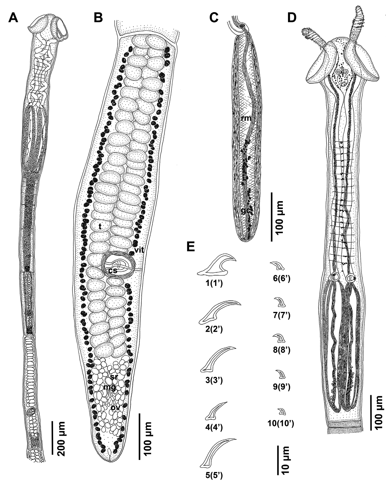
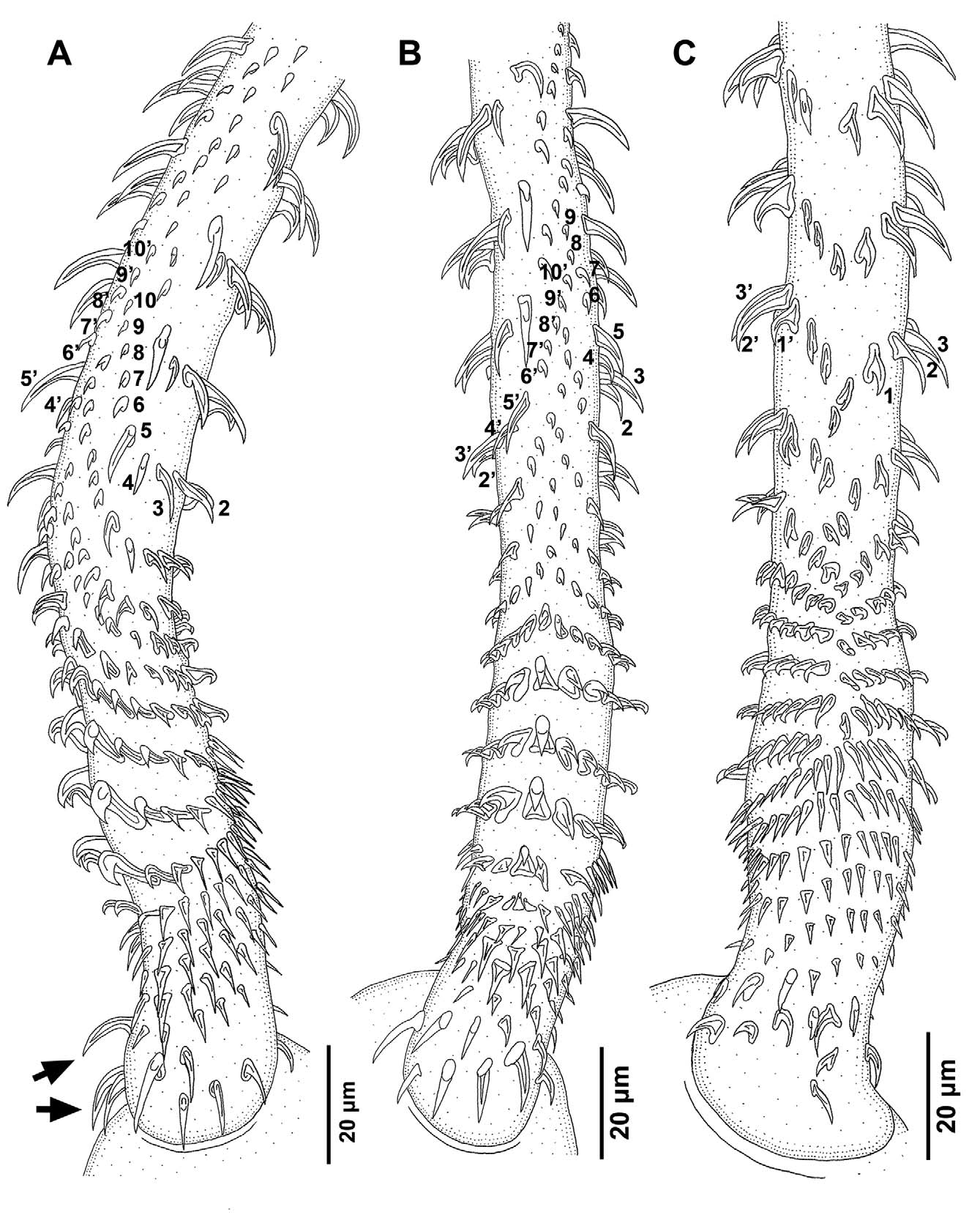
( Figs 9 View FIGURE 9 & 10 View FIGURE 10 )
Material examined: 35 whole-mounts.
Type-host: Taeniura lymma 1 (sensu Naylor et al. 2012) ( Rajiformes : Dasyatidae ) (KA-416).
Additional host: T. lymma (BO-80, BO-86).
Type-locality: Kunak, Sabah, Malaysia.
Additional localities: Semporna, Sabah, Malaysia; and Tanjung Batu, East Kalimantan, Indonesia.
Site of infection: Spiral intestine.
Deposited specimens: Holotype ( MZUM No 2012.01). Paratypes in ( MZUM Nos 2012.02–03; MZB Nos Ca 165–167; LRP Nos 7829–7834; SAM Nos AHC 35395–35399; and USNPC No 105183).
Etymology: This species is dedicated to Dr. Tomáš Scholz, who has contributed previously to trypanorhynch systematics.
Description: Cestodes small; mature specimens 3.6–6.0 (4.9 ± 0.8; n = 10) mm long with 7–9 (8 ± 1; n = 15) segments; gravid specimens 6.5 (n = 2) mm long with 9 (n = 2) segments. Scolex acraspedote, 950–1,465 (1,240 ± 153; n = 13) long ( Fig. 9 View FIGURE 9 A, D); maximum width at pars bothrialis, 280–330 (306 ± 18; n = 8) wide; pars bothrialis 150–220 (180 ± 21; n = 13) long; two sub-circular bothria, 175–200 (188 ± 10; n = 7) long, 205–240 (219 ± 10; n = 11) wide, with thick, free margins, notched posteriorly, V-shaped ridge present in some specimens ( Fig. 9 View FIGURE 9 A); pars vaginalis 450–920 (716 ± 138; n = 13) long; Pintner’s cells present in mid-line ( Fig. 9 View FIGURE 9 D); tentacle sheaths sinuous to almost straight, strongly coiled if contracted; transverse striations present in pars vaginalis ( Fig. 9 View FIGURE 9 A, D); pars bulbosa 470–590 (520 ± 38; n = 13) long, 165–225 (194 ± 18; n = 13) wide; prebulbar organs present; bulbs elongate, thick-walled, 455–570 (506 ± 38; n = 11) long, 60–80 (70 ± 5; n = 11) wide ( Fig. 9 View FIGURE 9 A, C, D), extend into pars proliferans in some specimens; bulb width: length ratio 1.0: 6.3–9.3 (7.4 ± 0.9; n = 11); retractor muscle inserts at base of bulb; gland-cells present, few, very small ( Fig. 9 View FIGURE 9 C, D), surrounding retractor muscle to mid-bulbar region, in some specimens into anterior third of bulb; scolex ratio (pars bothrialis: pars vaginalis: pars bulbosa) 1.0: 2.6–5.5: 2.4–3.7 (1.0: 4.0 ± 0.8: 2.9 ± 0.4; n = 13). Pars postbulbosa very short, up to 35 (9 ± 12; n = 15) long.
Fully everted tentacles 450–610 (536 ± 56; n = 9) long, with prominent basal swelling; diameter 24–25 (25 ± 1; n = 3) at base, 33–38 (35 ± 2; n = 4) at basal swelling, 25–28 (27 ± 1; n = 4) in metabasal region, 25–26 (25 ± 1; n = 3) distally. Metabasal armature heteroacanthous, heteromorphous; hooks hollow; hook rows consist of 10 hooks in posterior metabasal region ( Fig. 9 View FIGURE 9 E), decrease to 9 hooks per principle row anteriorly; rows start on antibothrial surface of tentacle, terminate on bothrial surface ( Fig. 10 View FIGURE 10 B, C); distinct space between hooks 1 and 1’ ( Fig. 10 View FIGURE 10 C); first four rows of principle rows with two smaller hooks posterior to hooks 1(1’) ( Fig. 10 View FIGURE 10 C); hooks 1(1’) uncinate, with elongate base, 11–14 (13 ± 1; n = 6) long, base 7–10 (8 ± 1; n = 6) long; hooks 2(2’) larger, falcate, with narrower base, 12–15 (14 ± 1; n = 7) long, base 4–6 (5 ± 1; n = 4) long; hooks 3(3’) slightly smaller, falcate, 12–15 (13 ± 1; n = 9) long, base 4–6 (4 ± 1; n = 9) long; hooks 4(4’) smaller, falcate, 8–10 (9 ± 1; n = 5) long, base 3–4 (3 ± 1; n = 5) long; hooks 5(5’) larger, falcate, 12–16 (14 ± 1; n = 6) long, base 4 (n = 6) long; hooks 6(6’) smaller, spiniform, 5–6 (5 ± 1; n = 6) long, base 2–3 (3; n = 6) long; hooks 7(7’) smaller, spiniform, 4–5 (4; n = 4) long, base 2 (n = 4) long; hooks 8(8’) spiniform, 4–5 (4 ± 1; n = 3) long, base 2 (n = 3) long; hooks 9(9’) slightly smaller, spiniform, 3–4 (4 ± 1; n = 2) long, base 2 (n = 2) long; hooks 10(10’) slightly smaller, spiniform, 3 (n = 2) long, base 2 (n = 2) long.
Characteristic basal armature present ( Fig. 10 View FIGURE 10 A–C); first rows of hooks enlarged on bothrial surface, falcate, 13–14 (14; n = 2) long, base 4–6 (5 ± 1; n = 2) long; basal hooks on antibothrial surface smaller, uncinate, 6–9 (7 ± 1; n = 3) long, base 4–5 (4 ± 1; n = 3) long; next 4–5 rows of hooks spiniform, bases reduced, 6–9 (7 ± 1; n = 8) long; basal swelling with 5 rows of hooks; increasing in size towards enlarged billhook on bothrial surface ( Fig. 10 View FIGURE 10 A, B); billhooks form single file ( Fig. 10 View FIGURE 10 B), largest billhooks in second and third row on basal swelling, 7 (n = 2) long, base 3 (n = 2) long, height 8 (n = 2); hooks on antibothrial surface spiniform, become falcate to almost uncinate anteriorly ( Fig. 10 View FIGURE 10 C); transition to metabasal armature occurs at about hook row 17 ( Fig. 10 View FIGURE 10 A, B).
Segments acraspedote; first segments wider than long; first mature segments elongate, 170–320 (247 ± 51; n = 8) long, 110–205 (147 ± 37; n = 8) wide ( Fig. 9 View FIGURE 9 A); late mature segments 985–2,020 (1,381 ± 336; n = 8) long, 130–230 (174 ± 38; n = 8) wide ( Fig. 9 View FIGURE 9 B); genital pores unilateral, in posterior half of segment ( Fig. 9 View FIGURE 9 A, B); cirrus sac oval to sub-circular, thick-walled, 68–105 (86 ± 13; n = 6) long, 70–100 (88 ± 12; n = 6) wide; internal and external vesicles absent; cirrus unarmed. Testes arranged in two columns, anterior to ovary, in intervascular space, 53–71 (62 ± 5; n = 14) in number, 41–57 (48 ± 5; n = 14) anterior to genital pore, 12–18 (15 ± 2; n = 14) posterior to genital pore, 48–63 (52 ± 4; n = 17) long, 33–39 (35 ± 2; n = 17) wide ( Fig. 9 View FIGURE 9 B). Vagina thin-walled, enters genital atrium from posterior aspect; seminal receptacle c. 20 (n = 2) in diameter; ovary at posterior extremity of segment, bilobed in dorso-ventral view; ovarian lobes elongate, 183–258 (207 ± 26; n = 8) long, 29–40 (33 ± 4; n = 8) wide ( Fig. 9 View FIGURE 9 B); Mehlis’ gland present between ovarian lobes, 25–38 (30 ± 6; n = 4) in diameter; vitelline follicles circumcortical, 20–23 (21 ± 1; n = 10) long, 13–15 (14 ± 1; n = 10) wide; uterus tubular, median, extends to anterior extremity of segment; uterine pore not observed.
Gravid segments 1,655–1,980 (1,845 ± 169; n = 3) long, 175–305 (227 ± 69; n = 3) wide; uterus extends to anteriormost extremity; cirrus sac sub-circular, 118 (n = 1) long, 108 (n = 1) wide; ovarian lobes 155–160 (158 ± 4; n = 2) long, 36–38 (37 ± 1; n = 2) wide; vitelline follicles 30–38 (35 ± 2; n = 10) long, 25–30 (27 ± 2; n = 10) wide.
Remarks: The present specimens are allocated to the genus Prochristianella , since they possess the typical, generic characteristics. The most significant features of this species are the smooth scolex, thick-walled bulbs with very few, tiny gland-cells, unilateral genital pores and a tentacular armature with enlarged basal hooks, rows of billhooks on the basal swelling and 10 hooks per half spiral row, decreasing in number to 9 hooks per row further anteriorly. The principle hook rows in the metabasal region have small hooks 4(4’), as described above for P. kostadinovae , which represents the most similar species within the genus. Prochristianella scholzi n. sp. differs from P. kostadinovae in its possession of enlarged basal hooks, two additional hooks posterior to hooks 1(1’) in the most proximal part of the metabasal armature, smaller hooks 1(1’)–6(6’), hooks 2(2’) and 3(3’) without a recurved tip, a much wider basal swelling than the remainder of the tentacle and a transition into the metabasal armature at about hook row 17. Moreover, P. scholzi n. sp. has smaller bothria, Mehlis’gland and vitelline follicles, but much more elongated ovarian lobes. Prochristianella scholzi n. sp. is described from stingrays of the genus Taeniura Müller & Henle from three localities in Sabah (Malaysian Borneo) and East Kalimantan (Indonesian Borneo).
No known copyright restrictions apply. See Agosti, D., Egloff, W., 2009. Taxonomic information exchange and copyright: the Plazi approach. BMC Research Notes 2009, 2:53 for further explanation.
|
Kingdom |
|
|
Phylum |
|
|
Class |
|
|
Order |
|
|
Family |
|
|
Genus |