
Macrobiotus sklodowskae, Michalczyk, Łukasz, Kaczmarek, Łukasz & Węglarska, Barbara, 2006
|
publication ID |
https://doi.org/10.5281/zenodo.174916 |
|
DOI |
https://doi.org/10.5281/zenodo.5617295 |
|
persistent identifier |
https://treatment.plazi.org/id/F20B87F8-BA7D-FFDD-7B37-F910D66EFDCF |
|
treatment provided by |
Plazi |
|
scientific name |
Macrobiotus sklodowskae |
| status |
sp. nov. |
Macrobiotus sklodowskae sp. nov.
( Figs 1–32 View FIGURES 1 – 6 View FIGURES 7 – 13 View FIGURES 14 – 19 View FIGURES 20 – 28 View FIGURES 29 – 32 )
Description
Adult (measurements of the holotype): Body length 569.0. Body transparent/white, eyes present. Cuticle smooth and without pores. Fine, regular granulation present on all legs, developed better on IV pair (DIC/PCM and SEM).
Bucco-pharyngeal apparatus of Macrobiotus - type ( Figs 1–4 View FIGURES 1 – 6 , 7–13 View FIGURES 7 – 13 ). Mouth anteroventral, surrounded by ring of 10 peribuccal lamellae. Oral cavity armature of harmsworthi - type with three bands of teeth ( Figs 7–13 View FIGURES 7 – 13 ). Teeth of the first band are smaller than those of the other two bands and are in the shape of small cones in SEM or granules in DIC/PCM. They are present in the anterior portion of the oral cavity just behind peribuccal lamellae. This band of teeth is continuous and looks the same on all oral cavity walls, usually consisting of 4–6 irregular rows of teeth. The second band of teeth: these are intermediate in size between those of the first band and those of the third band of teeth. They are in the shape of flat and irregularly serrated vertical partitions higher in their anterior portion in SEM, or small ridges parallel to the main axis of the buccal tube in DIC/PCM. They are positioned in the posterior portion of the oral cavity just behind the ring fold and just before the third band of teeth. This band of teeth is continuous and arranged in one row. Most teeth are uniform and regular, in the shape of partitions (SEM) or ridges (DIC/PCM), only sometimes joined one-by-one. Joined teeth are H-, V- and Wshaped. The third band of teeth: these are larger than those in the other two rows and there are usually six. They are in the shape of flat and irregularly serrated transverse partitions/ plates (SEM) or transverse ridges/baffles (DIC/PCM) and large cones (SEM) or granules (DIC/PCM). They are positioned in the rear of the oral cavity just behind the second band of teeth and just before the buccal tube opening. Usually this band is not continuous and is divided into two series: ventral and dorsal. Both series consist of one median and two lateral teeth. Sometimes ventro-lateral teeth may be joined with dorso-lateral ones and thus the third band becomes continuous (SEM). Dorsal teeth are higher (SEM), thinner and longer than ventral ones (SEM and DIC). The medio-ventral tooth may be broken into two or more smaller teeth in large specimens, thus there may be seven (or more) teeth in this band (SEM and DIC) ( Figs 10–13 View FIGURES 7 – 13 ).
Buccal tube 54.2 long and 10.5 [ 19.3] wide ( Figs 1–2 View FIGURES 1 – 6 ) with one bend in anterior part of tube (visible in lateral view). Stylet supports inserted on buccal tube at 46.1 [ 85.1]. Pharyngeal bulb slightly oval with apophyses, three macroplacoids and microplacoid ( Figs 1–4 View FIGURES 1 – 6 ). Pharyngeal apophyses distinct, rounded and forked posteriorly ( Fig. 3 View FIGURES 1 – 6 ). First macroplacoid thinner anteriorly, 8.1 [ 14.9] long, second oval, 7.6 [ 14.0] long, both without constriction. Third macroplacoid 9.5 [ 17.5] long, with distinct constriction in subterminal part. Microplacoid distinct, wide and with lateral ‘wings’ ( Fig. 4 View FIGURES 1 – 6 ), 4.3 [ 7.9] long and connected with third macroplacoid by thin cuticular ‘yarn’. ‘Yarn’ with drop-like thickening in subterminal portion. Macroplacoid row 29.5 [ 54.4] long. Placoid row 38.0 [ 70.2] long.
Claws of hufelandi - type, stout ( Figs 5–6 View FIGURES 1 – 6 ). Primary branches with distinct accessory points. Smooth and well developed lunules present on all legs, larger on IV pair of legs. Primary branch (pb) with basal claw of I pair of legs 13.3 [ 24.6] long, secondary branch (sb) 10.5 [ 19.4] long, II pb. 14.3 [ 26.3], sb. 11.4 [ 21.1]; III pb. 14.3 [ 26.3], sb. 11.4 [ 21.1]; IV pb. 16.2 [ 29.9], sb 12.4 [ 22.9]. Bars and other cuticular thickenings on legs absent.
Egg (measurements of a paratype): White, laid freely ( Figs 14–32 View FIGURES 14 – 19 View FIGURES 20 – 28 View FIGURES 29 – 32 ). Spherical, areolated, with 10 processes on circumference (22 on hemisphere) ( Figs 14–15 View FIGURES 14 – 19 ). Diameter without processes 96.0, with processes 112.0. Processes in shape of spherically terminated cones ( Figs 20–32 View FIGURES 20 – 28 View FIGURES 29 – 32 ). Processes consist of double wall with transverse supporting walls forming ‘cells’ visible in DIC/PCM as fine reticular design ( Fig. 20 View FIGURES 20 – 28 ). External walls of processes smooth or wrinkled (wrinkles in form of rings around a process) ( Figs 29–32 View FIGURES 29 – 32 ). Internal walls strongly porous (visible in SEM only). Processes 16.0–17.5 high and 20.5–23.5 wide at the base. Surface between processes areolated. Each areola with irregular thickened, porous central portion (pores are not always visible in DIC/PCM) ( Figs 14–19 View FIGURES 14 – 19 ). Ridges delimiting areolae (brims) continuous with processes and therefore also double-walled (visible as covered with reticular design in DIC/PCM) ( Fig. 20 View FIGURES 20 – 28 ).
Number of areolae around a process depending on the number of processes surrounding the particular process ( Figs 16–19 View FIGURES 14 – 19 ). Usually there are two areolae for each neighbour process, but sometimes only one develops. Therefore, if five processes surround a particular process, 8–10 areolae are present ( Figs 16–17 View FIGURES 14 – 19 ). If six processes surround a particular process, 10–12 areolae are present ( Figs 18–19 View FIGURES 14 – 19 ).
Remarks
Adults: Results of simple statistical analysis of measurements and pt values of selected morphological structures for nine randomly chosen specimens are given in Table 1 View TABLE 1 . The measurements of the smallest and largest specimen are provided in Table 2.
Eggs: Measurements for all measurable eggs are provided in Table 3 View TABLE 3 . Processes have the same type of structure but limited variability of shapes and sizes of some characters was noted and is shown in Figs 20–32 View FIGURES 20 – 28 View FIGURES 29 – 32 . The variability in the shape of processes is noteworthy – they always appear as cones, but may be terminated in a few ways. The most common termination is slightly hemispherical ( e.g., Figs 20–28 View FIGURES 20 – 28 ), but there are also processes terminated with a sharp point ( Fig. 27 View FIGURES 20 – 28 ) or strongly rounded ( Fig. 28 View FIGURES 20 – 28 ).The second variable character is the surface of processes – it may be smooth, slightly and strongly wrinkled (ring folds) ( Figs 29–32 View FIGURES 29 – 32 ).
CHARACTER N RANGE MEAN SD
µm pt µm pt µm pt Type depositories
Holotype and 27 paratypes ( 14 adults and 13 eggs) are preserved at the Zoological Museum of the Jagiellonian University, ul. Ingardena 6, 30-060 Kraków, Poland; 18 paratypes (adults and eggs) are preserved in the collection of Ł. Michalczyk; the remaining paratypes (adults and eggs) are preserved at the Department of Animal Taxonomy and Ecology, A. Mickiewicz University, Poznań.
CHARACTER N MIN MAX MEAN SD
Diameter of egg without processes 4 88.4 96.9 94.1 3.9
Diameter of egg with processes 4 104.3 115.0 110.7 4.5
Processes height 12 15.2 19.0 16.5 1.2
Processes base width 12 18.1 29.5 23.2 3.7
Number of processes on the circumference of egg 4 9 11 10.0 0.8
Number of processes on the hemisphere of egg 6 20 26 22.0 2.3 Etymology
The name ‘ sklodowskae ’ is given in recognition of the outstanding Polish scientist Maria Skłodowska-Curie ( 1867–1934) who co-discovered radium, polonium and natural radiation, the first woman to win the Nobel Prize and the first scientist to win two Nobel Prizes ( 1903 and 1911).
Differential diagnosis
Macrobiotus sklodowskae sp. nov. is the fifteenth described species in the richtersi group. The species within the group are usually most easily distinguished using characters of the egg shell. M. sklodowskae sp. nov. belongs to the subgroup of species within the richtersi group with the vanescens - type areolation (central portion of areolae thickened and sculptured). Other species of the richtersi group with the vanescens - type areolation are M. alekseevi , M. corgatensis , M. danielisae , M. gerlachae , M. lorenae , M. magdalenae , M. priviterae and M. vanescens .
The new species is most similar in the egg shell appearance to M. vanescens Pilato et al., 1991 (from Africa) and M. gerlachae Pilato et al., 2004 (from Seychelles). However, egg processes in M. vanescens have a slight sub-terminal constriction and are flat on top with irregular indentations while they have no constriction, no indentations and are rounded or (rarely) spiky on top in the new species. Moreover, adults of M. sklodowskae sp. nov. differ from adults of M. vanescens by having a large, distinct microplacoid (very small, sometimes almost invisible in M. vanescens ) and slightly narrower buccal tube ( 7.1–11.9 in M. sklodowskae sp. nov. and about 13.7 in M. vanescens ). Eggs of M. sklodowskae sp. nov. differ from eggs of M. gerlachae by lacking a small cap-like structures on top of the processes (not mentioned in the original description, but noticed by us in the paratypes, Figs 35–36 View FIGURES 33 – 36 ). Adults of M. gerlachae also have a more slender, tearshaped microplacoid without lateral wings ( Fig. 33 View FIGURES 33 – 36 ), lacking granulation on the first three pairs of legs and slightly slender claws with smaller lunules ( Fig. 34 View FIGURES 33 – 36 ) than the new species.
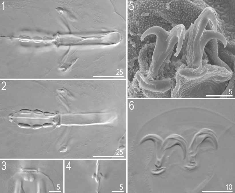
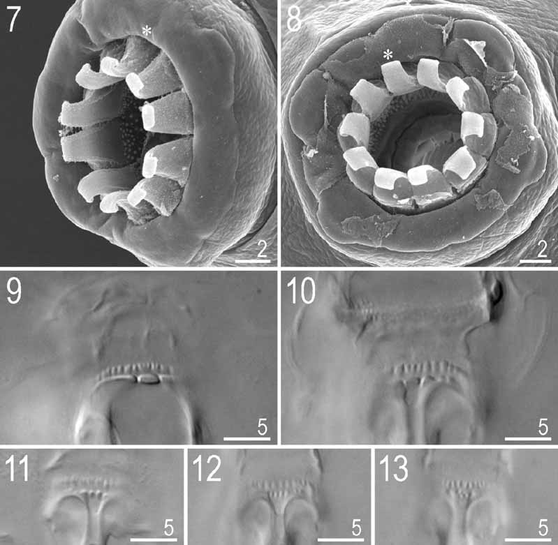
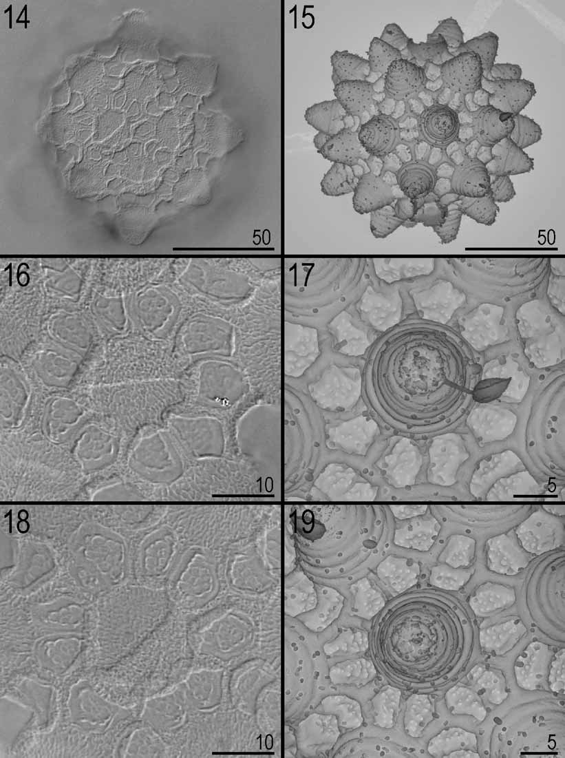
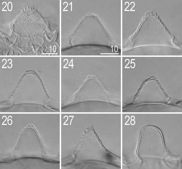
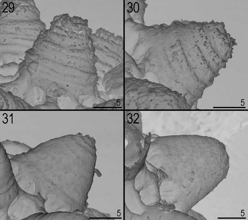
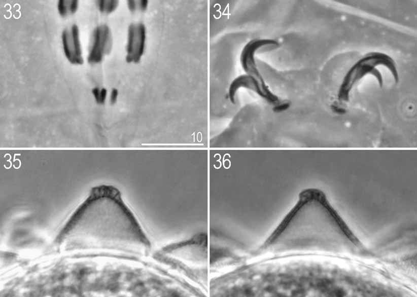
TABLE 1. Measurements [in µm] of selected morphological structures of specimens of Macrobiotus sklodowskae sp. nov. mounted in Hoyer’s medium (RANGE refers to the smallest and the largest structure found among all measured specimens; N — number of specimens / structures measured, SD — standard deviation).
| Body | 9 | 346.8 – 612.8 | 869.0 – 1057.4 | 486.0 | 972.9 | 87.1 | 58.0 |
|---|---|---|---|---|---|---|---|
| Buccal tube | 9 | 39.9 – 58.0 | - | 49.7 | - | 6.8 | - |
| Stylet support insertion point | 9 | 33.3 – 49.4 | 81.8 – 85.2 | 41.8 | 84.0 | 6.0 | 1.1 |
| Buccal tube external width | 9 | 7.1 – 11.9 | 17.0 – 20.5 | 9.4 | 18.9 | 1.7 | 1.1 |
| Macroplacoid 1 | 9 | 5.7 – 9.0 | 14.3 – 16.0 | 7.5 | 15.2 | 1.1 | 0.6 |
| Macroplacoid 2 | 9 | 4.8 – 8.6 | 11.4 – 14.8 | 6.7 | 13.4 | 1.4 | 1.1 |
| Macroplacoid 3 | 9 | 6.7 – 10.0 | 16.7 – 18.0 | 8.7 | 17.4 | 1.2 | 0.5 |
| Microplacoid | 9 | 2.9 – 4.8 | 6.8 – 8.2 | 3.9 | 7.7 | 0.7 | 0.5 |
| Macroplacoid row | 9 | 19.0 – 31.4 | 47.6 – 54.5 | 26.0 | 52.0 | 4.8 | 3.0 |
| Placoid row | 9 | 24.7 – 41.8 | 61.4 – 72.1 | 33.8 | 67.5 | 6.3 | 4.0 |
| Claw 1 - primary branch | 6 | 10.5 – 14.3 | 23.7 – 26.1 | 12.9 | 24.9 | 1.3 | 0.8 |
| Claw 1 - secondary branch | 8 | 7.6 – 11.4 | 18.2 – 24.6 | 10.2 | 20.7 | 1.6 | 2.2 |
| Claw 2 - primary branch | 6 | 10.9 – 14.3 | 23.7 – 28.6 | 12.9 | 26.6 | 1.4 | 1.7 |
| Claw 2 - secondary branch | 8 | 8.1 – 12.4 | 19.4 – 27.8 | 11.1 | 22.7 | 1.8 | 2.6 |
| Claw 3 - primary branch | 4 | 11.4 – 14.3 | 23.7 – 28.6 | 13.1 | 26.7 | 1.2 | 2.2 |
| Claw 3 - secondary branch | 7 | 8.1 – 12.8 | 19.4 – 27.8 | 11.1 | 22.6 | 1.9 | 2.7 |
| Claw 4 - primary branch | 5 | 11.9 – 16.2 | 28.5 – 34.6 | 14.7 | 30.7 | 1.8 | 2.3 |
| Claw 4 - secondary branch | 7 | 8.6 – 14.3 | 20.6 – 31.1 | 12.7 | 25.4 | 1.9 | 3.6 |
TABLE 3. Measurements [in µm] of selected morphological structures of eggs of Macrobiotus sklodowskae sp. nov. mounted in Hoyer’s medium.
| CHARACTER µm | pt | µm | pt |
|---|---|---|---|
| Body 346.8 | 869.0 | 612.8 | 1057.4 |
| Buccal tube 39.9 | - | 58.0 | - |
| Stylet support insertion point 33.3 | 83.3 | 49.4 | 85.2 |
| Buccal tube external width 7.1 | 17.9 | 11.9 | 20.5 |
| Macroplacoid 1 5.7 | 14.3 | 9.0 | 15.6 |
| Macroplacoid 2 4.8 | 11.9 | 8.6 | 14.8 |
| Macroplacoid 3 6.7 | 16.7 | 10.0 | 17.2 |
| Microplacoid 2.9 | 7.1 | 4.8 | 8.2 |
| Macroplacoid row 19.0 | 47.6 | 31.4 | 54.1 |
| Placoid row 24.7 | 61.9 | 41.8 | 72.1 |
| Claw 1 - primary branch? | ? | 14.3 | 24.7 |
| Claw 1 - secondary branch 7.6 | 19.0 | 11.4 | 19.7 |
| Claw 2 - primary branch 11.4 | 28.6 | ? | ? |
| Claw 2 - secondary branch 8.6 | 21.6 | 12.4 | 21.4 |
| Claw 3 - primary branch 11.4 | 28.6 | ? | ? |
| Claw 3 - secondary branch 8.6 | 21.6 | 12.4 | 21.4 |
| Claw 4 - primary branch? | ? | ? | ? |
| Claw 4 - secondary branch? | ? | 13.3 | 23.0 |
No known copyright restrictions apply. See Agosti, D., Egloff, W., 2009. Taxonomic information exchange and copyright: the Plazi approach. BMC Research Notes 2009, 2:53 for further explanation.
|
Kingdom |
|
|
Phylum |
|
|
Class |
|
|
Order |
|
|
Family |
|
|
Genus |