
Benthimermis platyptera, Miljutin, 2004
|
publication ID |
https://doi.org/ 10.5281/zenodo.5393181 |
|
DOI |
https://doi.org/10.5281/zenodo.5469327 |
|
persistent identifier |
https://treatment.plazi.org/id/F21C1022-F57B-6B76-FCEA-FF51FCDE6492 |
|
treatment provided by |
Marcus |
|
scientific name |
Benthimermis platyptera |
| status |
sp. nov. |
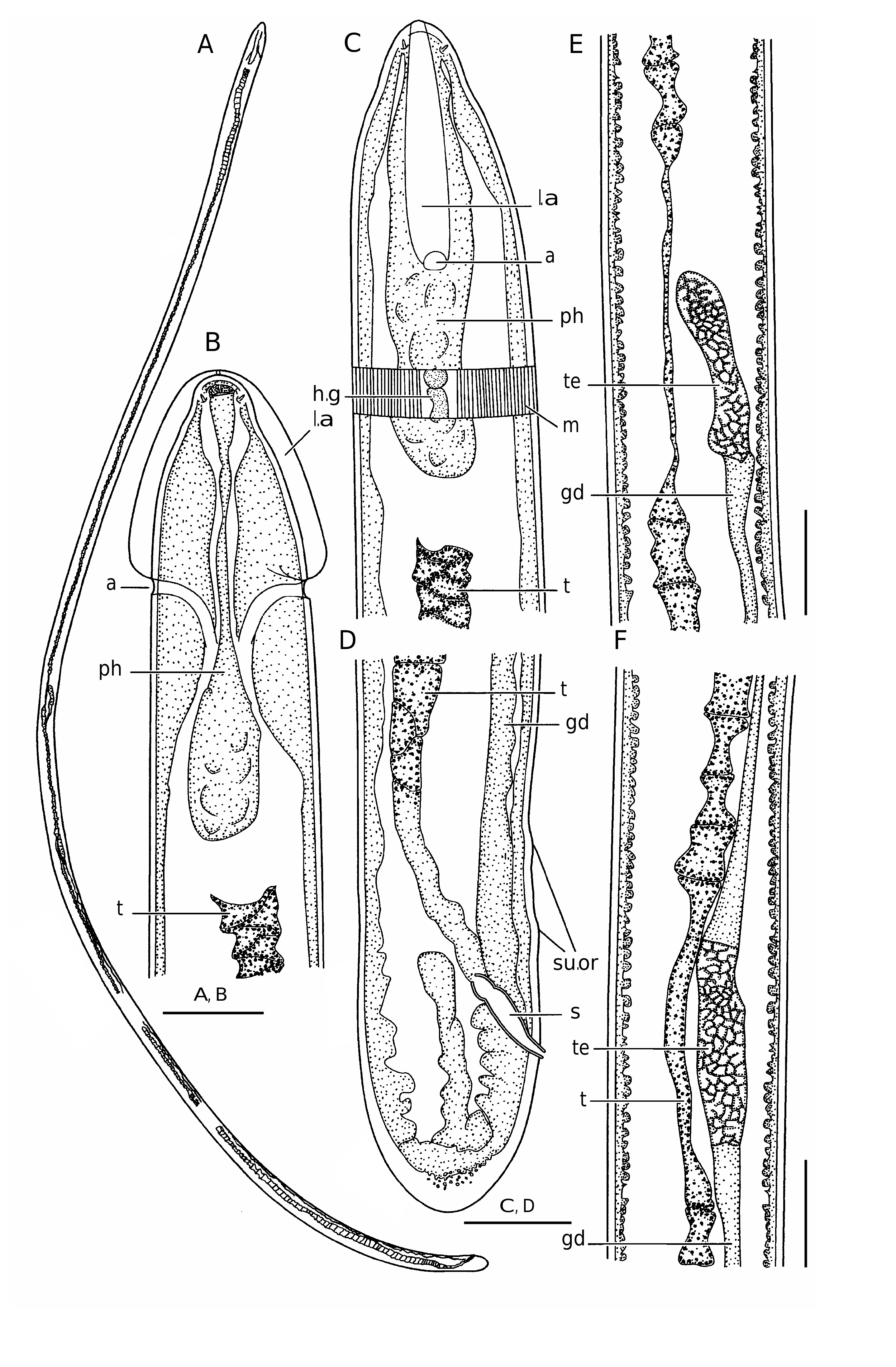
Benthimermis platyptera n. sp.
( Fig. 10 View FIG )
TYPE MATERIAL. — Holotype: Western Atlantic , cruise DEMERABY, stn KG 25, 10°22.41’N, 46°46.74’W, 4800 m, 27.IX.1980, mature ( MNHN 331 About MNHN BF). GoogleMaps
ETYMOLOGY. — From Greek platyptera (wide-wing).
DIAGNOSIS. — The male of B. platyptera n. sp. differs from all other described males of the genus Benthimermis in the fewest number of supplementary organs (2 vs 4-68 in other species), the shortest spicules (19 vs 28-160 µm in other species), and the smallest body length (2953 vs 3300-15000 µm in other species).
DESCRIPTION
Measurements: L = 2953 µm; “a” = 84.4; “c” = 101.8. Maximal body diameter = 35 µm. Diameter at level of: cephalic sensilla = 12 µm; amphids = 32 µm; midbody = 35 µm; anus = 33 µm. Distance from anterior end to amphid = 43 µm. Spicules 19 µm long.
Body thread-like, cylindrical. Anterior end shaped as a rounded cone. Posterior end rounded. Four mediolateral cephalic setae about 1.5 µm long, inserted in small pits. Amphids non-spiral. Amphidial aperture present as pore of 4 µm in diameter. Amphidial fovea tubiform. Cuticle width 1.5 µm at anterior part, 1.5 µm at midbody, and 6 µm at extremity of posterior end. Cervical alae of 4.5 µm maximal width, beginning right ahead amphids and reaching cephalic apex. Right and left alae join each other at cephalic apex. Mouth opening vestigial, present as an apical light trace in cuticle. Pharynx looking like a non-muscular string devoid of an internal lumen with a thickening at distal end. Midgut being a trophosome consisting of one row of big cells. At midbody, trophosome looking like a backbone. Rectum and anus present. Male reproductive system of 1700 µm length consisting of two testes arranged one after the other and connected by a short spermaduct. Spicules straight, without a gubernaculum. Two very small supplementary organs anterior to anus.
No known copyright restrictions apply. See Agosti, D., Egloff, W., 2009. Taxonomic information exchange and copyright: the Plazi approach. BMC Research Notes 2009, 2:53 for further explanation.
|
Kingdom |
|
|
Phylum |
|
|
Class |
|
|
Order |
|
|
Family |
|
|
Genus |