
Filispirifer hamadae, Schemm-Gregory, Mena, 2008
|
publication ID |
https://doi.org/10.5281/zenodo.181444 |
|
DOI |
https://doi.org/10.5281/zenodo.5657819 |
|
persistent identifier |
https://treatment.plazi.org/id/F25A3A7E-FF9D-4C4E-FF6D-FB9DFC6A4CFA |
|
treatment provided by |
Plazi |
|
scientific name |
Filispirifer hamadae |
| status |
sp. nov. |
Filispirifer hamadae new species
( Figs 2–4 View FIGURE 2 View FIGURE 3 View FIGURE 4 , Pl. 1 Figs 1 View FIGURE 1 –9)
v 2001 Filispirifer cf. merzakhsaiensis .—Jansen: Unter-Devonische Brachiopoden Dra Ebene: 36, 38, tab. 2. v 2004 b Filispirifer cf. merzakhsaiensis .—Jansen et al.: Pragian at Assa: 64, pl. 1 fig. 6.
v 2007 Filispirifer cf. merzakhsaiensis .—Jansen et al.: Neritic-pelagic correlation: 19, fig. 4.20.
Holotype: Internal mould of ventral valve, stored in the Forschungsinstitut und Naturmuseum Senckenberg under the inventory number SMF 65248b (Pl. 1 Figs 1 View FIGURE 1 A–D). Length 26.3 mm and width 38.2 mm.
Derivation of name: After the Arabian word “ hamada ” for stony desert in which the type locality is situated.
Type locality: El Ayoun, c. 12 km Southeast of Tata, southwestern Anti-Atlas Mountains, Dra Valley ( Morocco, North Africa).
Type horizon: Merzâ-Akhsaï Formation (‘Rich 2’), exact position unknown, Middle/Upper Siegenian (middle Lower Devonian).
Stratigraphic distribution: Assa Formation (‘Rich 1’) to Merzâ-Akhsaï Formation (‘Rich 2’), upper Lower Siegenian to Middle/Upper Siegenian (middle Lower Devonian).
Geographic distribution: southwestern Anti-Atlas Mountains, Dra Valley ( Morocco, North Africa).
Material. Locality Assa I. 4 articulated shells: SMF 65204, 66539, 66543, 66578; 8 articulated internal moulds: SMF 66527, 66528, 66542, 66545, 66547, 66585–66587; 2 isolated ventral shells: SMF 66548, 66549; 14 ventral external shells: SMF 65206, 66529–66531, 66533, 66537, 66538, 66540, 66541, 66544, 66547, 66575–66577; 1 ventral internal shell: SMF 66579; 8 ventral internal moulds: SMF 65022, 65065a, b, 65204, 66196, 66534, 66545, 66580; 7 dorsal internal moulds: SMF 66532, 66535, 66536, 66546, 66581, 66582, 66584; several shell fragments and fragments of internal moulds of both valves.
Locality Assa II. 1 ventral internal mould: SMF 65218; 1 dorsal internal mould: SMF 66583.
Locality El Ayoun. 2 ventral internal moulds: SMF 65248a, b ( holotype).
Locality Foum el Hassane. 13 articulated shells: SMF 66556–66563, 66565–66567, 66570, 66572, 66590; 1 articulated internal mould: SMF 66564; 5 ventral external shells: SMF 66550, 66551, 66554, 66568, 66573; 1 ventral internal shell: SMF 66555; 1 ventral internal mould: SMF 66552; 1 dorsal external shell: SMF 66571; 1 dorsal internal shell: SMF 66569; several ventral internal moulds: SMF 66553.
Locality Timziline. 1 ventral external mould: SMF 66589; 1 ventral internal mould: SMF 59649; 1 dorsal external mould: SMF 66588.
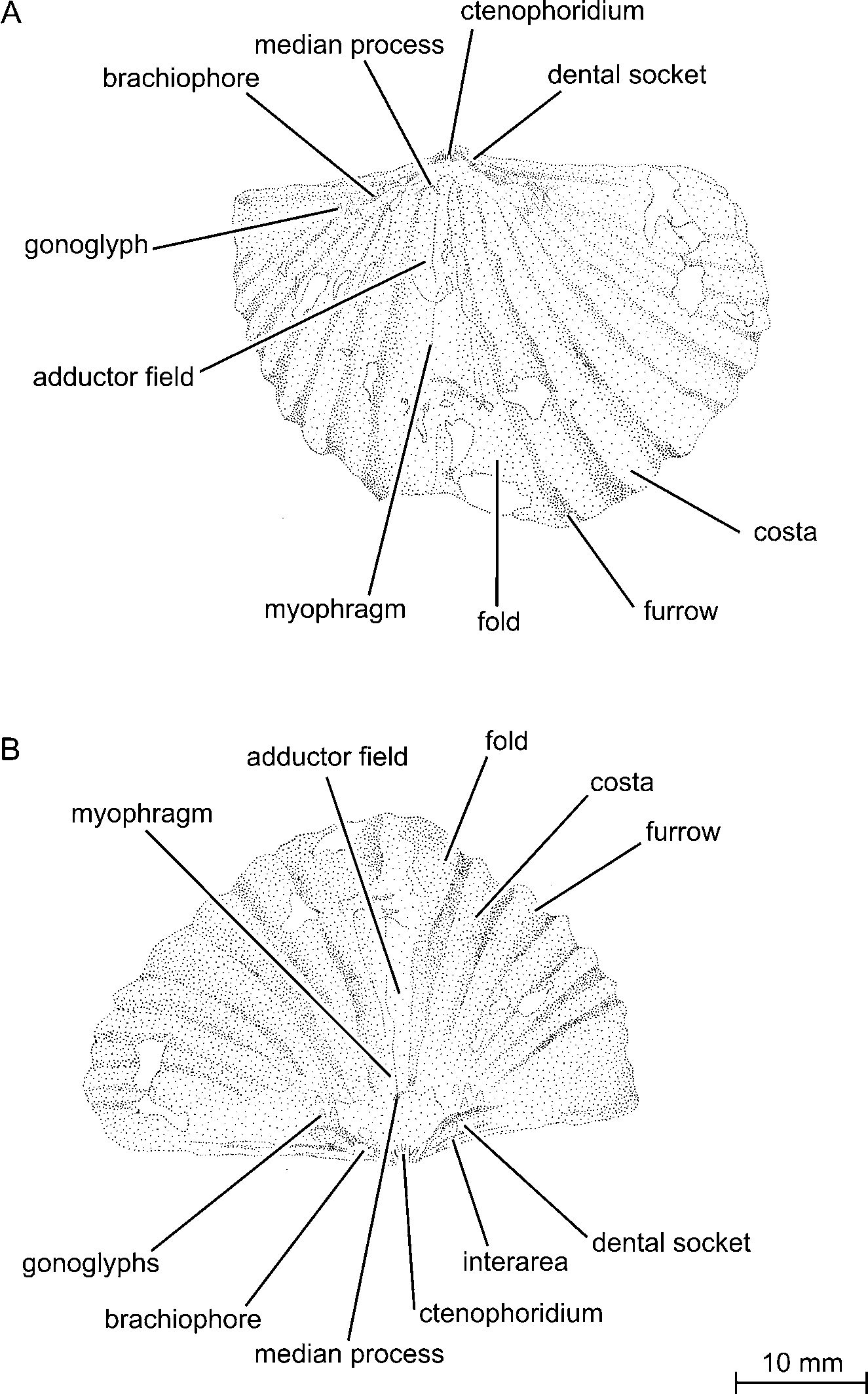
Diagnosis. Medium to large-sized Filispirifer with 8 to 11 coarse and simple costae on each external flank, a moderately high ventral interarea, a small ventral process, and an inconspicuous dorsal adductor field. The ventral muscle field is strongly imbedded, subelliptic to diamond-shaped in outline, and indented by a small ventral process. Median costa in sulcus may be lacking.
Description of the holotype (SMF 65248b)
Form and size. The ventral internal mould is transverse, semielliptic in outline, and brachythyrid without mucronations. The specimen is strongly convex in longitudinal section.
Exterior of ventral valve. The ventral umbo extends clearly to posterior over the hinge line. The ventral interarea is moderately high, apsacline, and curved. The delthyrium is open, restricted by a pair of thin deltidial lamellae that are not combined posteriorly.
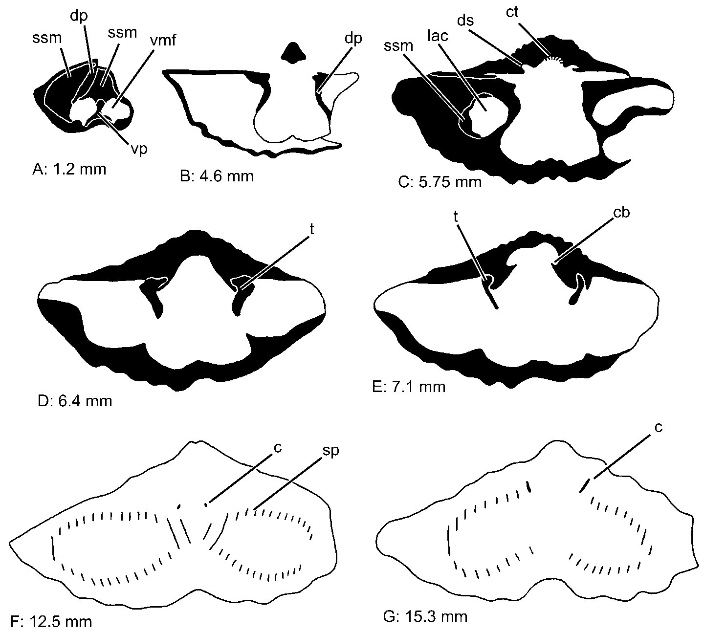
Interior of ventral valve. Ventral muscle field extends to posterior over the hinge line. Lateral apical cavities are almost completely filled by secondary shell material and not extending to posterior over the hinge line. A small septal pillow is developed dorsal of the ventral muscle field. Both sides of the septal pillow are bordered by a thin subdeltidial furrow on the internal mould of which only one is preserved. Ventral process weakly developed leaving a very small groove on the internal mould from the septal pillow to the beginning of the ventral myophragm. The myophragm is thick leaving a broad furrow through the posterior half of the ventral muscle field on the internal mould. The ventral muscle field is broad, elongated, almost diamondshaped in outline, and deeply impressed into the shell. On each side, 2 costae are gently impressed on the internal mould of the ventral muscle field. Diductor scars are impressed as subradial striae on the internal mould. Adductor scars situated next to the myophragm and enclosed completely by the diductor field; posterior and anterior pairs clearly differentiable and clearly impressed into shell. The anterior part is lancet-shaped and broad, the posterior part elongated and thin. Muscle bounding ridge broad and clearly developed, leaving a deep furrow on the internal mould as the lateral border of the ventral muscle field. Anteriorly, in the sulcus, the muscle bounding ridge is only weakly impressed. Free portions of dental plates short, leaving small wedge-like slits on either side of the ventral muscle field on the internal mould. Posteriorly they are imbedded into secondary shell material. Its anterior parts pass into the muscle bounding ridge. No teeth preserved. Due to the strong development of secondary shell material, a platform is formed on both sides of the ventral muscle field which is covered by numerous gonoglyphs preserved as tubercles on the internal mould. Sulcus smooth, conspicuous, and shallow, at base flattened in cross section. Each flank of the specimen is covered by 6 coarse and simple costae that are impressed anterior of the platform on the internal mould. The sulcus bounding costae are slightly weaker than the adjacent pair of costae and included into the sulcus. No impressions of growth lamellae.
Description of paratypes
Form and size.Shells medium to large-sized, compact to slightly transverse, and subcircular to subelliptic in outline. In longitudinal section biconvex, mostly equibiconvex to dorsibiconvex, brachythyrid to megathyrid without mucronations. Sometimes small ears are developed. Largest specimen 31.0 mm long and 46.3 mmm wide.
Exterior of ventral valve. The ventral umbo extends to posterior over the hinge line. The ventral interarea is moderately high, apsacline, and curved. In the lower part of the ventral interarea growth lamellae parallel to the hinge line are weakly preserved. The delthyrium is open. It is slightly constricted by a pair of deltidial lamellae that are not fused posteriorly. A deltidium is not developed. In the posterior part of the ventral umbo a deep and narrow sulcus begins. It is angular in cross section and with steep flanks. The sulcus tongue is long to short, its outline at the anterior margin is rounded to angular. A weak median costa is often developed that is rounded in cross section.
Exterior of dorsal valve. The dorsal umbo is small and extends slightly posterior over the hinge line. The dorsal interarea is very low and anacline to orthocline. The notothyrium is open, chilidial lamellae are not developed. In the posterior part of the umbo a steep and elevated fold begins that is angular in cross section.
Macro-ornamentation. The external ventral valve is covered by 8 to 11 coarse and simple costae that do not multiply by bifurcation or intercalation and are angular in cross section. The first pair of costae is included into the sulcus and also more weakly developed than the second pair. The external dorsal valve is covered by one or two costae less than on the ventral shell. In some cases, the first pair of costae is situated on the flanks of the fold. The costae on both valves are coarse, simple, do not multiply by bifurcation or intercalation, and are angular in cross section. They are separated by furrows that are angular in cross section and of the same size as the costae. At the anterior margin, coarse and concentric growth lamellae are developed. The edges of the growth lamellae overhang on the next younger lamellae.
Micro-ornamentation. The micro-ornamentation is capillate with 9 to 10 capillae per mm. Capillae are arranged in radial lines running over the whole shell. At the intersection of capillae and growth lamellae very small nodes are developed. (Pl. 1 Figs 9B, C).
Interior of ventral valve. The ventral muscle field extends posterior of the hinge line ( Fig. 4 View FIGURE 4 A). Lateral apical cavities are almost completly filled by secondary shell material, resulting in a deep imbedding of the ventral muscle field into the shell. Dorsal of the ventral muscle field, a small septal pillow (= coussinet septal sensu Gourvennec 1989: 27, fig. 13) is developed but hardly extending further posteriorly than the ventral muscle field. Both sides of the septal pillow are bordered by a very thin subdeltidial furrow (= sillon hypodeltidial sensu Gourvennec 1989: 23, 30, fig. 7) on the internal mould. Medially, the septal pillow is grooved by a fine furrow on the internal mould that is interpreted as a remnant of the small ventral process that leaves a small indentation in the posterior margin of the impression of the ventral muscle field ( Fig. 4 View FIGURE 4 A). A broad myophragm originates from the ventral process leaving a distinct furrow through the first half to two thirds of the ventral muscle field on the internal mould. The ventral muscle field is broad and generally elongated, always slightly longer than wide; subelliptic to rarely diamond-shaped in outline. Two to three costae are weakly impressed on the internal mould of the ventral muscle field. Diductor scars leave subradial striae on the internal mould. Adductor scars elongate, posterior and anterior pairs clearly differentiable, and situated admedian next to the myophragm. The posterior pair of adductor scars is thin and elongate. The anterior pair is elongate and lancet-shaped. Both pairs slightly imbedded into the shell. Muscle bounding ridge strongly to weakly developed leaving a fine to coarse furrow on the internal mould starting at the anterior end of the dental plates and diminishing towards the sulcus. Free portions of dental plates moderately short and knob-like, situated on the lateral border of the sulcus and leaving divergent broad slits on the internal mould. Even in juvenile specimens, dental plates are already imbedded into secondary shell material, in adult specimens they sometimes leave only a small indentation on the internal mould. Teeth are very small and knob-like ( Figs 4 View FIGURE 4 D, E; Pl. 1 Figs 1 View FIGURE 1 C, D, 2C). Due to the strong development of secondary shell material, a platform is developed next to the muscle field that increases in size in adult specimens. On this platform, gonoglyphs are preserved as narrow small tubercles on the internal mould (Pl. 1 Fig. 1 View FIGURE 1 B). Sulcus small and shallow, at base rounded to flattened in cross section. Anterior of the platform, each flank is covered by 6 to 10 well developed impressions of simple costae that are mostly rounded in cross section. Costae are separated by furrows of the same width that are angular in cross section. The sulcus bounding costae are included into the sulcus and slightly smaller than the second pair of costae. Impressions of growth lamellae are not recognisable.
Interior of dorsal valve. Dorsal filling of the umbo very small and extending a short distance over the hinge line. Notothyrial shelf lacking, in rare cases weakly indicated. Ctenophoridium situated posterior to the filling of the umbo perpendicular to commissural plane. Dorsal median process very weakly developed, situated dorsally of the ctenophoridium. It is followed by a fine myophragm that extends through the entire adductor field. Dental sockets thin, long, and rounded in cross section pointing in apical direction. Brachiophores long, relatively broad, and curved over the dental sockets (Pl. 1 Fig. 3 View FIGURE 3 D). Crural bases individualised as thin lamellae ( Fig. 4 View FIGURE 4 E). Up to 16 whorls are hanging on each crus ( Fig. 4 View FIGURE 4 F). Delimitation of adductor field inconspicuous. Its anterior margin developed as a ridge on the fold of the internal mould. Very few gonoglyphs are preserved as small tubercles next to the filling of the dorsal umbo. Fold smooth, moderately to highly elevated, rounded on top in cross section with steep flanks. 6 to 8 impressions of simple costae are developed on each flank and are separated by furrows of same size. Costae are rounded, furrows are angular in cross section. Impressions of growth lamellae are not preserved.

| SMF |
Forschungsinstitut und Natur-Museum Senckenberg |
No known copyright restrictions apply. See Agosti, D., Egloff, W., 2009. Taxonomic information exchange and copyright: the Plazi approach. BMC Research Notes 2009, 2:53 for further explanation.
|
Kingdom |
|
|
Phylum |
|
|
Class |
|
|
InfraClass |
Lower |
|
Order |
|
|
Family |
|
|
Genus |