
Pseudouroctonus brysoni Ayrey et Soleglad, 2017
|
publication ID |
https://doi.org/ 10.18590/euscorpius.2017.vol2017.iss237.1 |
|
publication LSID |
lsid:zoobank.org:pub:104A1B7E-73C5-48F3-9426-7CDAF3E5C97A |
|
DOI |
https://doi.org/10.5281/zenodo.4891026 |
|
persistent identifier |
https://treatment.plazi.org/id/CD9B7984-886F-4A46-B5AA-66AEB799C8B0 |
|
taxon LSID |
lsid:zoobank.org:act:CD9B7984-886F-4A46-B5AA-66AEB799C8B0 |
|
treatment provided by |
Carolina |
|
scientific name |
Pseudouroctonus brysoni Ayrey et Soleglad |
| status |
sp. nov. |
Pseudouroctonus brysoni Ayrey et Soleglad View in CoL , sp. nov.
( Figs. 1–13 View Figure 1 View Figures 2–9 View Figure 10 View Figure 11 View Figure 12 View Figure 13 ; Table 1 View Table 1 )
http://zoobank.org/urn:lsid:zoobank.org:act:CD9B79
84-886F-4A46-B5AA-66AEB799C8B0
REFERENCES:
Uroctonus apacheanus: Gertsch & Soleglad, 1972: 576 , 577 (in part).
Pseudouroctonus apacheanus: Bryson et al., 2013: 6 View in CoL , figs. 1, 2.
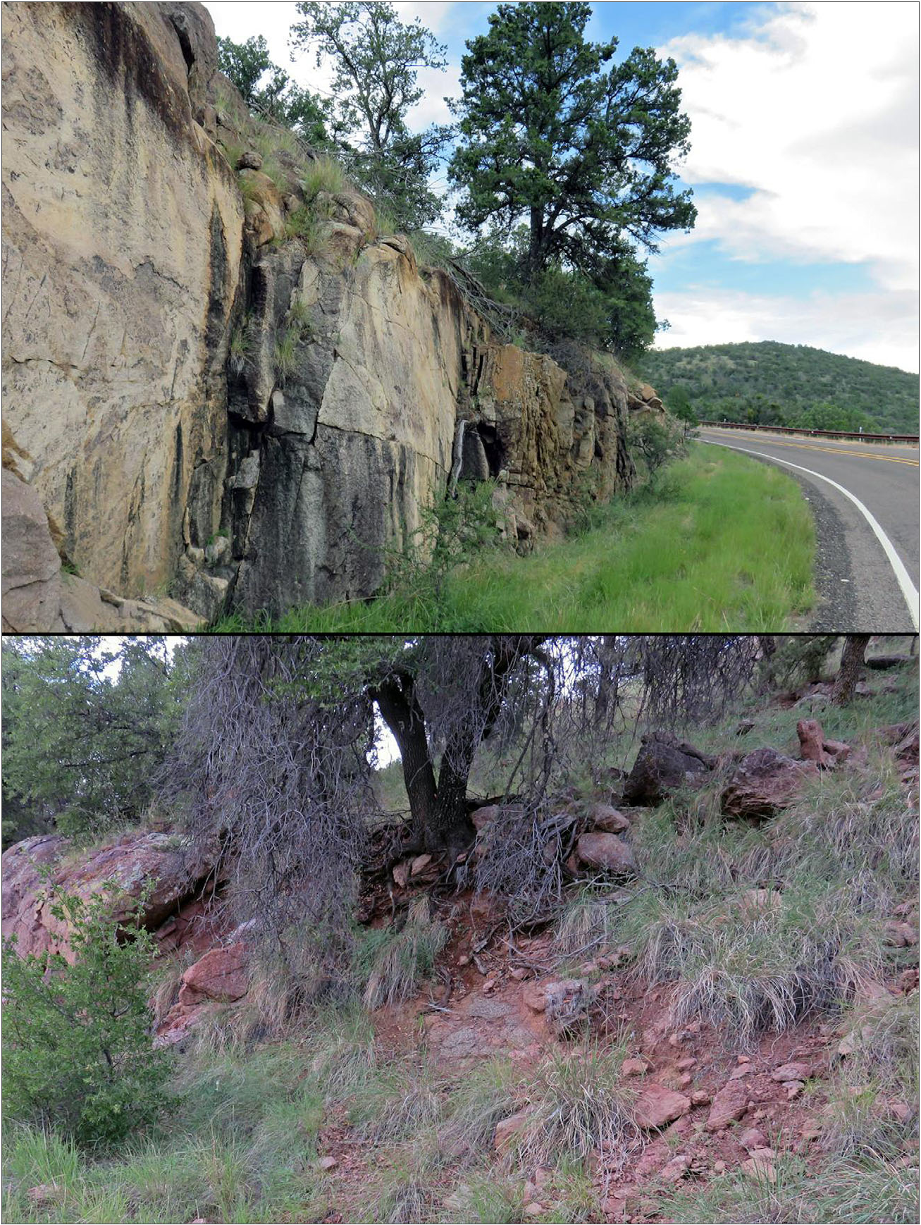
Type material. Holotype ♂, Musquiz Canyon, Hwy 118 N Alpine, Jeff Davis County, Texas, USA (30.53603, - 103.84914, 1470 m), 6 August 2016. leg. R. Bryson , (specimen RA2415 deposited in USNM) GoogleMaps . Paratypes: ♀, ♂, same locality, 6 August 2016, leg. R. Bryson , (specimens RA2412 deposited in USNM, RA2414 MES) GoogleMaps ; 2 ♂, Hwy 118, south of turn to McDonald Observatory , Jeff Davis County, Texas, 5 August 2016, leg. R. Bryson, (specimen # RA2422 deposited in USNM, RA2223 MES) ; 2 ♂, Madera Canyon , Hwy 118, Jeff Davis County, Texas, USA, 5 August 2016, leg. R. Bryson, (specimen RA2416 deposited in USNM, RA2419 MES) .
Diagnosis. Small species with heavy chelae, 20–27 mm. Pectinal tooth counts 10– 12 males, 10 females; metasoma segment V stocky, length to width ratio 1.97–2.06 in males; telson vesicle depth and width to fixed finger length ratio is 0.57–0.65 and 0.74–0.80 in males; fixed finger MD counts 48–55 for males and 52–53 for females; hemispermatophore lamina terminus with distal crest; mating plug brace-A and brace-B without projections.
Distribution. Jeff Davis County, Texas, USA. See map in Fig. 14 View Figure 14 .
Etymology. This species is named in honor of Robert W. Bryson, Jr. for his contributions to the biogeography of scorpions. Dr. Bryson also kindly provided the specimens used in this study as well as photographs of their locality.
MALE. The following description is based on holotype male from the Musquiz Canyon , Jeff Davis County, Texas, USA. Measurements of the holotype male and six paratypes are presented in Table 1 View Table 1 . See Figure 1 View Figure 1 for photographs of live paratype male and female specimens .
COLORATION. Carapace and mesosoma brown. Metasoma brown with darker carinae; telson vesicle orangebrown. Pedipalps brown with darker reddish carinae. Sternopectinal area, sternites, and legs light brown.
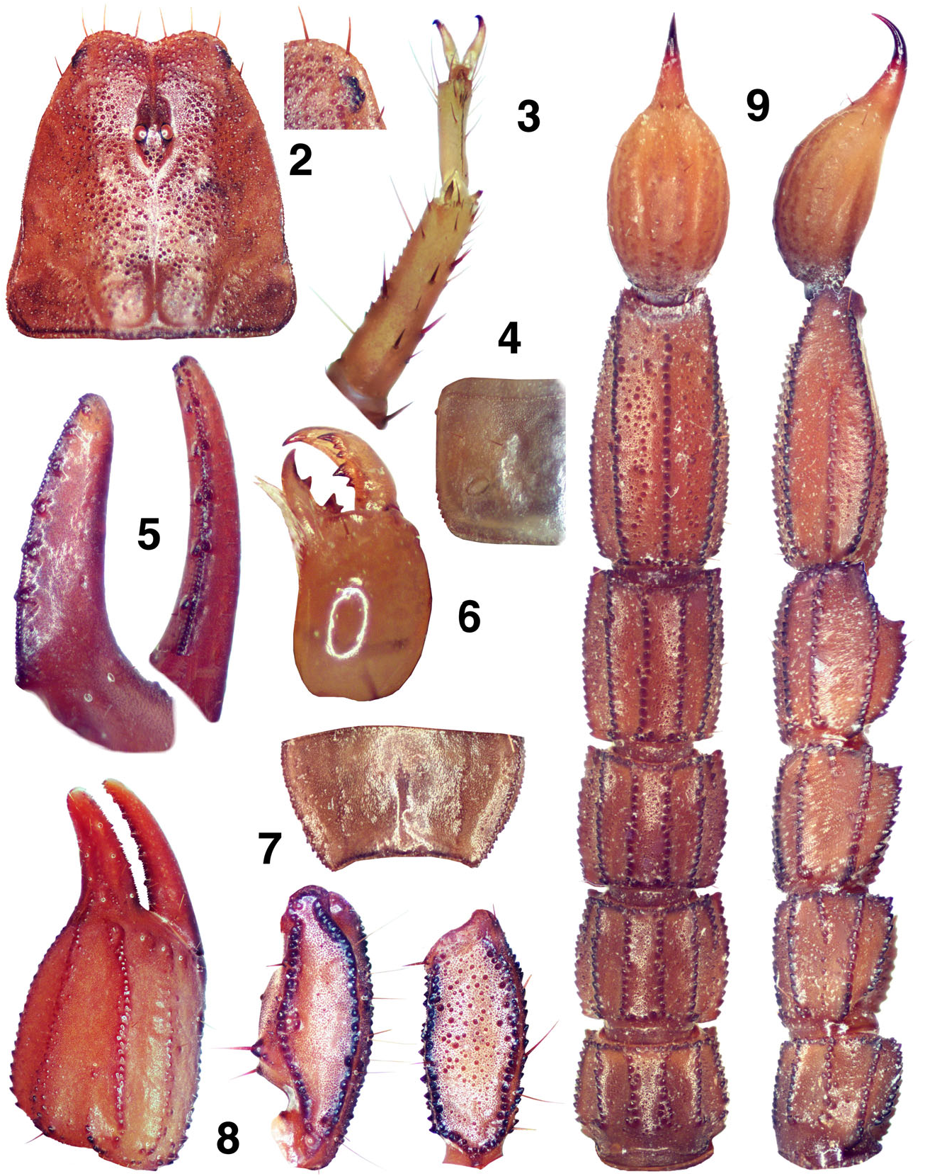
CARAPACE ( Fig. 2 View Figures 2–9 ). Anterior edge with a conspicuous narrow median indentation, providing a ratio of 0.040 when its depth is compared to the carapace’s length; edge with six primary setae visible; entire median surface densely covered with medium sized granules. Three lateral eyes are present, the posterior eye considerably smaller. Median eyes and tubercle of medium size, positioned anterior of middle with the following length and width ratios: 0.337 (anterior edge to medium tubercle middle / carapace length) and 0.182 (width of median tubercle including eyes / width of carapace at that point).
MESOSOMA ( Figs. 4, 7 View Figures 2–9 , 10 View Figure 10 ). Tergites I–VII densely covered with small granules, heaviest posteriorly; tergite VII lateral and median carinae strong and crenulate. Sternites III–V smooth, VI–VII with small dense granules on posterior lateral aspects; sternite VII with weak irregularly granulated lateral carinae and obsolete median carinae ( Fig. 7 View Figures 2–9 ). Stigmata ( Fig. 4 View Figures 2–9 ) are small to medium in size and elliptical in shape.
METASOMA ( Fig. 9 View Figures 2–9 ). Segments I–III wider than long. Segments I–IV: dorsal and dorsolateral carinae serrated; dorsal and dorsolateral (I–III) carinae terminate with an enlarged spine; lateral carinae serrated on I, serrated on posterior two-thirds on II and posterior one-fifth on III, obsolete on segment IV; ventrolateral and ventromedian carinae serrated. Dorsolateral carinae of segment IV terminate slightly above the articulation condyle. Segment V: dorsolateral carinae rounded and granulate to serrate; lateral carinae serrated for two-thirds of posterior aspect; ventrolateral and single ventromedian carinae serrated; ventromedian carina not bifurcated, terminating in straight line. Anal arch with 15 small granules. Intercarinal areas of segment V ventral surface scattered with minute granules.
TELSON ( Fig. 9 View Figures 2–9 ). Vesicle fairly robust with some lowprofile granules located on the ventral surface; slight setation on ventral surface. Aculeus with medium curve, well delineated from the vesicle when viewed ventrally. The subaculear setal pair is located on the vesicle/aculeus juncture. A vesicular linear patch on the dorsal surface is absent. Vesicular tabs with a single small curved spine.
PECTINES ( Fig. 10 View Figure 10 , paratype male). Well-developed segments exhibiting length / width ratio 2.429 (length taken at anterior lamellae / width at widest point including teeth). Sclerite construction complex, three anterior lamellae and seven middle lamella; fulcra of medium development. Teeth number 11/11. Sensory areas developed along most of tooth inner length on all teeth, including basal tooth. Scattered setae found on anterior and middle lamellae, fulcra, and distal pectinal tooth. Basal piece large, with well developed wide indentation along anterior edge, length / width ratio 0.550.
GENITAL OPERCULUM ( Fig. 10 View Figure 10 , paratype male). Sclerites triangular, wider than long, separated, genital papillae protruding posteriorly.
STERNUM ( Fig. 10 View Figure 10 , paratype male). Type 2, posterior emargination present, well-defined convex lateral lobes, apex shallow; sclerite wider than long, in ratio 0.636.
CHELICERAE ( Fig. 6 View Figures 2–9 ). Movable finger dorsal edge with two subdistal (sd) denticles; ventral edge smooth with well developed serrula on distal half (with 27 tines). Ventral distal denticle (vd) longer than dorsal (dd). Fixed finger with four denticles, median (m) and basal (b) denticles conjoined on common trunk; no ventral accessory denticles present.
PEDIPALPS ( Figs. 5, 8 View Figures 2–9 , 11 View Figure 11 ). Well-developed chelae, with short fingers, carinae well developed, no scalloping on the fingers. Planes formed by carinae D1 | D3 | D4 and V1 | V2 | V3 are essentially parallel. Femur: Dorsointernal and ventrointernal carinae heavily serrated, dorsoexternal crenulated, and ventroexternal with scattered rounded granules. Dorsal and ventral surfaces scattered with granules, internal surface scattered with large granules, and external surface smooth. Patella: Dorsointernal, ventrointernal, dorsoexternal and ventroexternal carinae heavily serrated, and exteromedian carina singular, strong and crenulated. Dorsal and ventral surfaces essentially smooth, rough but with no granulation; external surface with serrated exteromedian carina and 2–3 small granules in proximity of trichobothrium est; internal surface smooth with medium sized DPS and small VPS. Chelal carinae: Complies to the “10-carinae configuration”. Digital (D1) carina strong and serrated; subdigital (D2) essentially obsolete, composed of two small granules; dorsosecondary (D3) flat lined with delicate granules; dorsomarginal (D4) medium to strong with scattered granulation; dorsointernal (D5) medium with large granules; ventroexternal (V1) strong and serrated, terminating at external condyle of movable finger; ventromedian (V2) essentially obsolete; ventrointernal (V3) strong with scattered granulation; external (E) medium, lined with small granules. Chelal finger dentition ( Fig. 5 View Figures 2–9 ): Median denticle (MD) row groups aligned in a straight line, 6 on the fixed and movable fingers; 6/6 ID s on fixed finger and 7/7 ID s on movable finger; 5/5 OD s on fixed finger and 6/6 OD s on movable finger. No accessory denticles present. The number of MD s on the fixed finger for each row: right finger: md1 = 5, md2 = 5, md3 = 8, md4 = 7, md5 = 8, md6 = 17: total = 50; left finger: md1 = 4, md2 = 8, md3 = 7, md4 = 6, md5 = 8, md6 = 15: total = 48. Trichobothrial patterns ( Fig. 11 View Figure 11 ): Type C, orthobothriotaxic. Trichobothria ib -it located basally, ib on the palm and it on the fixed finger base; chelal V 4 is located on the V1 carina; Db is located external to the D1 carina; Dt is positioned well on the proximal half of the palm; patellar v 3 is located slightly posterior to et 3.
LEGS ( Fig. 3 View Figures 2–9 ). Both pedal spurs present on all legs, lacking spinelets; tibial spurs absent. Ventral surface of the tarsus with a median row of short spinules terminating distally with two pairs of spinules. Unguicular spine well-developed and pointed.
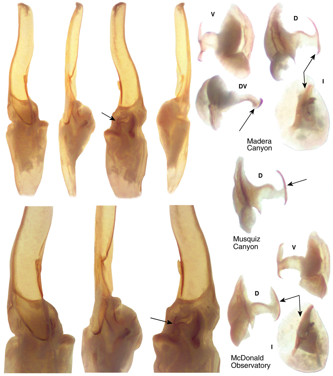
HEMISPERMATOPHORE ( FIG View Figure 12 . 12, three paratype males). Lamina edges subparallel, terminus truncated with a subtle distal crest on the dorsal side (also visible from the ventral side due to the structure’s translucency). Lamellar hook elongated, distinctly bifurcated, and originating from the dorsal trough. A secondary lamellar hook and basal constriction are absent. Right hemispermatophore length is 4.20 mm, lamina length is 2.50 mm, lamellar hook length is 1.05 mm, and trough difference is 0.50 mm. Lamellar hook length to lamina length ratio is 0.420 and trough difference to lamellar hook length ratio is 0.476. Mating plug with a smooth barb, its distal edges essentially straight, thus not “crescent-shaped”. Projections from brace-A and brace-B of the base are absent. A stem projection is present at its base.
Male and female variability. Of the seven specimens available for this study, only one is a female and deemed here to be subadult. Therefore, our comparison of the two genders is limited. Though the male has a slightly thinner metasoma in segments II–V, the MVDs (L/W) only range from 1.0 to 3.1 %. The chela fixed finger, however, is definitely shorter in the male when compared to the metasomal segment lengths which are relatively longer, in particular segments II–V, exhibiting sizable MVDs ranging from 22.4 to 25.5 %. The number of median denticles (MD) on the chelal fixed finger are essentially the same in both genders, male 48–55 (52.167) [12] and female 52–53 (52.500) [2]. Pectinal tooth counts in males exceed those of females by only one tooth, male 10–12 (11.083) [12] and female 10–10 (10.000) [2]. The genital operculum sclerites are fused medially except for the posterior one-third in the female, whereas they are separated along their entire length in the male, exposing developed genital papillae. See Appendix A for further statistical information.
Comparison of Species
The three western Texas populations of P. brysoni studied for this paper are located in close proximity, only 30 kilometers separate all three. The altitude of the three localities only varies 390 m. Each population is represented by two adult males each and, morphometrically, exhibit consistency in all ratio comparisons. When all possible ratios were calcuated for comparison with the other three species (351 ratios in all), the coefficient of variability (i.e., sdev/mean) for P. brysoni was quite small, ranging only 0.002–0.059 (0.030) for the six males. This implies total consistency across the six males representing three locations for all morphometric ratio comparisons.
The four species currently placed in “ apacheanus ” group of Pseudouroctonus , including new species P. brysoni described in this paper, are essentially structured the same. All roughly the same size, adult males 21–27 and females 29–32 mm in length, all have the same general carinal structure development with heavy granulation, and all with the same number of pectinal tooth counts, 10– 12 males and 9– 11 females, etc. Trichobothrial patterns are essentially identical, all exhibiting the same diagnostic positions considered in the genus. Except for the unusual brace projections exhibited on the hemispermatophore mating plug in species P. santarita , there are no exceptional structural differences between the four species. Therefore, morphometric ratio comparisons are relied upon heavily to separate these four species. Also, since ratio comparisons indicate the relative length of the chelal fingers, we have also relied on the counts of median denticles (MD), in particular important for species with proportionally short or long chelal fingers as indicated by morphometric comparisons. Following is a key to the four species of the “ apacheanus ” group of Pseudouroctonus , reflecting the molecularbased phylogeny suggested by Bryson et al. (2013: fig. 3).


| R |
Departamento de Geologia, Universidad de Chile |
| USNM |
Smithsonian Institution, National Museum of Natural History |
No known copyright restrictions apply. See Agosti, D., Egloff, W., 2009. Taxonomic information exchange and copyright: the Plazi approach. BMC Research Notes 2009, 2:53 for further explanation.
|
Kingdom |
|
|
Phylum |
|
|
Class |
|
|
Order |
|
|
Family |
|
|
Genus |
Pseudouroctonus brysoni Ayrey et Soleglad
| Ayrey, Richard F. & Soleglad, Michael E. 2017 |
Pseudouroctonus apacheanus:
| BRYSON 2013: 6 |