
Corellamysis eltanina
|
publication ID |
https://doi.org/ 10.11646/zootaxa.3780.2.6 |
|
publication LSID |
lsid:zoobank.org:pub:BFC5B41E-8154-4C24-8AE0-EB1C4AD1A380 |
|
DOI |
https://doi.org/10.5281/zenodo.6126095 |
|
persistent identifier |
https://treatment.plazi.org/id/F41D3E76-8172-B13E-1D80-FDEDFCB49780 |
|
treatment provided by |
Plazi |
|
scientific name |
Corellamysis eltanina |
| status |
|
Corellamysis eltanina San Vicente sp. nov.
( Figs. 1–7 View FIGURE 1 View FIGURE 2 View FIGURE 3 View FIGURE 4 View FIGURE 5 View FIGURE 6 View FIGURE 7 )
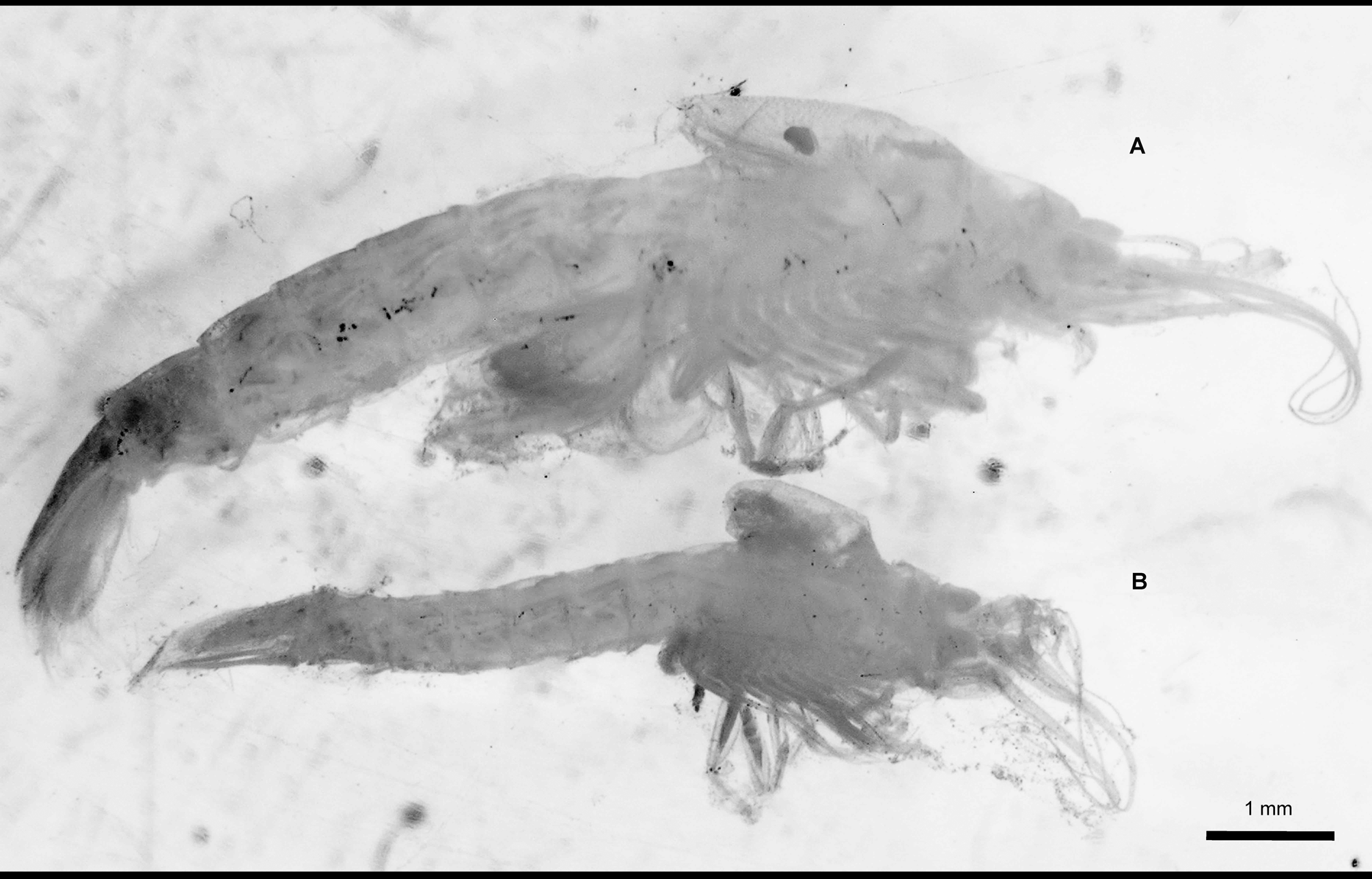
Material examined. Holotype: One brooding female 9.3 mm TL (catalogue number: ICMM 13102501). “Eltanin” survey USA. 54°31’S – 159°00’ E, 110 m depth. 18/06/1968. Dissected, one vial.
Paratypes: One mature male 5.6 mm TL (catalogue number: ICMM 13102502); dissected, one vial; from the same locality as holotype. One immature female 5.8 mm TL (catalogue number: ICMM 13102503); not dissected; one vial; from the same locality as holotype. One juvenile 3.4 mm TL (catalogue number: ICMM 13102504); not dissected; one vial; from the same locality as holotype. One juvenile 3.4 mm TL (catalogue number: ICMM 13102505); not dissected; one vial; from the same locality as holotype.
The material comes from the Muséum National d’Histoire Naturelle, Paris. Individuals were found inside the branchial sacs of three specimens (among 56 examined) of Corella brewinae Monniot 2013 (Ascidiacea, Phlebobranchia, Corellidae ) collected in the same trawl. The holotype and one juvenile paratype were removed from one C. brewinae specimen, the immature female paratype and one juvenile paratype from another ascidian specimen and the mature male paratype from a third ascidia .
Etymology. This species is named after the U.S. Navy Ship “Eltanin”, operated by the National Science Foundation from 1957 to 1972; specimens were collected in the 1968 cruise and sent to the Muséum National d’Histoire Naturelle for identification and study.
Description. The description below refers to both sexes, unless otherwise stated.
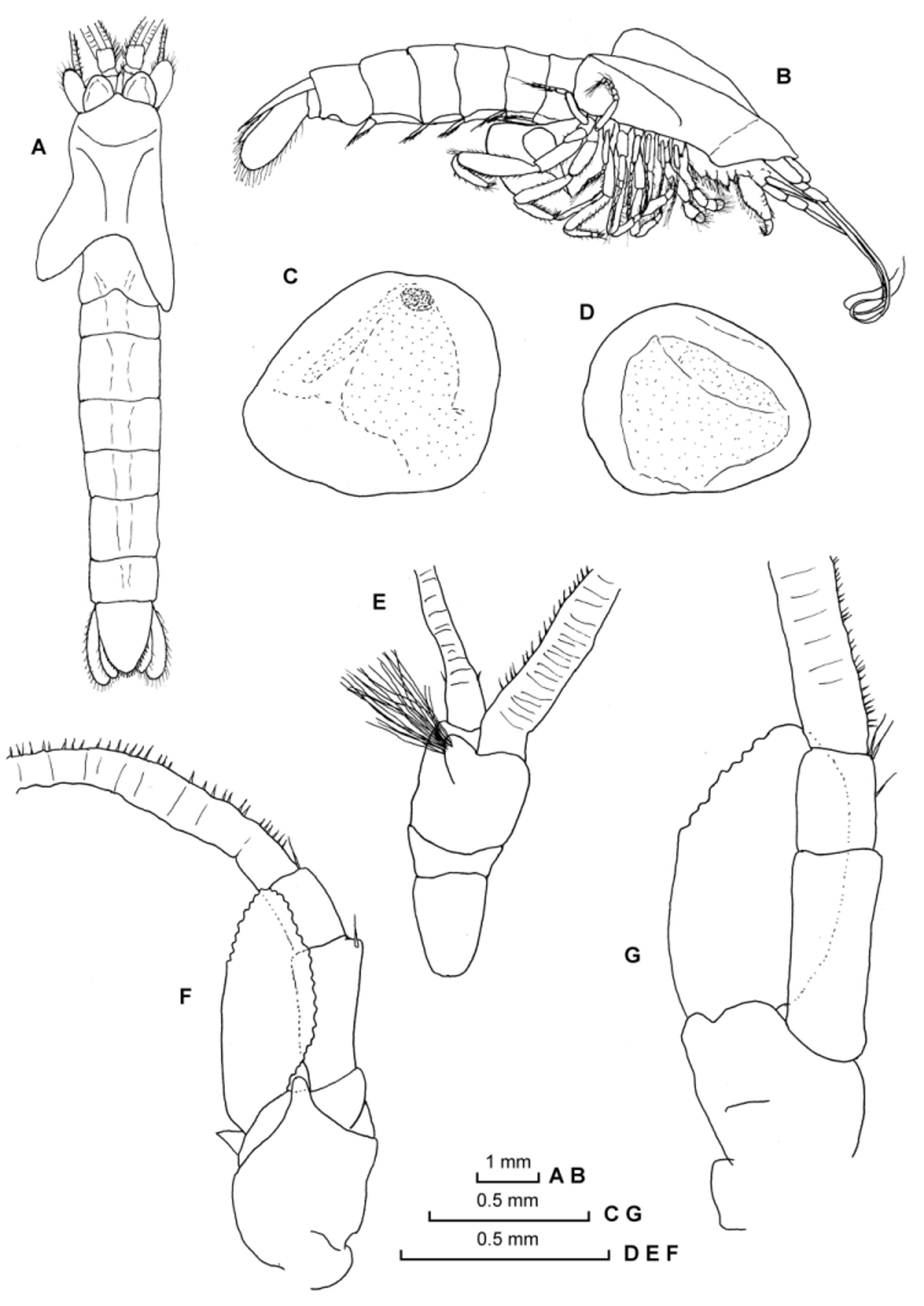
General body form moderately robust ( Figs. 1 View FIGURE 1 A–B, 2A–B). Carapace with anterior margin evenly rounded, not forming definite rostral projection; posterior margin dorsally emarginated, leaving last thoracic somite partially uncovered; posterolateral lobe covering anterior abdominal somite. Abdomen robust, as wide as middle portion of carapace, not tapering posteriorly; all abdominal somites subequal in length.
Eyes without definite eyestalks ( Figs. 2 View FIGURE 2 C–D), flattened and subquadrangular with rounded corners, visual elements imperfectly developed and without trace of pigment in preserved individuals, laterally not extending beyond carapace limits.
Antennular peduncle ( Figs. 2 View FIGURE 2 A, E) extending slightly beyond antennal scale. First article longer than wide; second article shortest, half as long as broad; third article as long as broad; in male, small sub-spherical and hirsute appendix masculina present on ventral side of third article.
Antennal sympod ( Figs. 2 View FIGURE 2 F–G) with outer distal dorsal angle rounded and ventral distal middle finger-like protuberance. Peduncle not extending beyond scale; first article short, as long as broad, inner margin rounded; second article twice as long as broad, inner distal margin armed with one simple seta; third article half shorter than second one, distal inner margin armed with one or two simple setae. Antennal scale two-three times longer than maximum width, not extending beyond antennular peduncle; margins convex, setose on its inner margin and on the distal one-third of its outer margin, proximal two-thirds of outer margin entire; without apical suture.
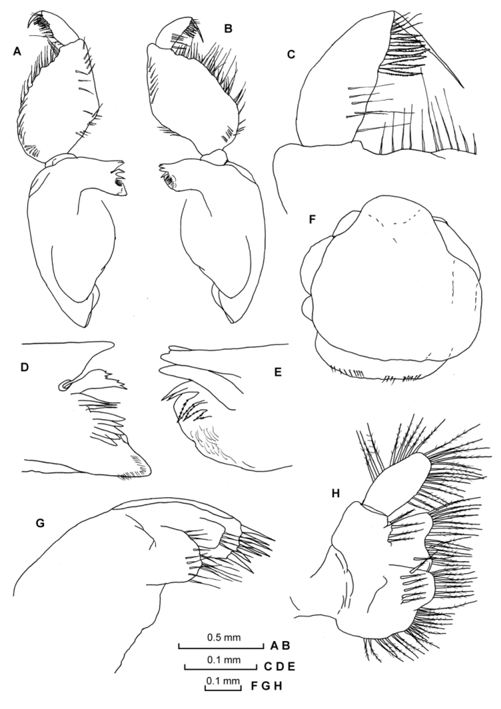
Labrum ( Fig. 3 View FIGURE 3 F) more or less symmetrical, as long as wide; without frontal spiniform process; posterior margin with short irregularly distributed thin simple setae.
Mandibles ( Figs. 3 View FIGURE 3 A–E) well developed. Three-segmented palp, first article shortest; second article slightly longer than wide, with simple setae on both margins; third article about twice as long as broad, armed on distal third of inner margin with 6 or 7 ventral serrated setae and 2 or 3 distal simple setae; one distal large conspicuous seta. Mandibles with well developed incisor and reduced molar process; left mandible plus row of 2 simple spines and 3 serrated spines; counterpart on right mandible comprising 8 entire spines; right lacinia mobilis well developed ( Figs. 3 View FIGURE 3 D–E).
Maxillule ( Fig. 3 View FIGURE 3 G) apex of outer lobe armed with 7 strong cuspidate setae and single row of 4 simple setae on ventral surface; inner lobe with 12 simple setae.
Maxilla ( Fig. 3 View FIGURE 3 H) apparently without exopod; distal article of endopod oval, longer than wide, margins armed with about 23 pappose setae on inner and outer distal two-thirds; inner margin of coxal endite armed with 2 rows of pappose setae; inner margin of bilobulate basal endites also armed with pappose setae.
Thoracopods grouped in three different shape groups. First and second thoracic appendages formed as maxillipeds, third to sixth endopods with carpopropodus 2-segmented, and seventh and eighth with endopods specialized as gnathopods.
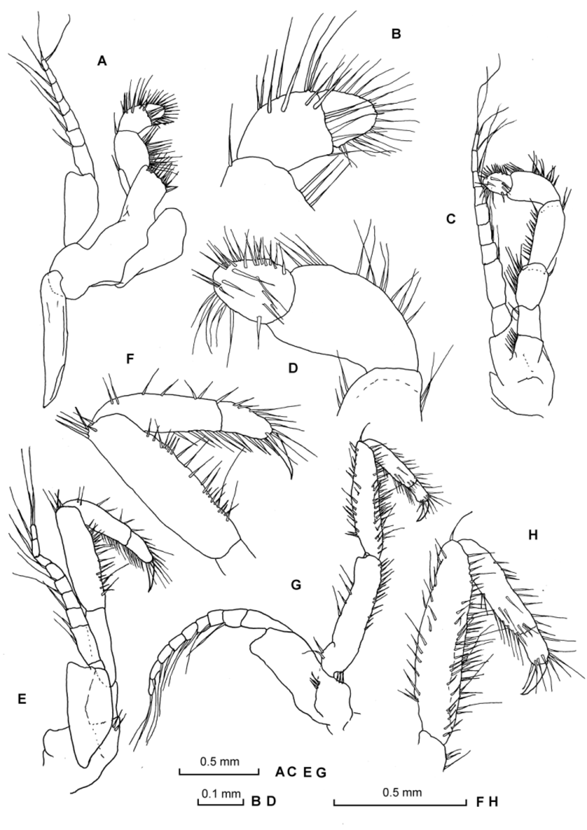
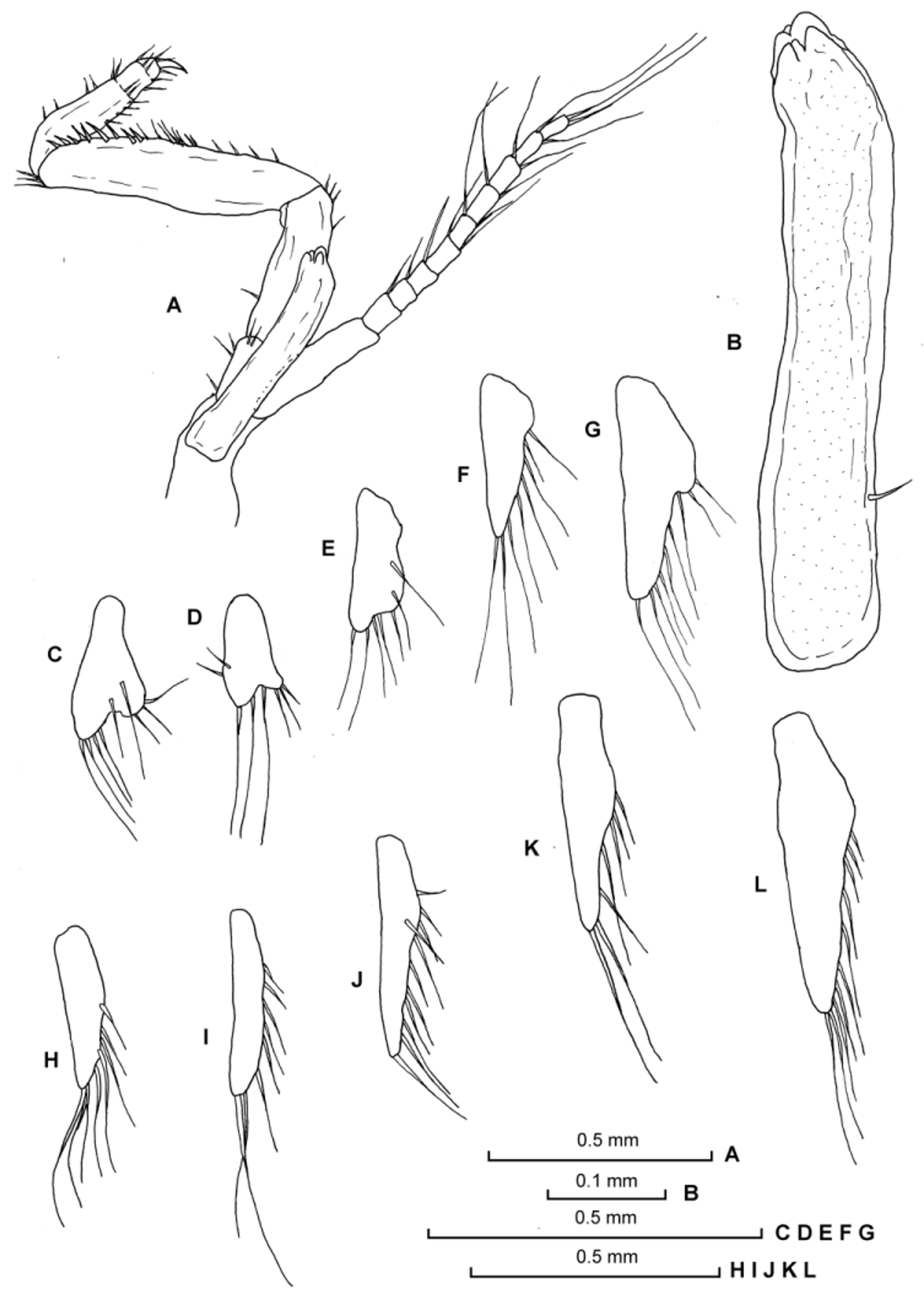
First thoracopod ( Figs. 4 View FIGURE 4 A–B) short, with unarmed epipodite. Endopod with merus longer than carpopropodus; dactylus sub-spherical densely setose, bearing a series of simple setae on both margins. Exopod longer than endopod, with 7-segmented flagellum.
Second thoracopod ( Figs. 4 View FIGURE 4 C–D) slightly longer than first one, with small endite on the basis. Endopod with preischium and ischium subequal in length; merus subequal to carpopropodus; dactylus sub-spherical, densely setose with a series of simple setae on both margins. Exopod longer than endopod, with 8-segmented flagellum.
Third to sixth thoracopods ( Figs. 4 View FIGURE 4 E–H, 5A–B) with endopod longer than exopod; ischium and merus subequal in length; carpopropodus 2-segmented, shorter than merus; inconspicuous segmentation between the carpopropodus and dactylus; strong distal nail present. Exopod with 9-segmented flagellum. Sixth thoracic appendages of female with rudimentary oostegites, armed with 3 simple setae on its distal margin, 1 on outer margin and 2 on inner margin ( Fig. 5 View FIGURE 5 A)
Seventh and eighth thoracopod endopods quite different from the rest, with undivided carpopropodus and inconspicuous segmentation between carpopropodus and dactylus; merus armed along its inner margin with strong cuspidate setae forming powerful subchela ( Figs. 5 View FIGURE 5 C–F, 6A). Eighth thoracopod with enlarged endopod, much longer and larger than anterior ones; endopod longer than exopod; merus large, armed along its inner margin with 9 (male) or 25 (female) cuspidate setae; undivided carpopropodus; dactylus with strong distal nail present. Exopod with rudimentary basal plate and 8-segmented flagellum.
Oostegites of seventh thoracopods developed, smaller than those of eighth thoracopods ( Figs. 5 View FIGURE 5 C, E). Penes roughly cylindrical, reaching two-thirds of ischium length. Each penis with simple seta located at proximal onefourth of its length on outer margin; distal margin obscurely 6-lobate ( Figs. 6 View FIGURE 6 A–B).
Pleopods uniramous, reduced to unsegmented lobes in both sexes ( Figs. 6 View FIGURE 6 C–L), increasing in length from first to fifth pairs; fifth pleopod extending to posterior half of last abdominal somite ( Fig. 2 View FIGURE 2 B).
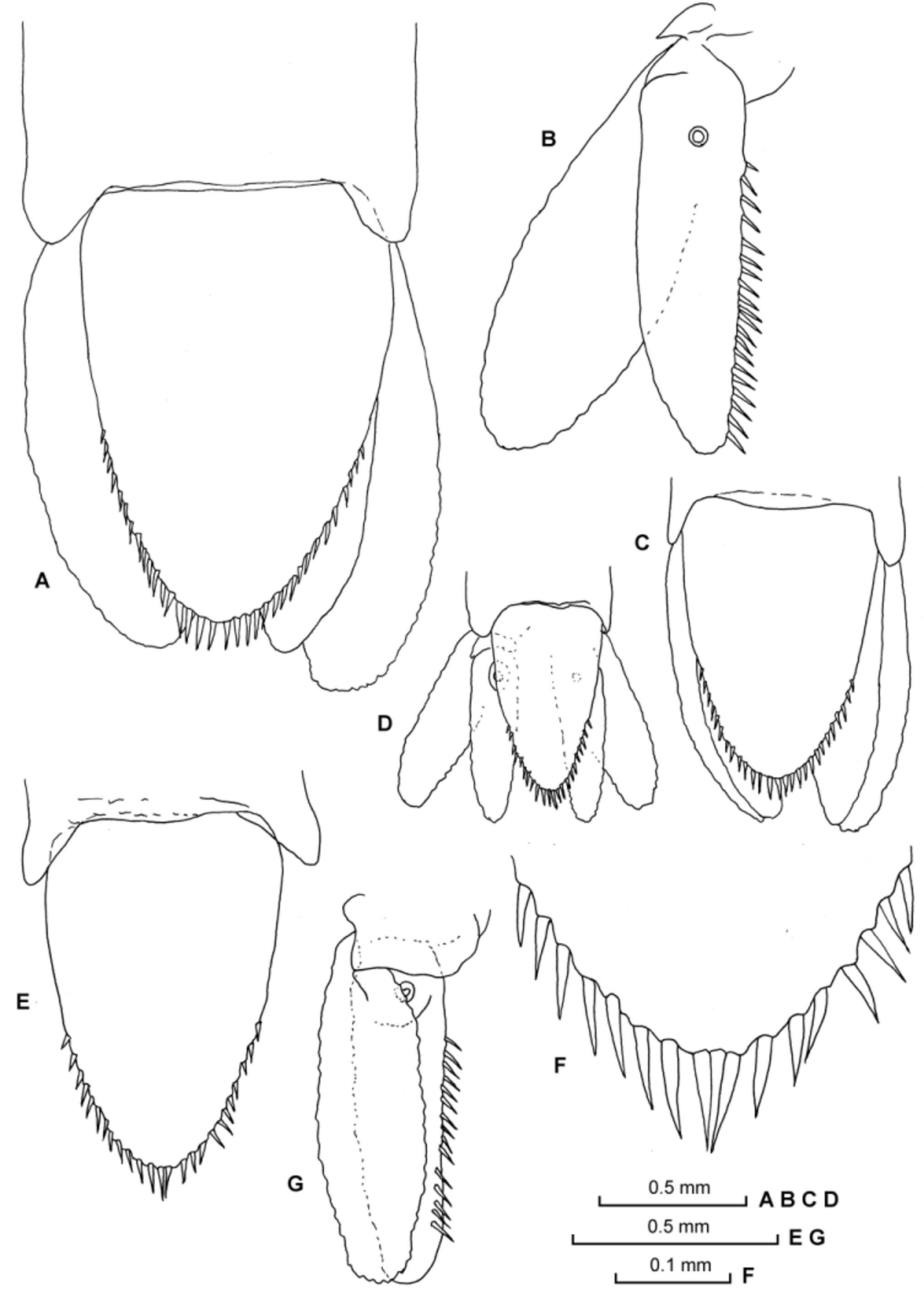
Uropodal endopod ( Figs. 7 View FIGURE 7 B, G) slender, extending slightly beyond apex of telson, inner margin armed with 15–20 short cuspidate setae extending from near statocyst to near apex. Female uropodal exopod slightly longer and broader than endopod (subequal in male and immature specimens), setose all round ( Figs. 7 View FIGURE 7 A–D, G).
Telson entire, linguiform, about two times as long as broad at base; distal half of lateral margins armed with 11 (juvenile) –17 (brooding female) cuspidate setae, increasing in size towards apex ( Figs. 7 View FIGURE 7 A, C–D, E–F). Colour (in long-time preserved specimens): almost transparent tegument with brown pigmentation distributed on the abdomen and some appendages.
Distribution and habitat. The known distributional area of the new Corellamysis species is at the moment limited to the Macquarie Island region (Southern Ocean), at 110 m depth. All individuals were collected in the branchial sacs of three ascidian individuals of Corella brewinae .

No known copyright restrictions apply. See Agosti, D., Egloff, W., 2009. Taxonomic information exchange and copyright: the Plazi approach. BMC Research Notes 2009, 2:53 for further explanation.