
Teloganodes (Teloganodes) dentatus Navás 1932
|
publication ID |
https://doi.org/ 10.11646/zootaxa.5244.6.3 |
|
publication LSID |
lsid:zoobank.org:pub:F7A8630A-2C58-4825-A309-B2FF35EFEFC3 |
|
DOI |
https://doi.org/10.5281/zenodo.7673103 |
|
persistent identifier |
https://treatment.plazi.org/id/F45787B3-730F-CE2E-FF39-5193FAFF5341 |
|
treatment provided by |
Plazi |
|
scientific name |
Teloganodes (Teloganodes) dentatus Navás 1932 |
| status |
|
Teloganodes (Teloganodes) dentatus Navás 1932 View in CoL
( Figs 73–105 View FIGURES 73–76 View FIGURES 77–81 View FIGURES 82–88 View FIGURES 89–92 View FIGURES 93–97 View FIGURES 98–105 )
Teloganodes dentata Navás 1932: 19 (J imago).
Teloganodes dentatus: Sartori, Peters & Hubbard 2008: 11 View in CoL View Cited Treatment (J imago).
Teloganodes sartorii Selvakumar, Sivaramakrishnan & Jacobus in Selvakumar, Sivaramakrishnan, Jacobus, Janarthanan & Arumugam 2014: 94 View Cited Treatment (larva), syn. n.
Material examined. INDIA, state Karnataka, border of Shivamogga and Udupi districts near Agumbe and Someswar , 11.I–1.II.2013, coll. N. Kluge & L. Sheyko: 2 L-S-IJ, 2 L-S-I ♀ /O, 1 I ♀, 4 larvae ( ZIN); state Tamilnadu: Theni district , Suruli Falls , 24–26.I.2016, coll. L. Sheyko & N. Kluge: L/SJ ( ZIN) .
Descriptions
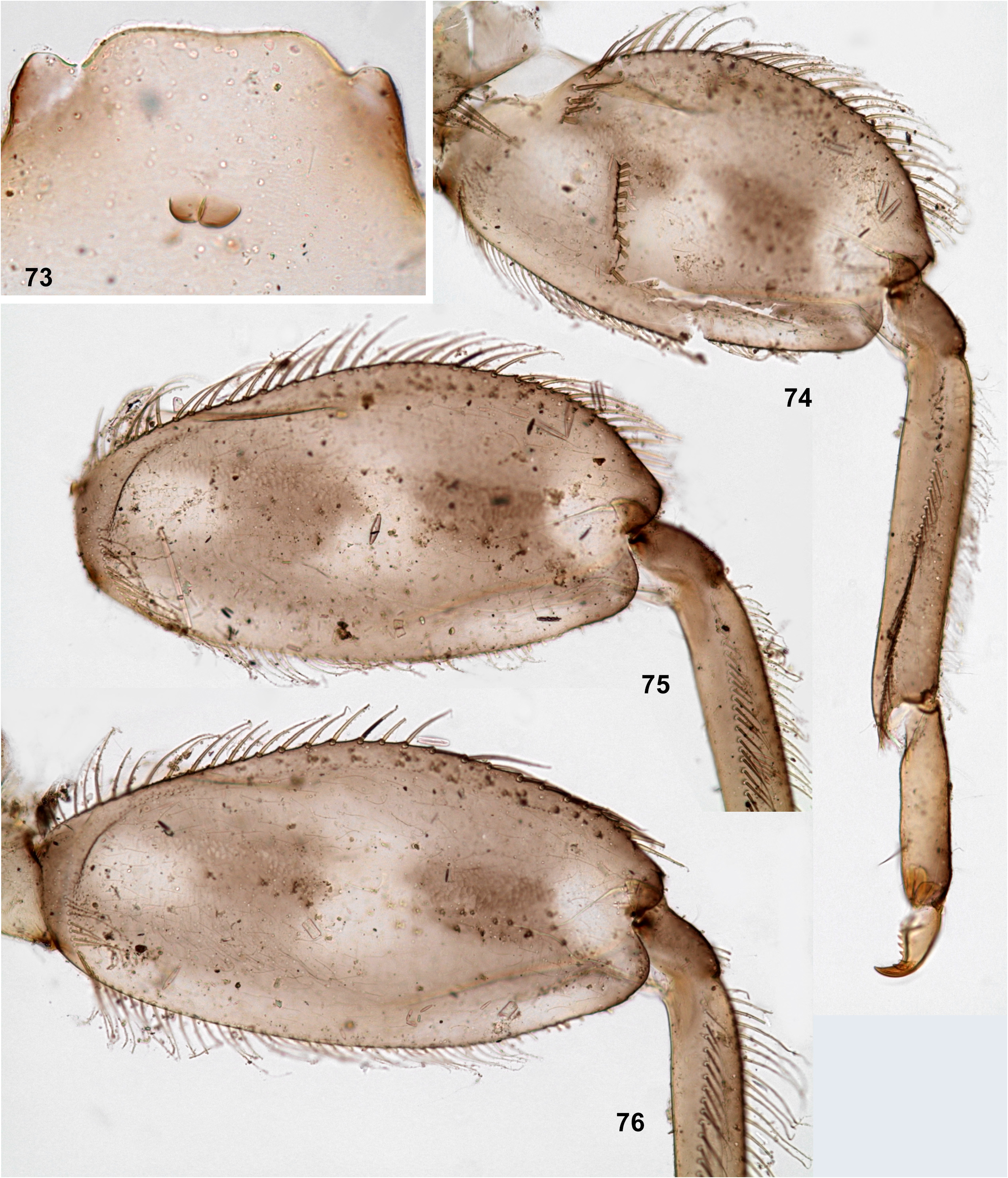
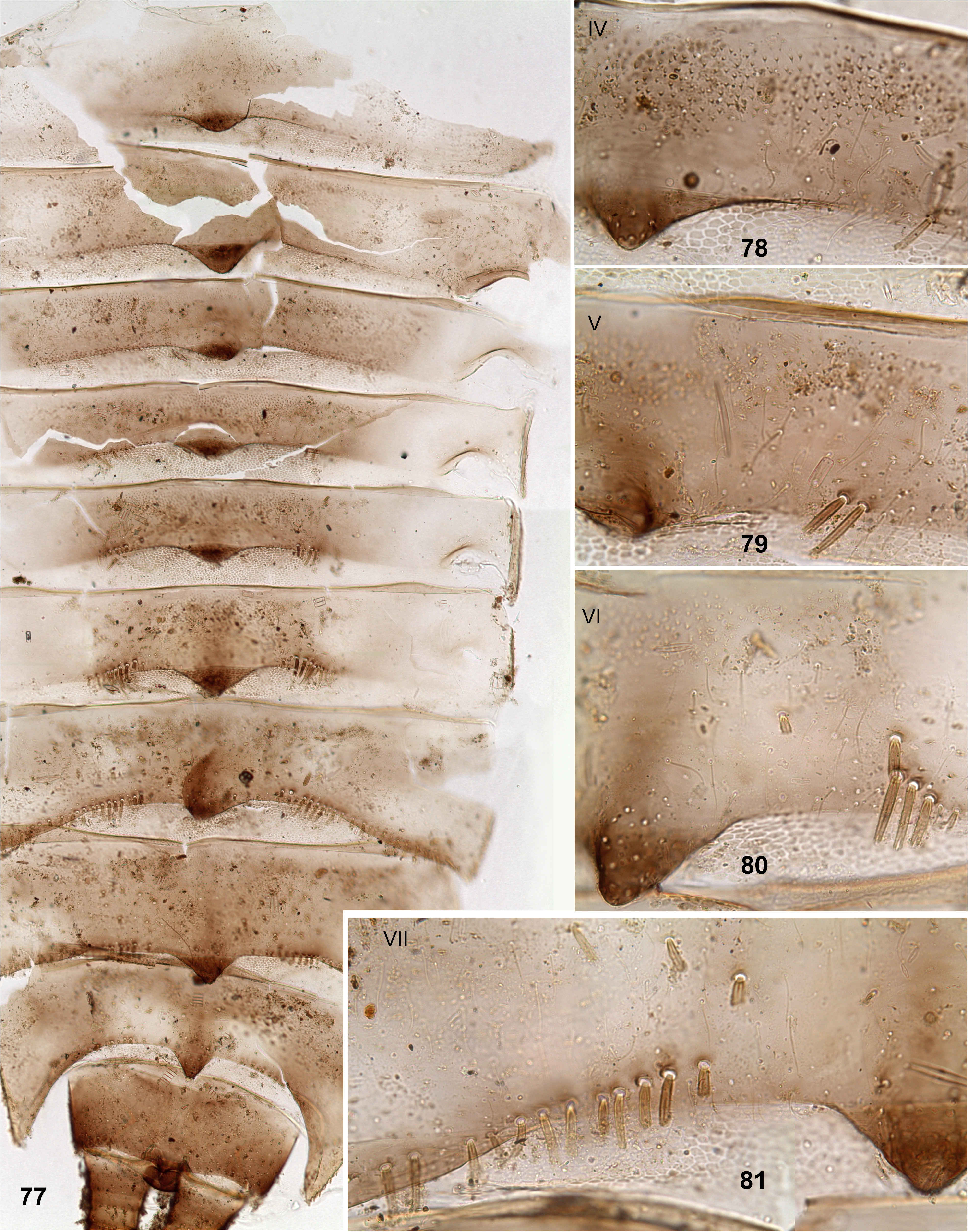
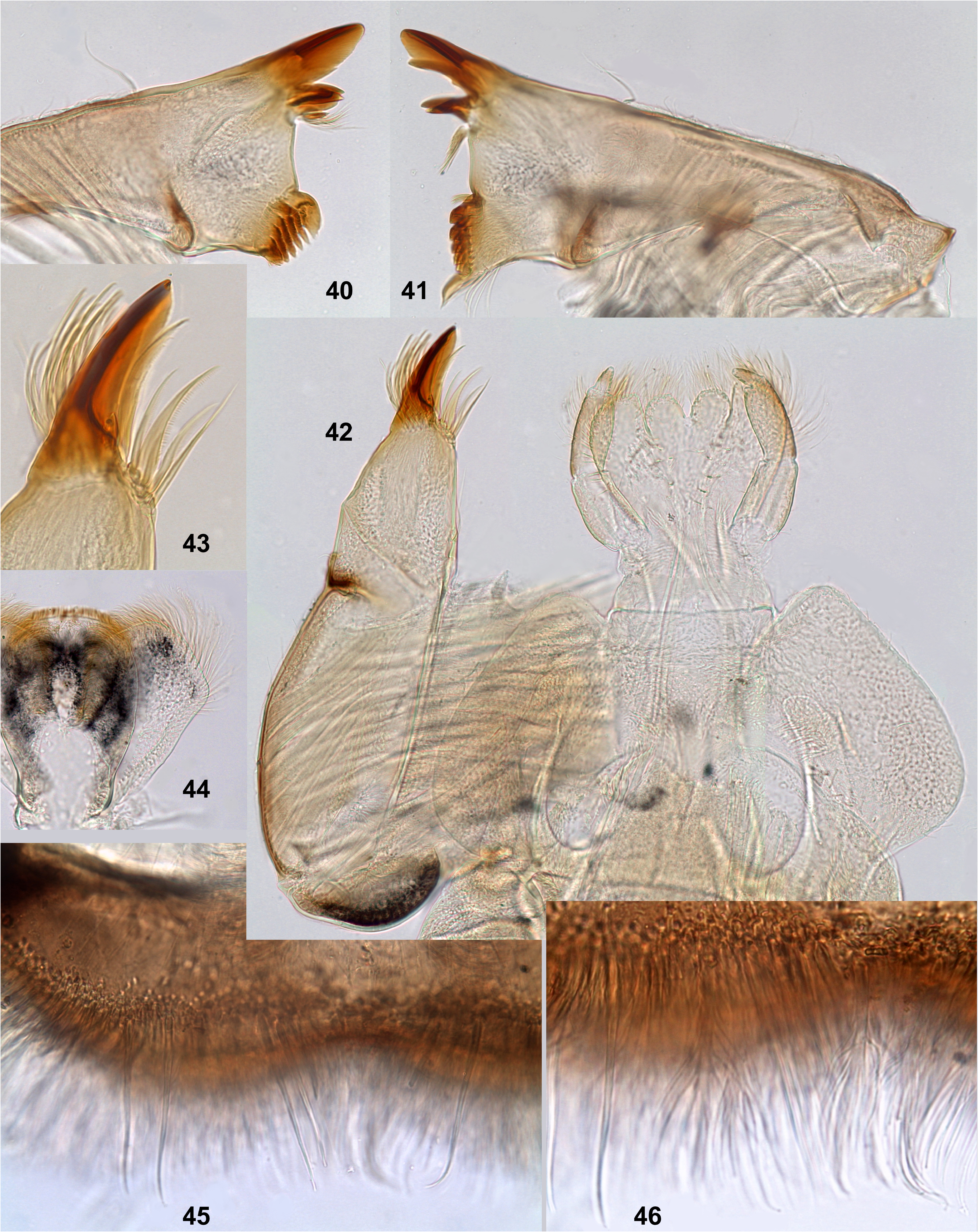
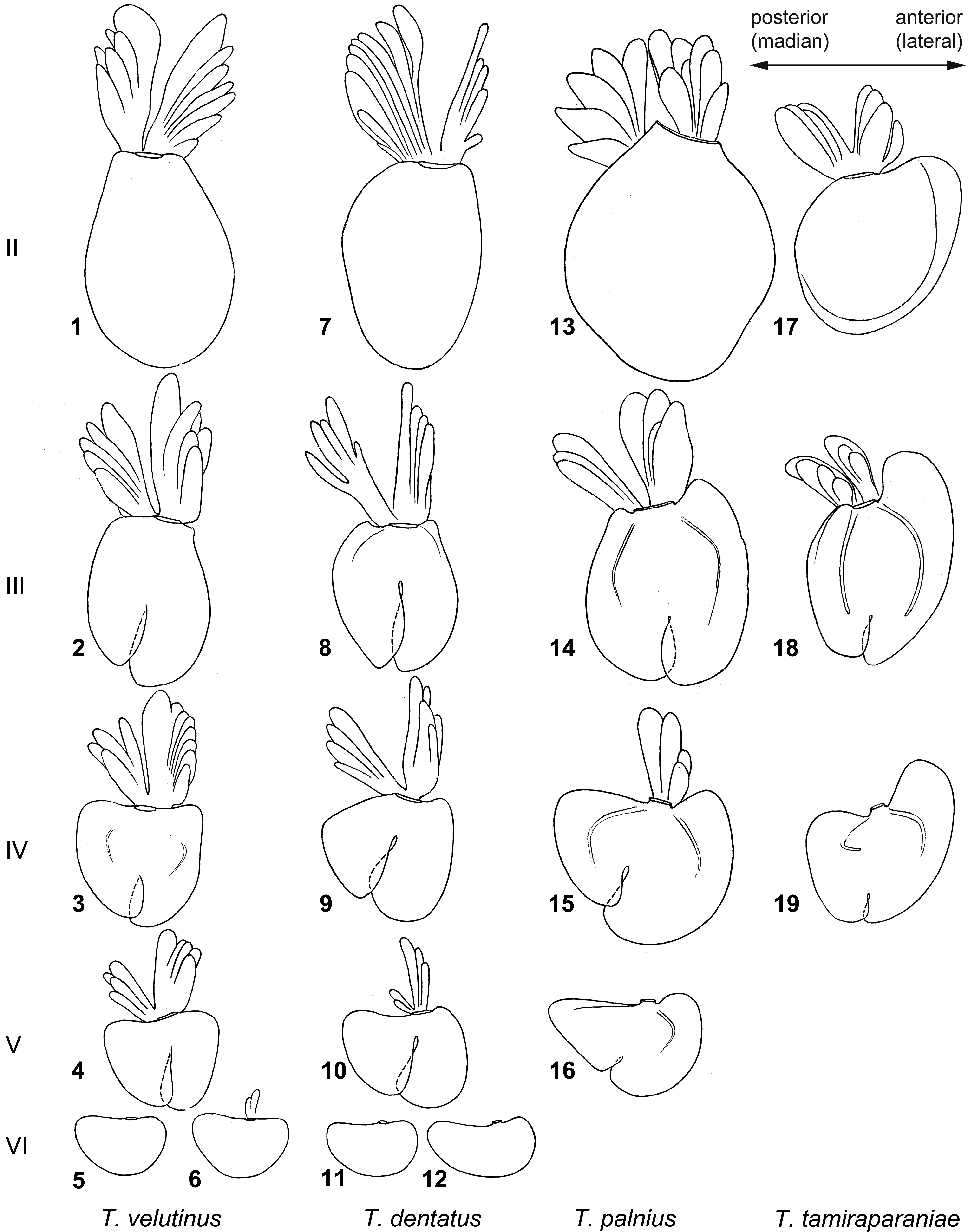
Larva. Described by Selvakumar et al. 2014. Additional characters: Labrum with simple and feathered setae forming transverse field all over labrum width (as in Figs 45–46 View FIGURES 40–46 ); smaller feathered setae located distad of this field. Abdominal terga I–III without setae on posterior margins; terga IV– VI with pair of short oblique rows of short, stout setae close to posterior margin ( Figs 78–80 View FIGURES 77–81 ); terga VII–VIII with such setae on most part of posterior margin ( Fig. 81 View FIGURES 77–81 ); posterior margin of tergum IX with smaller stout setae, like setae on other surface ( Fig. 77 View FIGURES 77–81 ); lateral sides of terga with dense, small, stout setae. Tergalii II–IV with two-branched ventral lobes; ventral branch of tergalius V consisting of well-developed anterior branch and vestigial posterior branch ( Fig. 10 View FIGURES 1–19 ); tergalius VI without ventral lobe ( Figs 11–12 View FIGURES 1–19 ). Posterior margin of sternum IX trapeze-like (in male with small protogonostyli by sides) ( Fig. 73 View FIGURES 73–76 ).
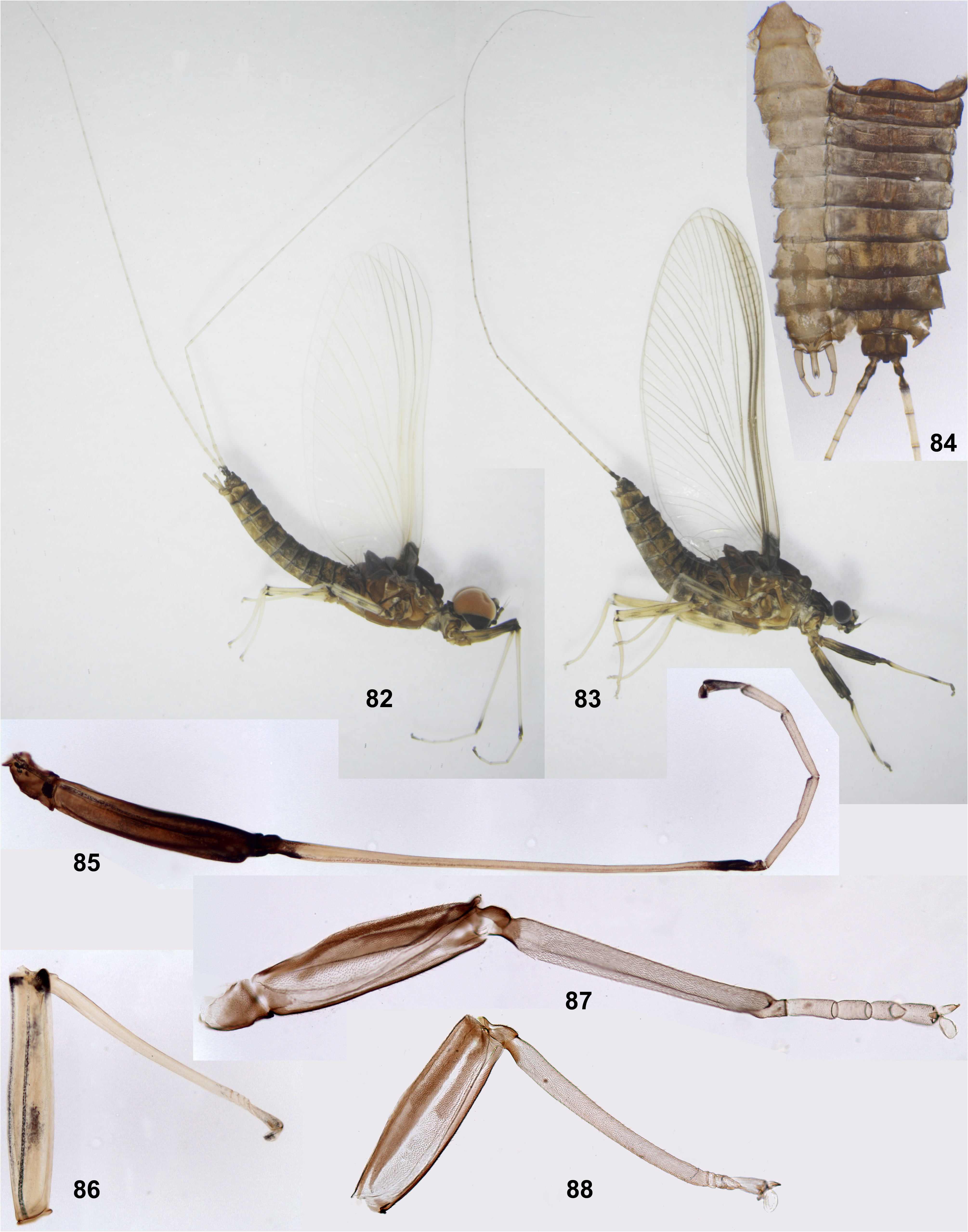
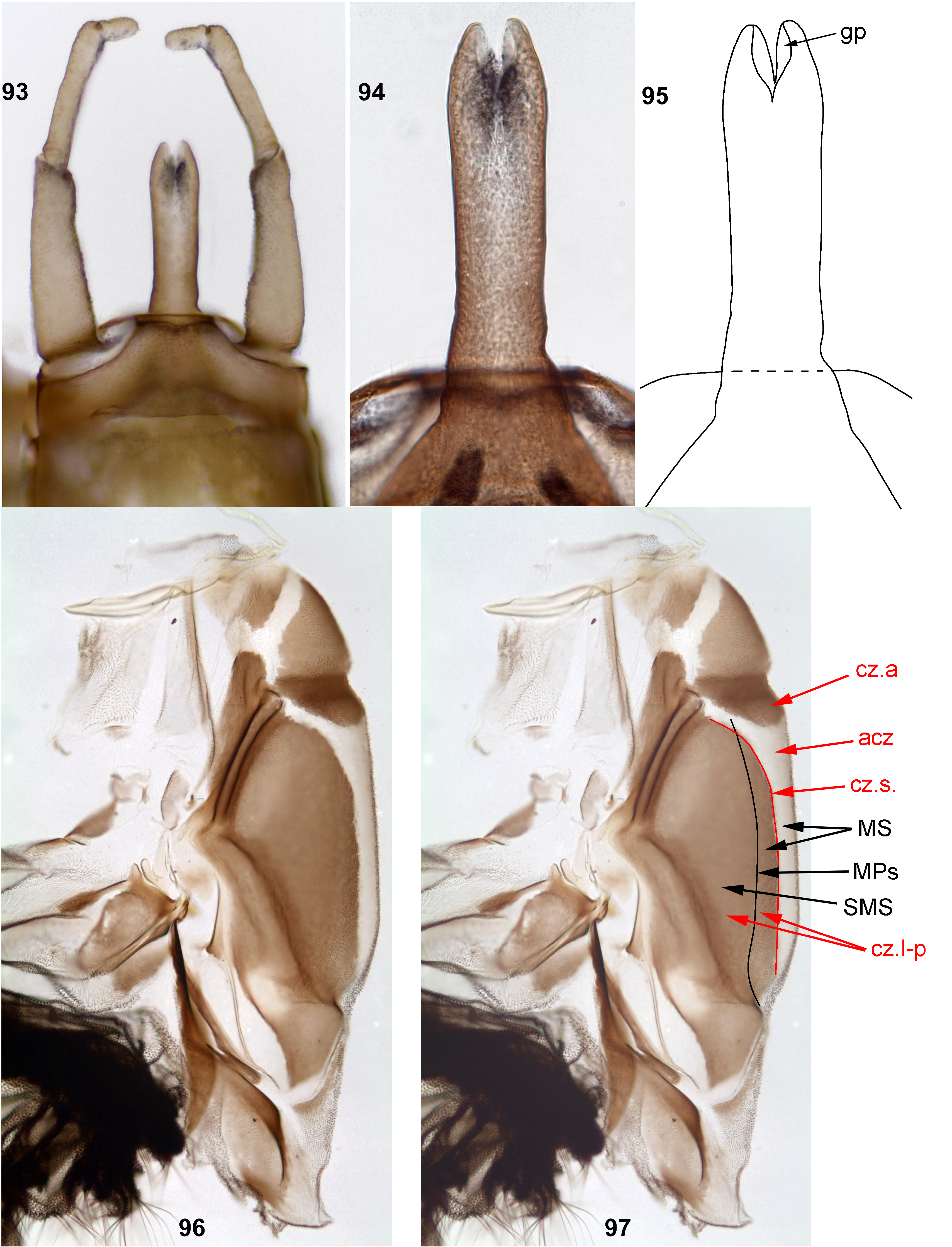
Subimago. CUTICULAR COLORATION. Pronotum brown. Mesonotum with anterior scutal chromozone and latero-posterior scutal chromozones dark brown, achromozone contrastingly colorless ( Figs 96–97 View FIGURES 93–97 ). Thoracic pleura and sterna with light brown and colorless areas. Wings brown. On all legs, femur with longitudinal dark brown and colorless bands; tibia with base brown ( Figs 87–88 View FIGURES 82–88 ). Abdominal terga brown, sterna lighter. Cerci light brown.
HYPODERMAL COLORATION. As in imago.
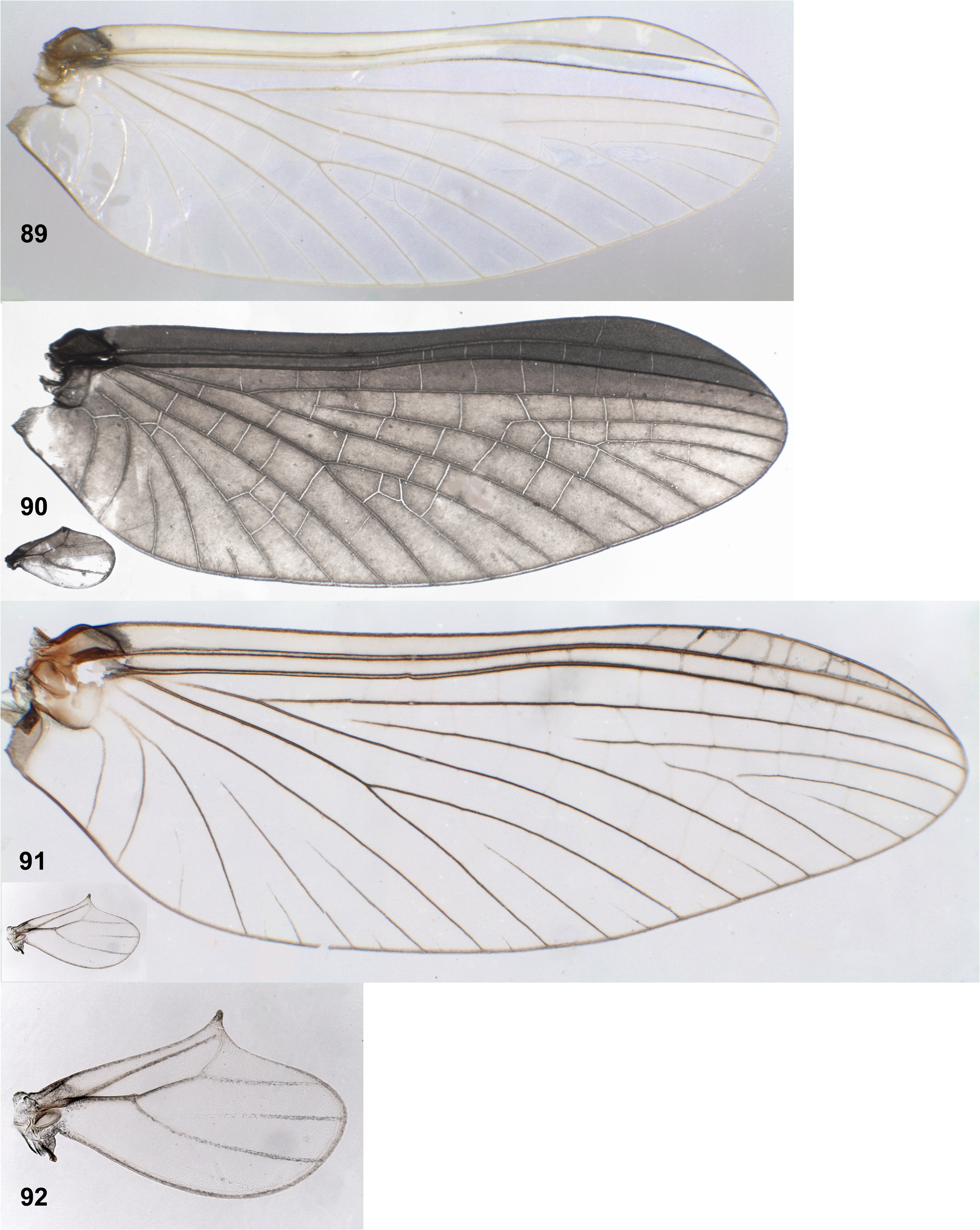
Imago, male ( Fig. 82 View FIGURES 82–88 ). Head brown. Dorsal eyes reddish. Thorax brown with membranes ochre. Base of fore wing including costal brace, dark brown; remainder membrane colorless, costal and subcostal fields whitish; veins C, Sc and RA partly colorless, partly bordered with dark brown, other longitudinal veins mostly ochre; crossveins colorless ( Figs 89 View FIGURES 89–92 ); crossveins of pterostigma simple and complete ( Fig. 90 View FIGURES 89–92 ). Hind wing widest in distal half, with costal projection near midlength; base dark brown, other membrane colorless, veins ochre (as in Fig. 92 View FIGURES 89–92 ). On foreleg, femur dark brown, proximally lighter; tibia mostly ochre, with dark brown base and apex; tarsus mostly ochre, with dark brown apex ( Fig. 85 View FIGURES 82–88 ). On middle and hind legs, femur mostly ochre with two diffusive, longitudinal brown markings and extreme apex dark brown; tibia entirely ochre; tarsus mostly ochre, with dark brown apex ( Fig. 86 View FIGURES 82–88 ). Abdominal terga mostly brown, with darker brown, light ochre and grayish maculae; paired ochre maculae largest on terga VI –VII ( Fig. 84 View FIGURES 82–88 ). Abdominal sterna ochre, with indistinctly expressed lighter sigilla. Styliger, gonostyli and penis ochre, bordered with brown. Penis longer than 1st segment of gonostylus, slightly widened subapically; apically V-shaped cleft with pair of gonopores exposed medially-dorsally ( Figs 93–95 View FIGURES 93–97 ). Cerci ochre, basally dark brown ( Fig. 82 View FIGURES 82–88 ).
Imago, female. Coloration of head, thorax, legs, abdomen and cerci as in male ( Fig. 83 View FIGURES 82–88 ). On fore wing, longitudinal veins brown; crossveins in pterostigma light brown, other crossveins colorless ( Fig. 91 View FIGURES 89–92 ).
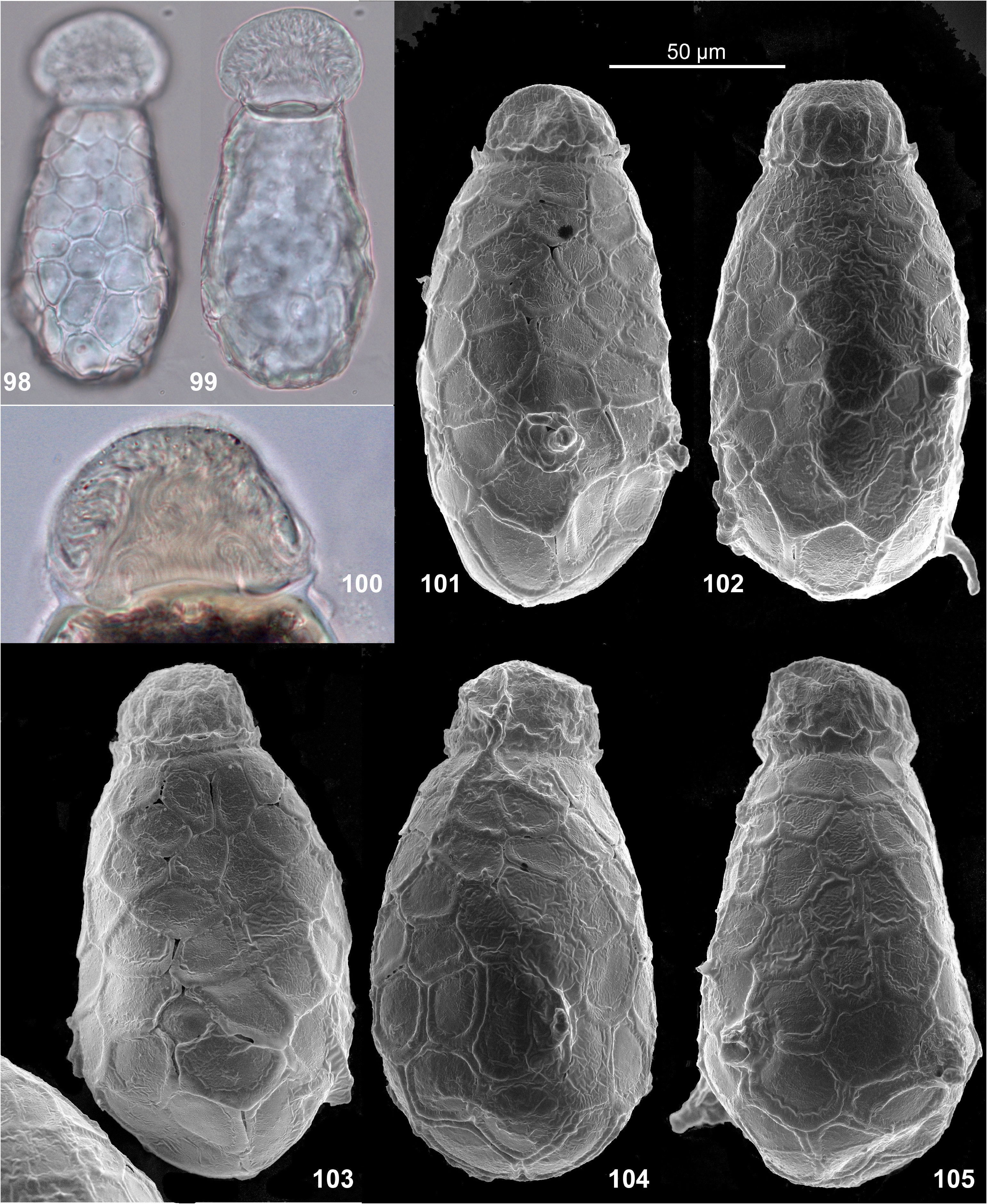
Egg ( Figs 98–105 View FIGURES 98–105 ). Oval, thinner near cap-bearing pole and thicker near cap-free pole. Whole surface except cap-bearing pole, covered with polygonal plates and round plates with vestiges of attachment structures in center; plates separated either by thickenings, or by grooves bordered by thickenings, mostly not overlapping one another. Polar cap surrounded by crown of low, pointed denticles; sometimes denticles sparse or absent.
Synonymy of T. dentata and T. sartorii . The original description of Teloganodes dentata was based on a male imago (Navás 1932); the holotype was redescribed ( Sartori et al. 2008). The original description of Teloganodes sartorii was based on larvae ( Selvakumar et al. 2014). Rearing imagines from larvae reveals that these imago and larvae are conspecific.
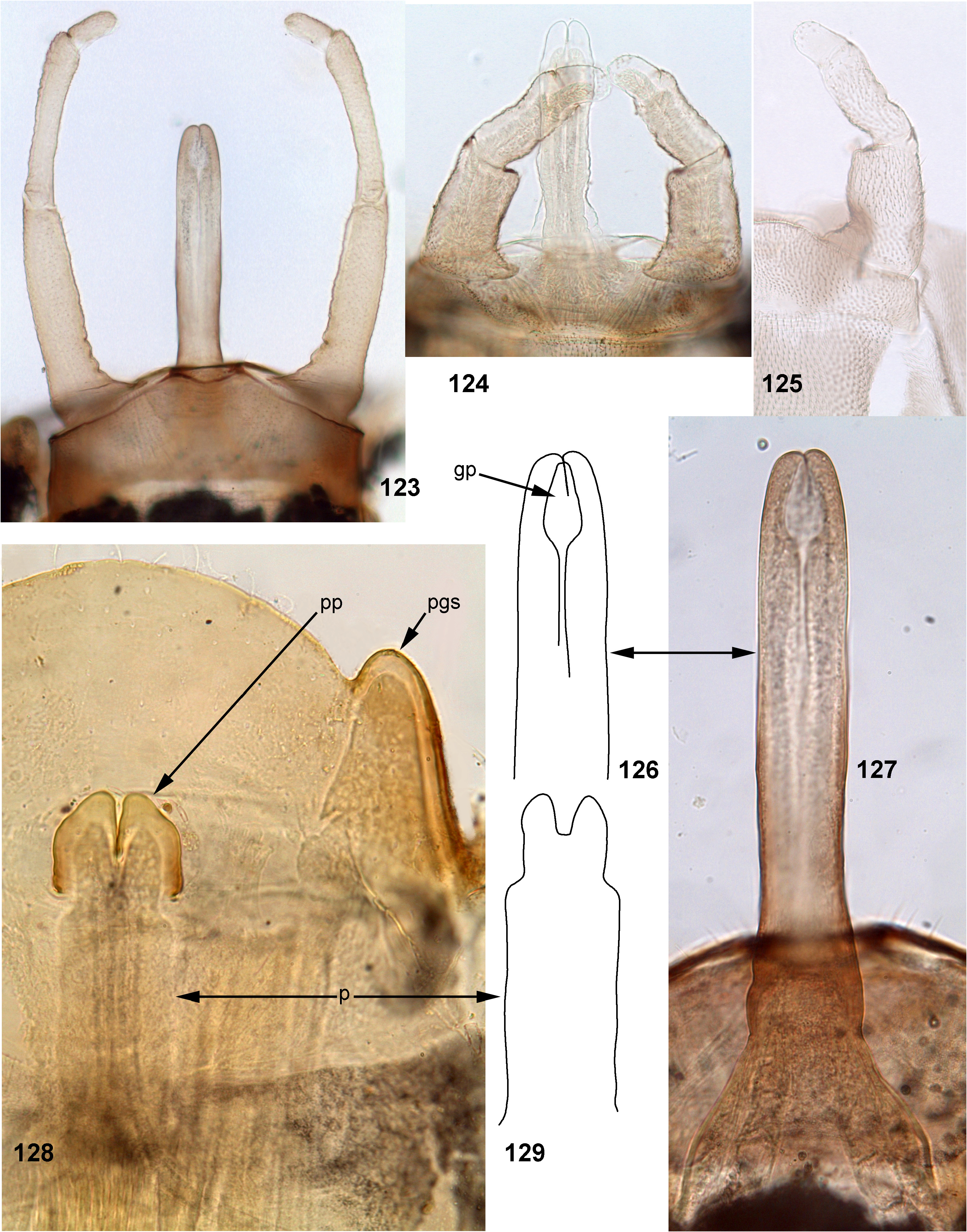
Comment: male genitalia in larval stage. As in other examined species of Teloganodes , larval protopenis is short, independently from size and shape of the imaginal penis ( Fig. 73 View FIGURES 73–76 ); developing subimaginal penis is located under the larval cuticle in such a method, that its small apical portion is located inside the cuticle of larval protopenis, and remainder part of penis is spread, located proximad of the protopenis and is sharply wider than the protopenis; this gives the developing penis a peculiar shape (as in Figs 128–129 View FIGURES 123–129 ) different from the shape of imaginal penis ( Figs 93–95 View FIGURES 93–97 ). Because of this, structure of larval genitalia does not help to associate larvae and male imagines.
| ZIN |
Russian Academy of Sciences, Zoological Institute, Zoological Museum |
| VI |
Mykotektet, National Veterinary Institute |
| V |
Royal British Columbia Museum - Herbarium |
No known copyright restrictions apply. See Agosti, D., Egloff, W., 2009. Taxonomic information exchange and copyright: the Plazi approach. BMC Research Notes 2009, 2:53 for further explanation.
|
Kingdom |
|
|
Phylum |
|
|
Class |
|
|
Order |
|
|
Family |
|
|
Genus |
Teloganodes (Teloganodes) dentatus Navás 1932
| Kluge, Nikita, Srinivasan, Pandiarajan, Sivaruban, T., Barathy, S. & Isack, Rajasekaran 2023 |
Teloganodes sartorii
| Selvakumar, C. & Sivaramakrishnan, K. G. & Jacobus, L. M. & Janarthanan, S. & Arumugam, M. 2014: 94 |
Teloganodes dentatus: Sartori, Peters & Hubbard 2008: 11
| Sartori, M. & Peters, J. G. & Hubbard, M. D. 2008: 11 |