
Rhinophis gunasekarai, Wickramasinghe & Vidanapathirana & Wickramasinghe & Gower, 2020
|
publication ID |
https://doi.org/ 10.11646/zootaxa.4810.1.3 |
|
publication LSID |
lsid:zoobank.org:pub:F7161842-821F-486A-B12D-FEDC44640CC3 |
|
persistent identifier |
https://treatment.plazi.org/id/F53287CA-FFB7-FFA7-D5BB-FDDE19C8FB67 |
|
treatment provided by |
Plazi |
|
scientific name |
Rhinophis gunasekarai |
| status |
sp. nov. |
Rhinophis gunasekarai sp. nov.
( Figs. 1–10 View FIGURE 1 View FIGURE 2 View FIGURE 3 View FIGURE 4 View FIGURE 5 View FIGURE 6 View FIGURE 7 View FIGURE 8 View FIGURE 9 View FIGURE 10 ; Table 1)
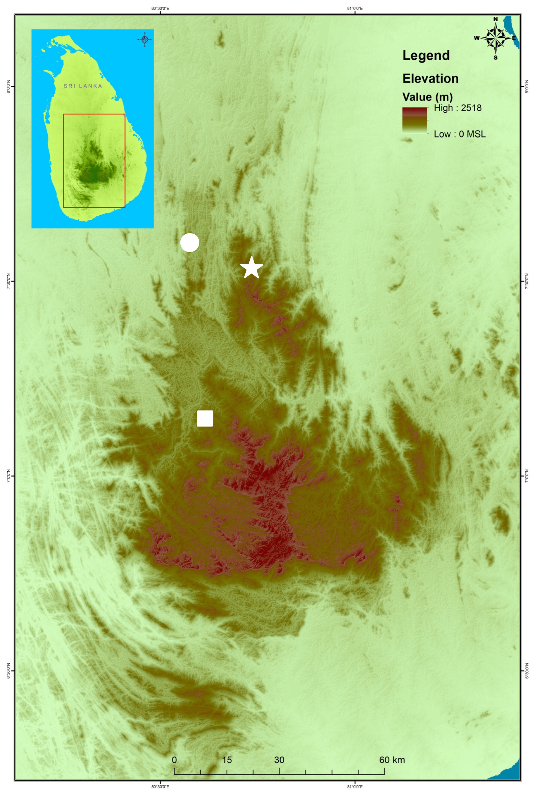
Holotype. NMSL-NH 2020.05.01, male, SVL 258 mm ( Figs. 2–4 View FIGURE 2 View FIGURE 3 View FIGURE 4 ), collected from Riverstone , Knuckles Massif, Matale District, Central Province, Sri Lanka (07°31’39” N, 80°44’01” E, elevation 1420 m), by L.J.M. Wickramasinghe and D. R. Vidanapathirana, on 28 September 2018. GoogleMaps
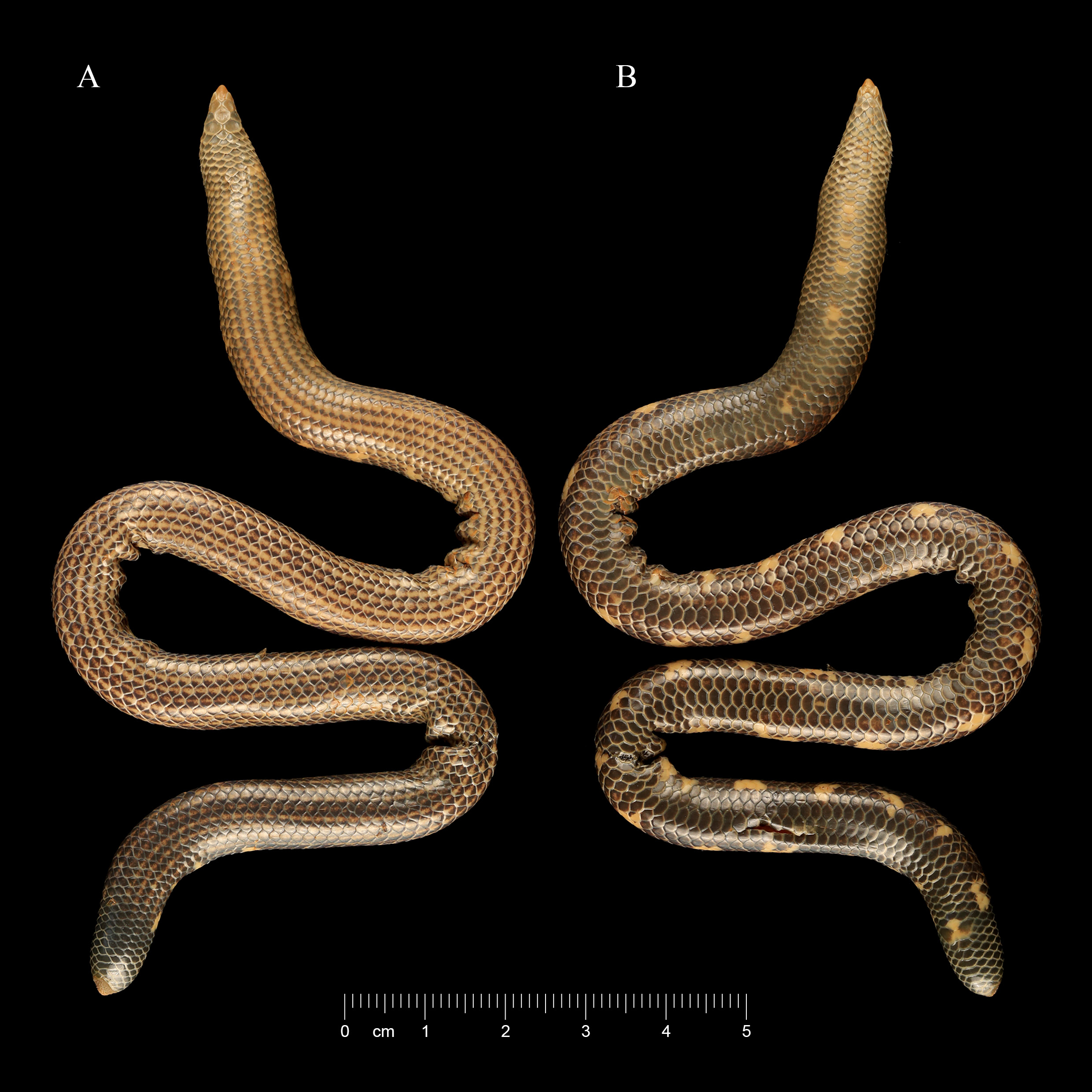
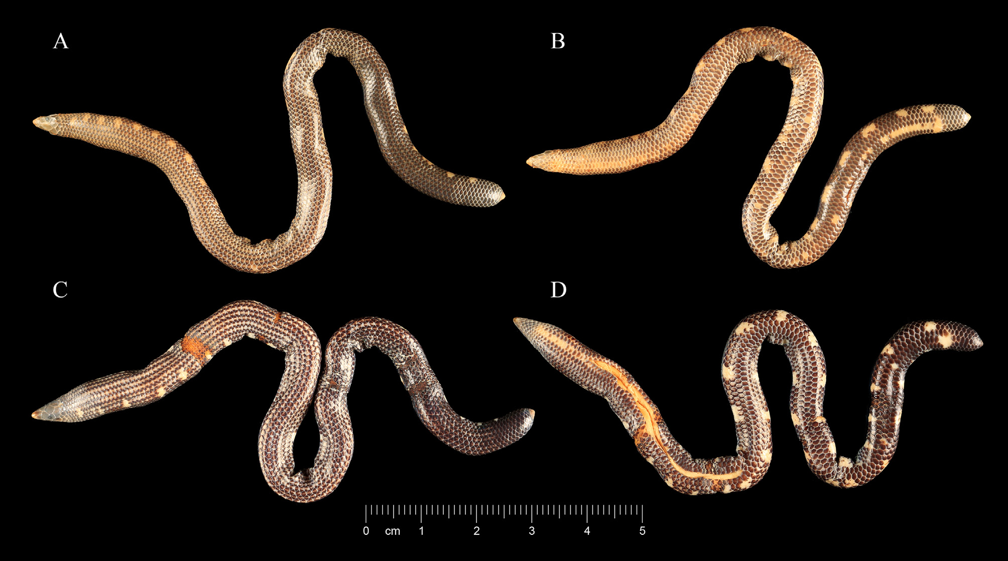
Paratypes (n =2). Males, NMSL-NH 2020.05.02, SVL 143 mm ( Fig. 5A&B View FIGURE 5 ); DWC 2020.05.01, SVL 154 mm ( Fig. 5C&D View FIGURE 5 ); collection details same as holotype .
Referred specimen (n =1). NMSL uncatalogued (previously Wildlife Heritage Trust) WHT 5786. This is considered a referred rather than type specimen because we are doubtful of the locality data recorded in the draft, handwritten WHT catalogue available in the NMSL. This specimen is recorded in this catalogue as a R. homolepis from Pussellawa collected by M. Bahir and Krishan in 2002, but Pussellawa is in the biogeographically distinct Central Hills rather than the Knuckles Massif ( Fig. 1 View FIGURE 1 ), and we doubt that the collectors would mistake the specimen for R. homolepis based on their very different tail shield and colour pattern. Furthermore, although the specimen is similar to the three types of R. gunasekarai sp. nov., it is likely a female (having only five rather than seven or eight subcaudals) but has a very similar number of ventrals (180) to the male types, whereas in the most similar congeners females typically have more ventrals than males.
Diagnosis. The new species is distinguished from its congeners by the combination of 17 dorsal scale rows at midbody; 177–182 ventrals ( Figs. 3B View FIGURE 3 , 5B&D View FIGURE 5 , 6 View FIGURE 6 ); a small to moderate tail shield covered with small homogenous spines in an approximately radial distribution; 17 yellow, longitudinal lines from slightly more than one head length behind head to level with vent, around body except on ventral scale row; dorsal, lateral and ventral regions of tail entirely blackish, without markings; a single irregular and uneven row of blotches on the ventrolateral surface of the body, visible in ventral view except on the anteriormost region of the body.
Rhinophis gunasekarai sp. nov. most closely resembles R. phillipsi ( Nicholls, 1929) , sharing dorsal yellow lines and lateral yellow blotches, and a relatively small tail shield. However, the new species differs from R. phillipsi in having 182 or fewer ventrals (vs 197 or more), lacking yellow lines on the tail (vs present), lacking two larger and notably horizontally projected spines on the tail shield (vs present), having more than 7 yellow lines along the body (vs 7 yellow lines), and in the yellow blotches on the body being visible in ventral aspect (vs not visible).
Among other Sri Lankan congeners, R. gunasekarai sp. nov., differs from R. saffragamus ( Kelaart, 1853) by having a small, domed tail shield (vs large and flat), nasal shields separated by rostral (vs nasal shields in contact, behind rostral), and by having dorsal scales in 17 rows (vs 19); from R. dorsimaculatus Deraniyagala, 1941 , R. oxyrhynchus ( Schneider, 1801) , R. porrectus Wall, 1921 , R. punctatus Müller, 1832 and R. zigzag Gower & Maduwage, 2011 , by having 200 or fewer ventrals (vs more than 207), and additionally from the former four species by having a moderate-sized tail shield (vs relatively much larger tail shield); from R. lineatus Gower & Maduwage, 2011 , by the presence of a row of blotches on the ventrolateral margin of the body (vs absent), absence of an irregular yellow lateral line on the tail (vs present), a relatively shorter distance between rostral and frontal scales (18–20% of rostral length vs 0–14%), and perhaps by having fewer ventrals (177–182 vs 180–195 for known material); from R. blythii Kelaart, 1853 , R. erangaviraji Wickramasinghe, Vidanapathirana, Wickramasinghe & Ranwella, 2009 , R. melanogaster (Gray, 1858) , and R. tricoloratus Deraniyagala, 1975 , in having 170 or more ventrals (vs fewer than 170) a moderate-sized tail shield (vs much larger tail shield), and from R. melanogaster by the absence of two notably large, horizontally projected spines on the tail shield (vs present).
The ventral scale count in R. gunasekarai sp. nov. is similar to or overlapping with that of R. drummondhayi Wall, 1921 , R. homolepis ( Hemprich, 1820) , and R. philippinus ( Cuvier, 1829) . However, the new species differs from R. drummondhayi in having (vs lacking) longitudinal narrow lines on the dorsum, lacking pale markings on the lateral surface of the tail (vs present), and in having a narrow (vs relatively much wider) tail shield; from R. homolepis by the absence (vs presence) of a conspicuous yellow band (1–2 scales wide) around the tail base and by a much less protuberant tail shield; and from R. philippinus by the presence (vs absence) of pale longitudinal lines on the upper surface of the body, and by its much smaller and less protuberant tail shield.
In addition to its colour pattern, Rhinophis gunasekarai sp. nov. differs from all five Indian species of Rhinophis by having a smaller and less protuberant tail shield. It differs further in having 177–182 ventrals (vs more than 200 in R. goweri Aengals & Ganesh, 2013 ; 195 in R. fergusonianus Boulenger, 1896 ; 150 or fewer in R. travancoricus Boulenger, 1893 ; 218–236 in R. melanoleucus Cyriac, Narayan, Sampaio, Umesh & Gower, 2020 ). Rhinophis gunasekarai sp. nov. also differs from R. sanguineus Beddome, 1863 , R. fergusonianus and R. melanoleucus by having 17 dorsal scale rows at or just behind midbody (vs 15).
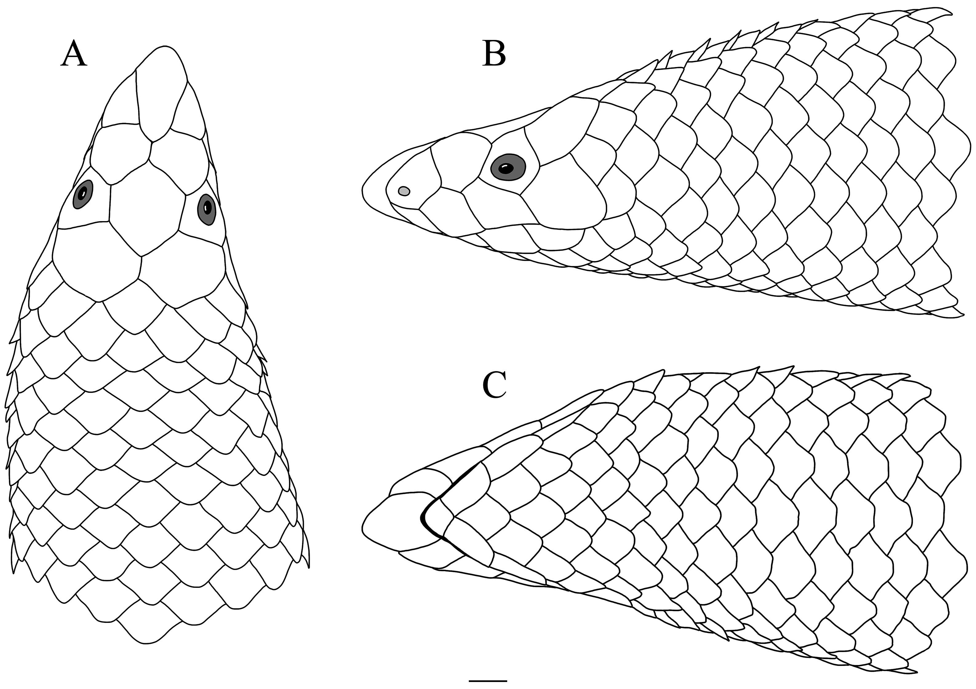
Description of holotype. A well preserved specimen in generally good condition. Somewhat dehydrated in parts including head. There is a small ventral incision into the coelom. Head small, snout pointed ( Fig. 4 View FIGURE 4 ). Rostral pointed, longer than wide, without dorsal crest, widest at level of anterior superior corner of first supralabials, 4.6 times longer (in dorsal view) than rostral-frontal gap. Frontal slightly longer and wider than rostral, irregularly hexagonal, longer than wide, lateral (ocular) margins slightly converging posteriorly, posterolateral margins straight, lateral (ocular) margin shortest, posterolateral edges longest ( Fig. 4A View FIGURE 4 ). A pair of nasals, separated from each other by posterior half of rostral; external naris small, subcircular, slightly countersunk within small depression, located in anteroventral corner of nasal; nasal in contact with first and second supralabials ( Fig. 4B View FIGURE 4 ). Prefrontals (for most of their length) in contact with each other along midline (left overlapping right), separating frontal from rostral. Prefrontals wider than long, shorter than frontal ( Fig. 4A View FIGURE 4 ). Four supralabials, first smallest, making least contribution to margin of mouth; fourth distinctly largest. Ocular in contact with third and fourth supralabials. Eye distinct, diameter approximately one third length of ocular, located near anteroventral corner of ocular, bulging slightly from ocular profile, pupil circular ( Fig. 4B View FIGURE 4 ). Paired parietals wider than long, shorter and wider than frontal, posteriorly broadly rounded, angle between posteromedial and posterolateral edges approximately 90°. Opposite parietals in brief midline contact, left overlapping right. Each parietal contacting four scales other than head shields ( Fig. 4A View FIGURE 4 ). No mental groove; mental wider than long, smaller than infralabials, contacting first infralabials and single postmental (= first ventral). Three pairs of infralabials, second largest, third smallest. First to fourth ventrals longer than wide, fifth approximately as long as wide, sixth and subsequent ventrals wider than long ( Fig. 4C View FIGURE 4 ). Six maxillary and five mandibular teeth on each side. Teeth simple, pointed, distinctly retrorse, straight, evenly spaced.
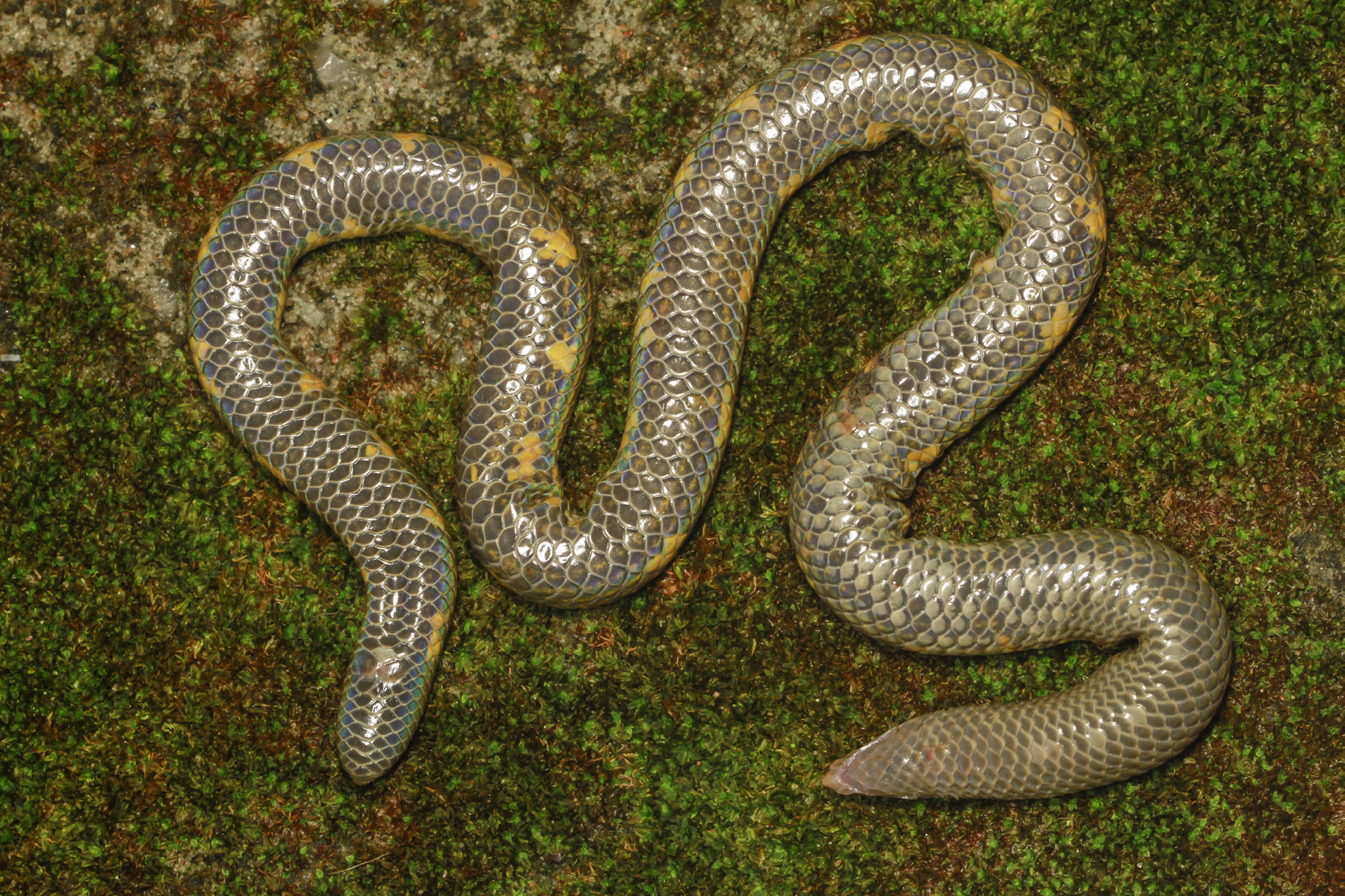
Body subcylindrical ( Figs. 2 View FIGURE 2 , 3 View FIGURE 3 & 6 View FIGURE 6 ). Body scales mostly evenly sized on dorsum and along body except for those involved in dorsal scale row reductions. Midline ventral scales between mental and anal of even size though anteriormost ones gradually narrow. Ventrals 182, posteriormost ventral notably smaller ( Figs. 3B View FIGURE 3 & 6 View FIGURE 6 ). Dorsal scale rows 19 anteriorly, reducing to 17 by level with 72 nd ventral and maintained along most of body; scale row reduction formula:
4 + 5 (72)
19 --------------- 17
4 + 5 (71)
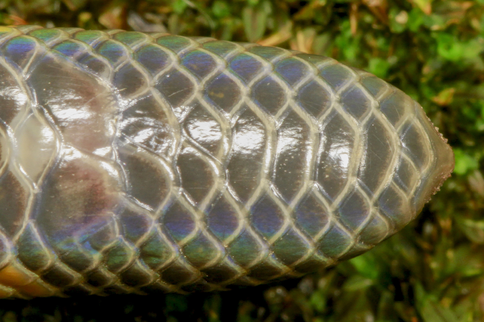
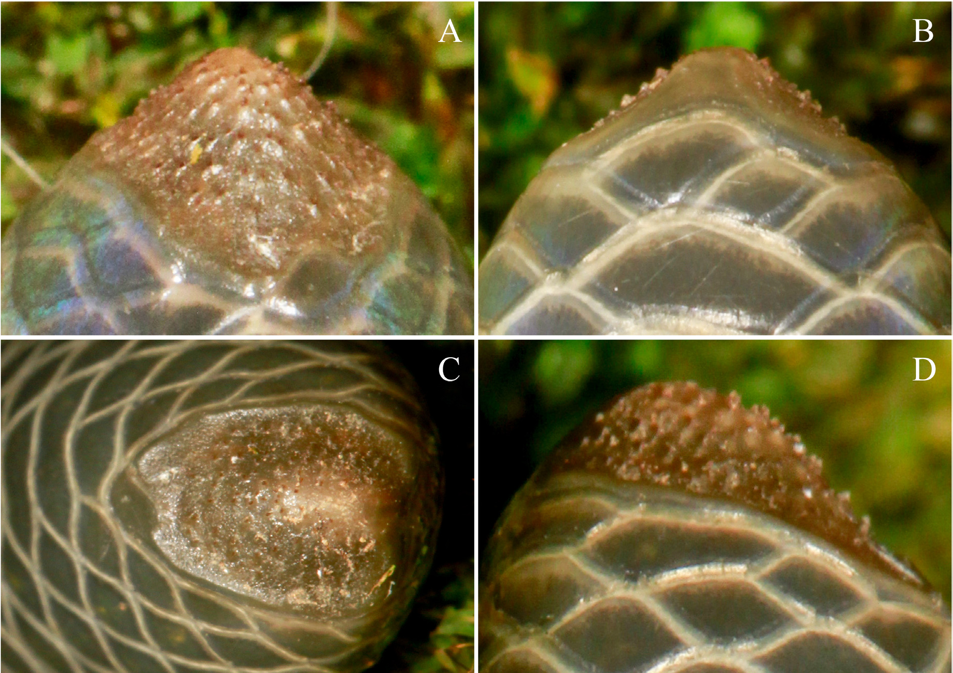
Dorsal scale rows approximately 12 at base of tail. Head, body and tail scales macroscopically smooth. Paired anal scales (right overlying left) considerably larger than posteriormost ventrals and subcaudals. Distal margin of each anal overlapping three other scales in addition to anteriormost subcaudals. Seven subcaudals, first, second and seventh paired ( Fig. 7 View FIGURE 7 ). Tail shield pointed in dorsal ( Fig. 8A View FIGURE 8 ) and ventral aspects ( Fig. 8B View FIGURE 8 ). In posterior view, tail shield suboval ( Fig. 8C View FIGURE 8 ), taller than wide ( Fig. 8D View FIGURE 8 ). Shield surface covered with small, more or less subequal spines in approximately radial distribution. Most of perimeter of shield with a thin, glossy, spineless margin ( Fig. 8 View FIGURE 8 ). Shield surrounded by 13 scales including last subcaudals.
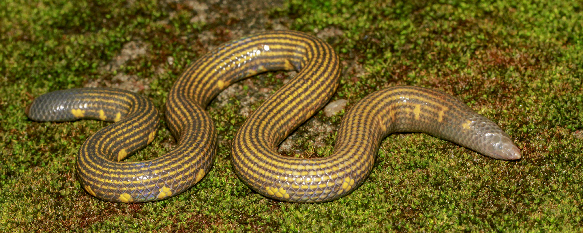
Coloration in life. Body background dark, blackish rather than the more greyish appearance of the somewhat dehydrated specimen at time of preservation. Seventeen yellow lines from approximately two head lengths behind snout tip to level of vent, distributed evenly around body excluding ventral scale row. Yellow lines narrow, running through centres of body scales ( Fig. 2 View FIGURE 2 ), most prominent dorsally and anteriorly, fading toward posterior and venter ( Figs. 6 View FIGURE 6 ). Single row of conspicuous yellow blotches on ventrolateral surface of body, beginning from two head lengths behind snout tip to level with vent. On right hand side (RHS), anteriormost blotch is on the 6 th and 7 th dorsal scale rows approximately level with 6 th vertebral scale; on the left hand side (LHS), anteriormost blotch is approximately level with the 7 th vertebral scale. Blotches irregularly arranged, uneven in size (1–3 scales wide; 2–3 scales tall), anteriormost two blotches more dorsally placed, gradually becoming more lateral in orientation (10 th blotch on the 4 th dorsal scale row, by mid-body on the 2 nd and 3 rd or on the 2 nd, 3 rd and 4 th, or on the 3 rd and 4 th dorsal scale rows). RHS with 35 blotches, 32 on LHS.
Five yellow blotches irregularly placed on anterior of venter midline, from 13 th to 36 th ventral scale ( Fig. 6 View FIGURE 6 ). Posteriormost ventral and centers of divided anal scales forming a conspicuous yellow mark. Dorsal ( Fig. 9 View FIGURE 9 ), lateral ( Fig. 10 View FIGURE 10 ) and ventral ( Fig. 7 View FIGURE 7 ) aspects of tail entirely dark with no pale markings. Tail shield brownish, paler towards apex.
Coloration in alcohol. The colouration in life is persists after 1.5 years in preservative, with a little fading ( Fig. 3 View FIGURE 3 ): with the blackish coloration becoming dark brown ( Fig. 3A View FIGURE 3 ), and yellow becoming off white ( Fig. 3B View FIGURE 3 ).
Variation among paratypes. See Table 1 for meristic and morphometric data. Condition generally good. DWC 2020.05.01, SVL 154 mm ( Fig. 5C&D View FIGURE 5 ), with some scales damaged by injury at three positions in region of mid-body (level with vertebral scales 36–41, 57–58 on LHS and 59–60 th on RHS) ( Fig. 5C View FIGURE 5 ). Longitudinal ventral incision into coelom from 20 th to 67 th ventral scale, to examine internal organs ( Fig. 5D View FIGURE 5 ) .
Both paratypes smaller than holotype. Scalation very similar to that of holotype ( Table 1); reduction from 19 to 17 dorsal scale rows occurring by 36 th ( DWC 2020.05.01) and 64 th ( NMSL-NH 2020.05.02) ventral. Colour pattern very similar to holotype, with minor variations. Paratypes less dehydrated than holotype, so body background colour more black than grey. DWC 2020.05.01 , has RHS:28 and LHS:25 blotches, the anteriormost blotch level with 5 th vertebral on RHS and 6 th on LHS ( Fig. 5C&D View FIGURE 5 ). NMSL-NH 2020.05.02 has RHS:31, LHS:29 blotches, the first blotch level with the 4 th vertebral on both sides ( Fig. 5A&B View FIGURE 5 ). Some yellow blotches in paratypes larger than in holotype, up to four scales wide. Some yellow blotches more dorsal than in holotype, up to dorsal scale rows 6–8 anteriorly on LHS in DWC 2020.05.01 .
Several anteriormost ventrals whitish in both paratypes ( Fig. 5B&5D View FIGURE 5 ), most of ventrals 6–14 in DWC 2020.05.01 ( Fig. 5B View FIGURE 5 ) and 4–26 in NMSL-NH 2020.05.02 ( Fig. 5D View FIGURE 5 ). All of right and part of left anal scale yellow in both paratypes. Last two and most of last 15 ventrals pale yellow in DWC 2020.05.01 and NMSL-NH 2020.05.02, respectively .
Etymology. The specific epithet is an eponym latinised as a noun in the genitive singular, honouring Samantha Gunasekara, a Sri Lankan conservationist, for his contributions to the field of biodiversity conservation, especially for his service in establishing a Biodiversity Protection Unit in the Department of Customs, Government of Sri Lanka.
Suggested vernacular names. Gunasekarage Thudulla, Gunasekaran nilakael pambu, Gunasekara’s sheildtail in Sinhala, Tamil, and English, respectively.
Distribution. We have encountered Rhinophis gunasekarai sp. nov. only from the type locality, within the Knuckles Conservation Forest, at 1,420 m elevation close to the top of a hill.
Ecology and natural history. In addition to the collection of the type specimens in September 2018, we made 12 other visits to the type locality from June 2018 to May 2019. During these visits we recorded eight additional sightings of R. gunasekarai sp. nov. in four visits in June, July and October (twice) 2018, within an area of less than 100 m 2. Rhinophis gunasekarai sp. nov. were found in areas of moist humus-rich soil under thin leaf litter in well-shaded areas of evergreen cloud forest densely wooded with trees (dominated by Syzigium sp. R.Br. ex Gaertn.) heavily covered in epiphytes. Three specimens were found within a circle of 50 cm radius on one visit. During the rains (in the months of May and June) the snakes were found mostly under rocks and logs, while in drier weather (from August to October) they were found by digging in soil at approximately 30–50 cm beneath the surface. Other fossorial reptiles seen during these visits to this locality were Aspidura ceylonensis (Günther, 1858) , A. desilvai Wickramasinghe, Bandara, Vidanapathirana & Wickramasinghe, 2019 , and Nessia bipes (Smith, 1935) .
Conservation. The type locality of R. gunasekarai sp. nov. lies within a protected area, which is a UNESCO World Heritage Site. However, the area is being impacted by the improvement of the Matale-Rattota-Riverstone- Ilukkubura (B 274) road from Rattota (10 km) to the Sera Ella Junction (35 km). The project includes surfacing the road with asphalt, construction of ‘U’ and ‘L’ type concrete drains, rehabilitation of damaged culverts and introduction of vehicle passing bays within the existing right of way. These may lead to further degradation of the habitat of this species. Beyond fragmentation of habitat, potential threats to R. gunasekarai s p. nov. related to road construction include pollution from bitumen, asphalt, cement and concrete, and the invasion of alien species such as Mimosa pigra which were not found in the area prior to the construction work.


No known copyright restrictions apply. See Agosti, D., Egloff, W., 2009. Taxonomic information exchange and copyright: the Plazi approach. BMC Research Notes 2009, 2:53 for further explanation.