
Enghoffosoma funda, Likhitrakarn, Natdanai, Golovatch, Sergei I. & Panha, Somsak, 2014
|
publication ID |
https://doi.org/ 10.11646/zootaxa.3811.4.4 |
|
publication LSID |
lsid:zoobank.org:pub:AE22B01B-B3FF-4B60-9452-A94DDDB20C2B |
|
DOI |
https://doi.org/10.5281/zenodo.6136570 |
|
persistent identifier |
https://treatment.plazi.org/id/F53487E6-FFCD-D80F-FF7E-FA06FC1A5C9C |
|
treatment provided by |
Plazi |
|
scientific name |
Enghoffosoma funda |
| status |
sp. nov. |
Enghoffosoma funda View in CoL sp. n.
Figs 14–16 View FIGURE 14 View FIGURE 15 View FIGURE 16
Holotype male ( CUMZ), Thailand, Sisaket Province, Kantharalak District, Sisaket Nature Resources Environment Office # 6, ca 180 m a.s.l., 14°39'20"N, 104°37'35"E, 0 8.10.2010, leg. N. Likhitrakarn.
Paratype. 1 female ( CUMZ), same data, together with holotype.
Name. To emphasize the slingshot-shaped solenomere; noun in apposition.
Diagnosis. This species seems to be especially similar to E. zebra sp. n., particularly in colour pattern, but differs in the width of the anterior segments, an entire, linguiform, sternal lobe between male coxae 4 and in the development of pleurosternal carinae (see also Key below).
Description. Length 29 mm (male) or 27 mm (female), width of midbody pro- and metazonae 2.95 and 3.5 mm (male) or 3.0 and 3.5 mm (female), respectively.
Coloration of live animals maroon to brownish with a pattern of contrasting light brown posterior halves of collum, metaterga, paraterga and epiproct, as well as light brown legs and antennae ( Fig. 14 View FIGURE 14 A); coloration in alcohol, after half a year of preservation, dark brown to brown, posterior halves of collum, metaterga, paraterga and epiproct mostly light brown to whitish ( Figs 14 View FIGURE 14 B–F & H), antennae, legs and venter light brown to whitish ( Figs 14 View FIGURE 14 B, C & E–J).
All characters as in E. zebra sp. n., except as follows.
Antennae long ( Fig. 14 View FIGURE 14 B), reaching body segment 4 (both sexes) when stretched dorsally. In width, head <segment 3 = 4 <2 <collum <segments 5–16 (male), or head <segment 2 = 3 = 4 <collum <segment 5–16 (female), gently and gradually tapering thereafter. Collum with three transverse rows of setae: 5+ 5 in anterior, 3+ 3 in intermediate, and 4+ 4 in posterior row; caudal corner of paraterga very broadly rounded, declined, extending behind rear margin ( Figs 14 View FIGURE 14 B & C).
Tegument smooth and shining, prozonae finely shagreened, metaterga smooth and leathery; surface below paraterga finely microgranulate ( Figs 14 View FIGURE 14 B–F & H). Postcollum metaterga with two transverse rows of setae traceable at least as insertion points when setae broken off: 4+ 4 in anterior (pre-sulcus), 5+ 5 in posterior (postsulcus) row, caudal row barely traceable as insertion points. Axial line visible. Paraterga rather well-developed ( Figs 14 View FIGURE 14 B–F & H), mostly slightly upturned, all lying faintly below dorsum, set at about half of midbody height, subhorizontal, caudal corner rounded, mostly produced beyond rear tergal margin until segment 15, increasingly rounded and smaller on following segments; posterior edge nearly straight. Paraterga 2 broad, anterior edge angular, lateral edge without incision ( Figs 14 View FIGURE 14 B & C). Transverse sulcus at most very faint and abbreviated ( Figs 14 View FIGURE 14 B–F). Stricture between pro- and metazonae rather narrow, deep, line-shaped, beaded at bottom down to base of paraterga ( Figs 14 View FIGURE 14 B–E). Pleurosternal carinae strongly developed, complete crests with a very sharp caudal tooth in segments 2–7 (male) or 2–4 (female), small sharp caudal tooth in segments 8–16 (male) or 5–11 (female), absent from segment 17 (male) or 11 (female) ( Figs 14 View FIGURE 14 C, E & H). Epiproct ( Figs 14 View FIGURE 14 F & H) conical, flattened dorsoventrally, with two small apical papillae; tip subtruncate; pre-apical papillae rather small, but visible, lying rather close to tip.
Sterna very densely setose, with a small cone caudally near each coxa; an entire, linguiform, sternal lobe between male coxae 4 ( Figs 14 View FIGURE 14 I & J). Legs moderately long and slender, midbody ones ca 1.2–1.3 (male) or 1.1–1.2 times (female) as long as body height; ventral brushes traceable on male tibiae and tarsi until legs of segment 8 and 10, respectively.
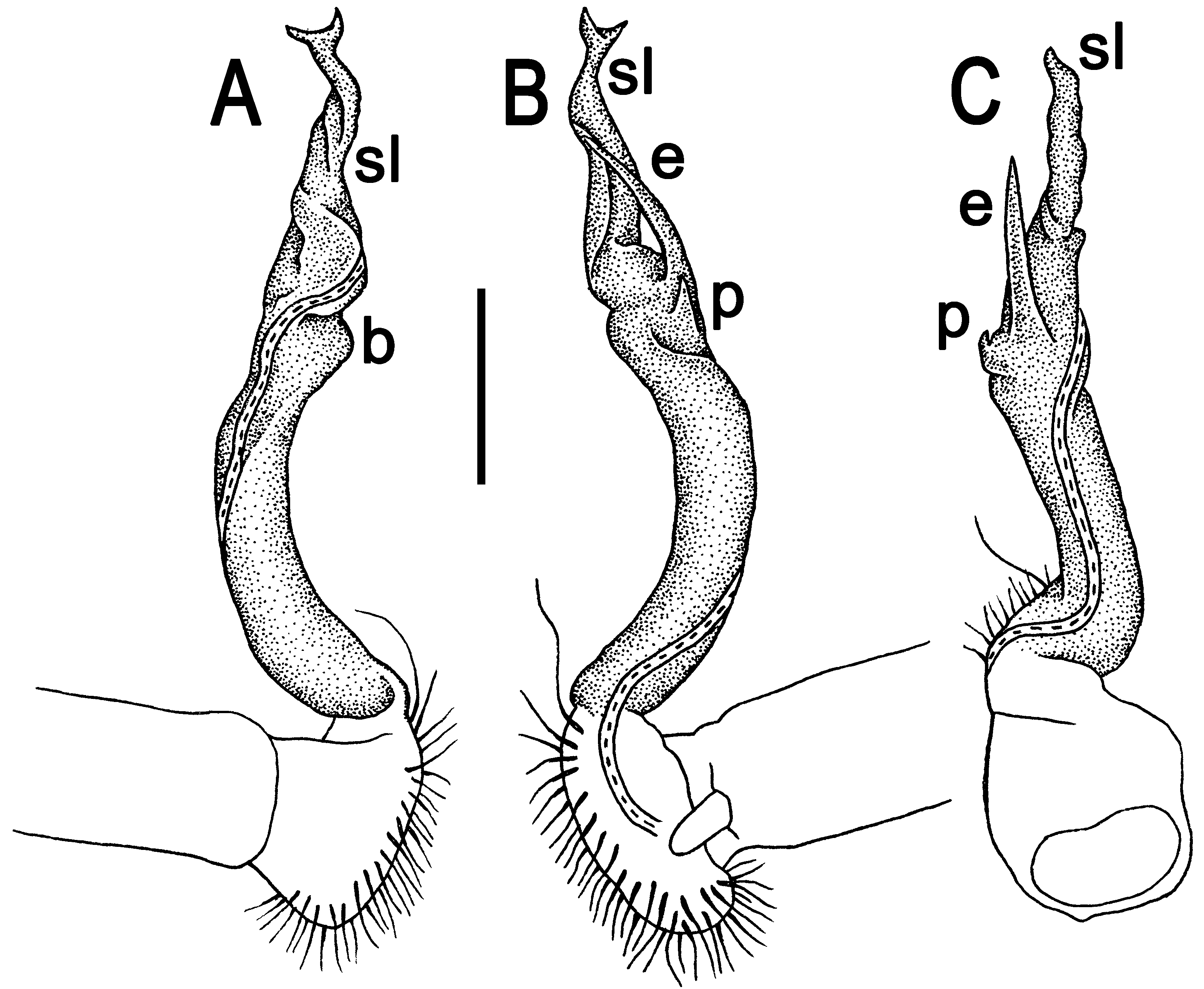
Gonopods ( Figs 15 View FIGURE 15 & 16 View FIGURE 16 ) rather simple. Femorite about 3 times as long as prefemoral (= strongly setose) part. Femorite long and slender, slightly curved; seminal groove first running mesally to turn laterad in distal half of femorite, with an evident apicoventral shelf (b) and a small, ventral, tooth-shaped spinicle (p) at base of e ( Figs 16 View FIGURE 16 B & C). “Postfemoral” portion demarcated by a distinct lateral sulcus. Process e spiniform, prominent, pointed, long and slender. Solenomere (sl) twisted, suberect, bifid, slingshot-shaped.


| CUMZ |
Chulalongkorn University Museum of Natural History |
No known copyright restrictions apply. See Agosti, D., Egloff, W., 2009. Taxonomic information exchange and copyright: the Plazi approach. BMC Research Notes 2009, 2:53 for further explanation.