
Enghoffosoma bispinum, Likhitrakarn, Natdanai, Golovatch, Sergei I. & Panha, Somsak, 2014
|
publication ID |
https://doi.org/ 10.11646/zootaxa.3811.4.4 |
|
publication LSID |
lsid:zoobank.org:pub:AE22B01B-B3FF-4B60-9452-A94DDDB20C2B |
|
DOI |
https://doi.org/10.5281/zenodo.6136574 |
|
persistent identifier |
https://treatment.plazi.org/id/F53487E6-FFD2-D80A-FF7E-FA21FAB35C9C |
|
treatment provided by |
Plazi |
|
scientific name |
Enghoffosoma bispinum |
| status |
sp. nov. |
Enghoffosoma bispinum View in CoL sp. n.
Figs 17–19 View FIGURE 17 View FIGURE 18 View FIGURE 19
Holotype male ( CUMZ), Thailand, Rayong Province, Mueang Rayong District, Kaosab temple, 9 m a.s.l., 12°36'48"N, 101°23'23"E, 10.11.2013, leg. S. Panha and N. Likhitrakarn.
Paratypes. 3 males, 3 females ( CUMZ), 1 male, 1 female ( ZMUC), 1 male, 1 female ( ZMUM), 1 male, 1 female ( NHMW), same data, together with holotype.
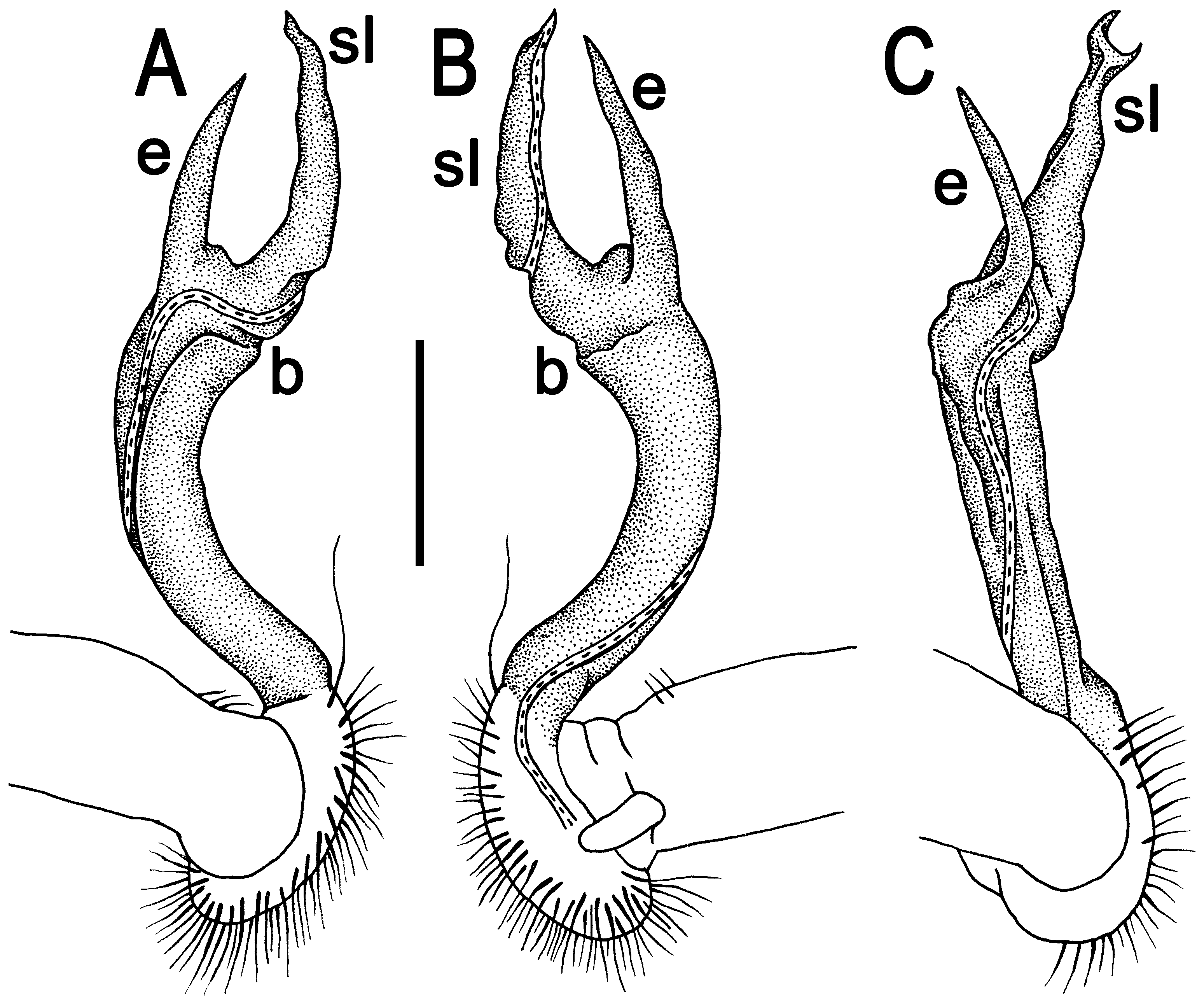
Name. To emphasize both gonopod process e and the solenomere (sl) being prominent and spiniform; adjective.
Diagnosis. Differs by mostly strongly developed paraterga, coupled with process e and the solenomere (sl) being prominent and spiniform (see also Key below).
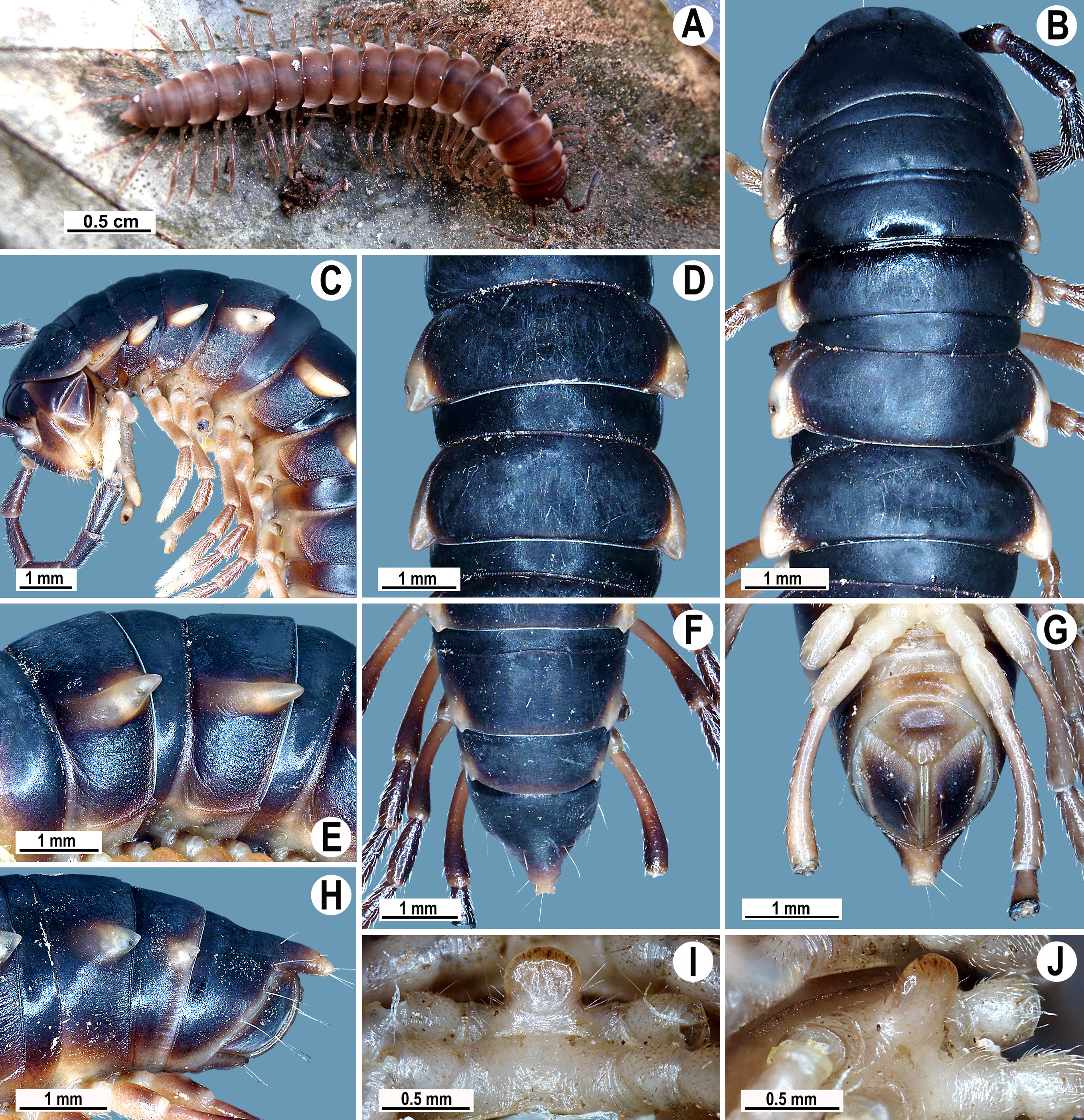
Description. Length 22–28 mm (male) or 26–33 mm (female), width of midbody pro- and metazonae 2.7–3.1 and 3.4–4.1 mm (male), 3.2–4.0 and 4.0– 4.8 mm (female), respectively.
Coloration of live animals blackish to brown with a pattern of contrasting light brown to whitish paraterga and epiproct ( Fig. 17 View FIGURE 17 A); coloration in alcohol, after half a year of preservation, blackish to light brown with a pattern of contrasting light brown to whitish paraterga and epiproct ( Figs 17 View FIGURE 17 B–F & H); antennae and legs blackish to light brown, but venter and a few basal podomeres light brown to whitish ( Figs 17 View FIGURE 17 B, C & F–J).
All characters as in E. zebra sp. n., except as follows.
Antennae long ( Fig. 17 View FIGURE 17 A), clavate, extending behind body segment 4 (male) or 3 (female) when stretched dorsally. In width, head <segment 3 = 4 <collum <segment 2 <5–14 (both sexes), gently and gradually tapering thereafter. Collum with three transverse rows of setae: 4+ 4 in anterior, 3+ 3 in intermediate, and 5+ 5 in posterior row; caudal corner of paraterga very broadly rounded, declined, extending behind rear margin ( Figs 17 View FIGURE 17 B & C).
Tegument smooth and leathery, prozonae finely shagreened, metaterga smooth and leathery, delicately rugulose; surface below paraterga finely microgranulate ( Figs 17 View FIGURE 17 B–F & H). Postcollum metaterga with two transverse rows of setae traceable at least as insertion points when setae broken off: 2+ 2 in anterior (pre-sulcus), 3+ 3 in posterior (post-sulcus) row, anterior row usually with retained setae, caudal row barely traceable as insertion points. Tergal setae long, strong, slender, about 1/3 as long as metaterga. Axial line visible on both pro- and metazonae. Paraterga very strongly developed ( Figs 17 View FIGURE 17 B–F & H), especially so in male, mostly slightly upturned, all lying below dorsum, set at about upper 1/3 of body height, subhorizontal, caudal corner almost or fully pointed, largely produced beyond rear tergal margin until segment 15, increasingly rounded and smaller on following segments; posterior edge concave ( Figs 17 View FIGURE 17 B–F). Paraterga 2 broad, anterior edge angular to rounded, lateral edge without incision ( Figs 17 View FIGURE 17 B & C). Transverse sulcus at most very faint and abbreviated ( Figs 17 View FIGURE 17 B, D & F). Stricture between pro- and metazonae narrow, deep, line-shaped, clearly beaded at bottom down to base of paraterga ( Figs 17 View FIGURE 17 B–F). Pleurosternal carinae strongly developed, complete crests with a very sharp caudal tooth in segments 2–13 (male) or 2–12 (female), a small sharp caudal tooth in segments 14–17 (male) or 13–16 (female), absent thereafter ( Figs 17 View FIGURE 17 C, E & H).
Sterna very densely setose, with an evident cone caudally near each coxa, rear cones being a little stronger than front ones; a rather large, linguiform, densely setose sternal lobe between male coxae 4 ( Figs 17 View FIGURE 17 I & J). Legs moderately long and slender, midbody ones ca 1.1–1.2 (male) or 0.9–1.0 times (female) as long as body height; ventral brushes traceable on male tibiae and tarsi until legs of segment 8 and 10, respectively.
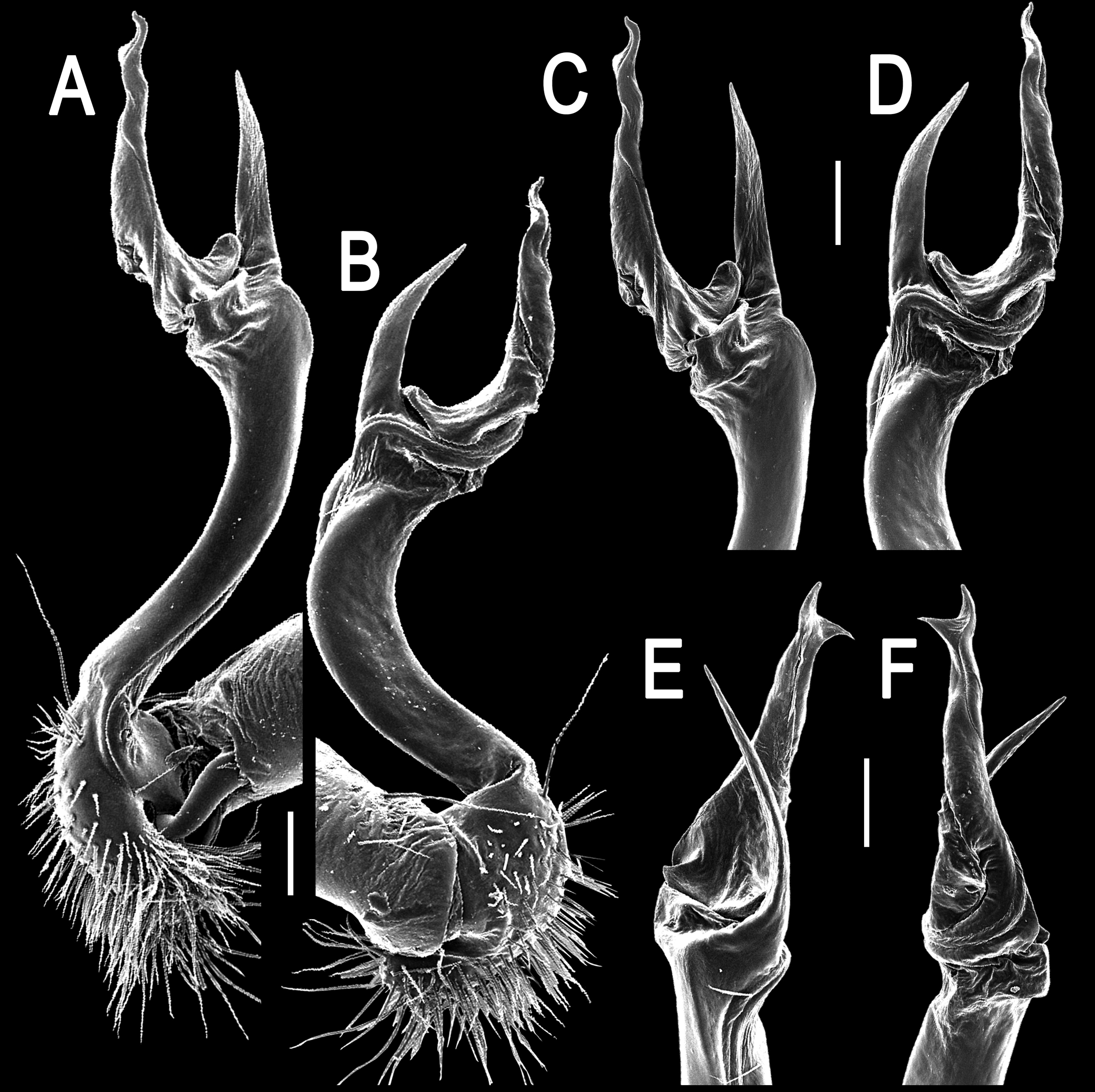
Gonopods ( Figs 18 View FIGURE 18 & 19 View FIGURE 19 ) simple. Femorite slender and long, slightly curved, with seminal groove running mesally, but turning laterad already in distal half of femorite; apicoventral shelf (b) evident, both process e and solenomere (sl) large, spiniform, slightly curved, but sl longer and bifid.
Remarks. The specimens were collected hidden under leaf litter, in a rotten log and even under roof tiles near a monk’s house in a temple. Some of the animals were still colourless and soft-bodied shortly after molting.
No known copyright restrictions apply. See Agosti, D., Egloff, W., 2009. Taxonomic information exchange and copyright: the Plazi approach. BMC Research Notes 2009, 2:53 for further explanation.